Marília Pacífico Lucisano(a) Paulo Nelson-Filho(a) Leslie Morse(b) Ricardo Battaglino(c)
Plauto Christopher Aranha Watanabe(d)
Raquel Assed Bezerra da Silva(a) Lea Assed Bezerra da Silva(a)
(a) Department of Pediatric Clinics, Preventive and Community Dentistry, School of Dentistry of Ribeirão Preto, Univ de São Paulo - USP, Ribeirão Preto, SP, Brazil. (b) Department of Physical Medicine and
Rehabilitation, School of Medicine, Harvard Univ, Boston, MA, USA.
(c) Department of Skeletal Biology, Forsyth Institute, Cambridge, MA, USA.
(d) Department of Morphology, Stomatology and Physiology, School of Dentistry of Ribeirão Preto, Univ de São Paulo - USP, Ribeirão Preto, SP, Brazil.
Corresponding Author: Paulo Nelson-Filho E-mail: [email protected]
Radiodensitometric and DXA analyses
for the measurement of bone mineral
density after systemic alendronate
therapy
Abstract: Precise techniques for the measurement of maxillary bone mineral density (BMD) are useful for the early diagnosis of systemic dis-eases. The aim of this study was to compare in vivo the eficacy of dual-energy x-ray absorptiometry (DXA) and radiographic densitometry for the measurement of BMD after systemic administration of sodium alen-dronate. Wistar rats were randomly allocated to a control group (n = 5), which received distilled water, and a sodium alendronate group (n = 8), which received two doses of chemically pure sodium alendronate (1 mg/ kg) per week. After 8 weeks, the animals were euthanized, the tibias were removed, and the BMD of the proximal tibial metaphysis was ana-lyzed radiographically and by DXA. The data were subjected to statisti-cal analysis by the Kruskal-Wallis test at a signiicance level of 5%. Both of the techniques revealed that the alendronate-treated group had a sig-niicantly higher BMD (p < 0.05) than the control group after 8 weeks of treatment. Comparing the groups with and without alendronate therapy revealed increases of 14.9% and 29.6% in BMD, as detected radiographi-cally and by DXA, respectively. In conclusion, both of the methods were able to detect an increase in BMD of the proximal tibial metaphysis after alendronate therapy.
Descriptors: Bone Density; Alendronate; Bone Remodeling.
Introduction
Osteometabolic disorders have recently been the subject of extensive research in the ield of bone biology. Special attention has been directed towards osteoporosis, which is one of the major health problems that af-fects postmenopausal women due to decreased estrogen levels secondary to the loss of ovarian function.1,2 The trabecular bone is compromised as a result of excessive resorption,3 and bone mass is reduced, thus increas-ing bone fragility and the risk of fractures.4
The complications of osteoporosis and other bone disorders may be prevented by early detection of the pathology1 and establishment of an adequate therapy, which justiies the large number of experiments ad-dressing disease progression and drug eficacy. Several studies have sug-gested a relationship between osteoporosis and oral diseases and have emphasized the potential utility of dentists issuing early warnings about Declaration of Interests: The authors
certify that they have no commercial or associative interest that represents a conflict of interest in connection with the manuscript.
Submitted: Sep 27, 2012
osteoporosis risk.5
The mineral density of long bones is widely used to evaluate alterations in the balance between osteo-blastic bone formation and osteoclastic bone resorp-tion, which could be suggestive of bone disorders.6 Bone densitometry facilitates the early diagnosis of metabolic bone diseases associated with intense bone resorption, such as osteopenic and osteoporot-ic conditions, and this method can be used to moni-tor treatment eficacy, thereby signiicantly decreas-ing the incidence of pathological fractures.7
Some non-invasive imaging technologies, such as magnetic resonance imaging (MRI), ultrasound, computed tomography (CT), dual-energy x-ray ab-sorptiometry (DXA)8 and radiographic densitom-etry,1 have been employed in medical diagnosis for different purposes. Furthermore, periapical and panoramic radiography techniques, which are com-monly used in dental practice, could also be useful for the detection of bone disorders.
DXA is currently the most widely applied meth-od used to evaluate bone mineral density (BMD) and bone mineral content (BMC) in a highly accurate, rapid and effective manner.6,9-13 However, due to the limited availability and cost implications of DXA devices in some regions, the diagnosis of bone dis-orders may be delayed until the clinical symptoms appear.1 Therefore, the use of alternative methods, such as radiographic densitometry, would facilitate patient access to early diagnosis of osteometabolic diseases and broaden the opportunities for animal studies, which could contribute to advances in the ield of bone biology.
Bisphosphonates are the major class of drugs that are currently employed in the treatment of osteopo-rosis and a number of other diseases characterized by increased bone resorption due to osteoclastic ac-tivity.14 Sodium alendronate belongs to the subgroup of nitrogen-containing bisphosphonates; it acts as a potent, speciic inhibitor of osteoclast-mediated bone resorption and increases BMD.15
Therefore, the purpose of this study was to com-pare, in vivo, the eficacy of DXA and radiographic densitometry for measuring BMD after systemic ad-ministration of sodium alendronate. The hypothesis tested was that both the DXA and radiographic
den-sitometry techniques could be similarly useful in the evaluation of BMD, and hence, dentists could play an important role in the early detection of systemic bone diseases.
Methodology
Thirteen male Wistar rats (Rattus norvegicus al-binus) (36 to 42 days old) weighing 200 to 230 g were selected for use after the study was approved by the institutional Ethics Committee for Animal Care and Research Use (Protocol number 10.1.468.53.5). The animals were housed in cages with natural lighting and a mean temperature of 24°C ± 0.5°C, and they were fed standard rat chow and water ad libitum during the entire experimental period.
light intensity was the proximal tibial metaphysis, as in most of the bone biology studies.8,16,17 The values that were obtained from each step of the aluminum stepwedge were used to calibrate the software and as references for the sample values. After three ra-diographs were obtained of each specimen, a mean BMD value was calculated for each group (sodium alendronate group and control group).
BMD analysis by DXA
After radiographic analysis of bone density, the samples were placed in Falcon tubes (Techno Plastic Products AG, Transadingen, Switzerland) with lids containing phosphate buffer saline, and the BMD of the proximal tibial metaphysis was analyzed by a dual-energy x-ray densitometer (Dual Lunar PIXI-mus; PIXImus Corp. Headquarters, Madison, USA) at the Department of Skeletal Biology of The For-syth Institute, Cambridge, USA. The tibias were examined with DXA to record the BMD in g/cm². Data were analyzed using the manufacturer-sup-plied software Lunar PIXImus, version 2.2 (Lunar PIXImus Corp. Headquarters, Madison, USA).
Statistical analysis
Radiographic densitometry and DXA data were statistically analyzed by the Kruskal-Wallis test at a 5% signiicance level using the SAS (Statistical Anal-ysis System) software for Windows version 9.1.3 (SAS Institute Inc., Cary, USA).
Results
The animals in both of the groups showed a similar pattern of gradual weight gain during the experiment (data not shown), which indicates that alendronate administration did not affect growth.
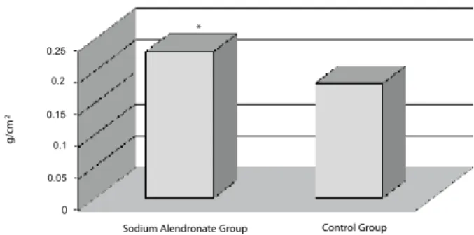
Figures 1 and 2 present the BMD values of prox-imal tibial metaphysis obtained by radiographic densitometry and DXA in the sodium alendronate and control groups, respectively, after 8 weeks of treatment. In Table 1, the results obtained with both of the methods are compared between the 2 groups. Both the radiodensitometric and DXA analyses revealed that the sodium alendronate–treatment group had a signiicantly higher BMD (p < 0.05) than the control group (distilled water) after 8 could be adjusted according to the body weight of
each animal.
The rats were killed after 8 weeks of treatment with a lethal injection of a mixture of ketamine hydrochloride (Ketamina Agener, União Química Farmaêutica Nacional S/A, Embu-Guaçu, Brazil; 300 mg/mL) and xylazine hydrochloride (Dopaser; Caleir S.A., Barcelona, Spain; 30 mg/mL). The tib-ias were surgically removed, and the BMD was eval-uated by radiographic densitometry and DXA.
BMD analysis by radiographic densitometry
The freshly removed tibias from both of the groups were dissected, cleaned and frozen at
-20°C. The sodium alendronate (n = 15) and con-trol (n = 10) tibias were arranged symmetrically on extraoral radiographic ilms (KodakMin-R S, Ko-dak Eastman Co., Rochester, USA) together with an aluminum stepwedge with 8 steps (NDT Mart Inc., Covington, USA), ranging from 2 to 16 mm, which served as a reference for densitometric analysis. Ra-diographic densitometry was performed based on the studies by Issa et al.7 and Erdogan et al.1 The tube of the x-ray unit (Weber x-ray unit; The Weber Dental Mfg. Co., Canton, USA) was placed perpen-dicular to the ilm, and a single radiographic expo-sure was made with expoexpo-sure parameters of 10 mA, 60 kV, 0.6 seconds and 40 cm focus-ilm distance. Radiographic processing was performed for 2 min-utes in the developing solution (27°C), 30 s in the intermediate phase, 4 minutes in the ixer solution, and 10 minutes in the inal rinse. All of these factors were monitored using a Victodreen NERO 6000B (Non-Invasive Evaluator Radiation Outputs, Mod-elo 6000B, Victoreen Inc., Everett, USA).
weeks of treatment. Comparing the groups with and without alendronate therapy revealed increases of 14.9% and 29.6% in BMD, as determined using radiographic densitometry and DXA, respectively. Briely, these results showed that when the DXA technique is inaccessible, radiographic densitometry is a valid method to detect alterations in the balance between bone formation and bone resorption.
Discussion
Sodium alendronate, which is a nitrogen-con-taining bisphosphonate, enters the mature osteo-clasts and inhibits the synthesis of farnesyl pyro-phosphate synthase, an enzyme of the mevalonate pathway. As a result, a cascade of events is initiated that causes toxic effects on the osteoclasts, including suppression of resorptive activity, loss of cytoskel-etal integrity and rufled borders and, ultimately, apoptotic cell death.14 Briely, sodium alendronate acts as a potent, speciic inhibitor of osteoclast-me-diated bone remodeling and increases bone mineral
density.
Bone densitometry is commonly used in medicine to measure BMD,7 and it is an important method for the early diagnosis of osteopenic and osteoporotic conditions, as well as for monitoring treatment evo-lution.18 In addition to radiographic densitometry, the contemporary methods of bone densitometry include single-photon absorptiometry, single-energy x-ray absorptiometry, dual-photon absorptiometry and DXA.7
The measurement of BMD in long bones or the spine by DXA is currently recognized as a well-es-tablished method for the diagnosis of osteoporosis.10 The operation of DXA devices is based on the prin-ciple that the bone and soft tissue have different at-tenuation properties as a function of photon energy. Therefore, DXA uses an x-ray source to produce a discrete energy beam that is attenuated as it travels through the patient.9,19 In the present study, DXA was highly effective in measuring the BMD of rat proximal tibial metaphysis and revealed a
signii-55 60 65 70
Me
a
n
value
of
lig
ht
int
e
nsit
y
Sodium Alendronate Group Control Group
*
Figure 1 - Bone mineral density values of proximal tibial metaphysis (mean value of light intensity) that were obtained by radiographic densitometry in the sodium alendronate (administration of sodium alendronate) and control (admin-istration of distilled water) groups. Asterisks indicate statisti-cally significant differences.
Figure 2 - Bone mineral density values of proximal tibial metaphysis (g/cm²) that were obtained by DXA in the sodium alendronate (administration of sodium alendronate) and control (administration of distilled water) groups. Asterisks indicate statistically significant differences.
Method
Sodium alendronate group
(n = 15) Control group(n = 10) p value*
M (Q1–Q3) M (Q1–Q3)
Radiographic
densitometry 66.8(64.1–72.6) 60.5(57.9–62.8) 0.0010
DXA 0.230 (0.197–0.252) 0.181 (0.176–0.185) 0.0001
* p value for the Kruskal-Wallis test; M: median; Q1: first quartile; Q3: third quartile. Table 1 - Comparison of bone
mineral density values in the proximal tibial metaphysis that were obtained by radiographic densitometry and DXA (g/cm²) in the sodium alendronate (administration of sodium alendronate) and control (administration of distilled water) groups.
0 0,05 0,1 0,15 0,2 0,25
g
/cm
2
Sodium Alendronate Group Control Group
0.25
0.2
0.15
0.1
0.05
cant difference between the 2 groups, with an al-most 30% increase in BMD in the animals that were subjected to alendronate therapy for 8 weeks.
DXA has recently been adapted to measure body composition in animals, such as rats and mice, us-ing specialized software in conjunction with clinical whole-body DXA machines.20 The Lunar PIXImus used in the present study is a fully integrated densi-tometer with an attached laptop computer and desk-top printer; it was designed for the estimation of BMD and body composition. The Lunar PIXImus stands out as an effective DXA device for measuring BMD in small lab animals for bone biology research.21-23
The measurement of BMD in long bones by DXA could be useful to predict BMD of the man-dibular body.Changes in the metabolism of bone tissue in the jaw region seem to be similar to those observed in long bones because both of these bones are subjected to strenuous muscle activity.24
Radiographic analysis is an important adjunc-tive tool for clinical evaluation because it is a rapid, non-invasive and low-cost method4 and can produce results similar to those of histological studies.25 The measurement of BMD by radiographic densitometry has been performed in a number of studies, and its eficacy has been conirmed.1,4,7,26 Radiographic den-sitometry may be considered as a minimally invasive and convenient method for diagnosing osteoporo-sis.1 According to Erdogan et al.,1 the radiological parameters can facilitate the establishment of a dei-nite diagnosis of osteoporosis.
In the present study, radiographic densitometry showed that the BMD values of proximal tibial me-taphysis differed between the sodium alendronate and control groups. BMD was signiicantly higher in the tibias of rats that were treated with sodium alendronate compared to those that received dis-tilled water. Comparing the two groups revealed
in-creases of 14.9% and 29.6% in BMD, as determined by radiographic densitometry and DXA, respective-ly. Although a higher percentage increase in BMD was determined by DXA, the results of the present study showed that radiographic densitometry is also an effective, accurate and safe method for measur-ing the BMD of long bones. Similarly, accordmeasur-ing to Grifith et al.27, although radiographic densitometry may not be the most sensitive method of measur-ing bone density, it is nevertheless a readily avail-able, though widely underused, determinant of bone quality. Furthermore, radiographic assessment is commonly used in everyday clinical practice.28
Because radiographic densitometry is more read-ily available to health professionals and more ac-cessible to the population due to its lower cost, this technique is useful for the early diagnosis of osteo-metabolic disorders, especially osteoporosis, which allows for the establishment of an adequate treat-ment strategy and improves the prognosis of these pathologies. Additionally, the use of radiographic densitometry could increase the number of studies in the ield of bone biology, thus contributing to ad-vances in the diagnosis, treatment and monitoring of bone diseases, which would increase the life qual-ity and life expectancy of patients.
Conclusion
Based on the results of this study, it may be con-cluded that both of the methods were effective in de-tecting an increase in the BMD of the proximal tibial metaphysis after systemic administration of sodium alendronate for 8 weeks. Additionally, the results suggest that when DXA is not available, radiograph-ic densitometry may be used as an auxiliary method in humans in the ield of bone biology to evaluate the balance between bone formation and bone re-sorption.
References
1. Erdogan O, Incki KK, Benlidayi ME, Seydaoglu G, Kelekci S. Dental and radiographic findings as predictors of osteo-porosis in postmenopausal women. Geriatr Gerontol Int. 2009 Jun;9(2):155-64.
2. Consensus Development Conference on Osteopororsis. Am J Med. 1993 Nov;95(5A):5S-16S.
4. Giro G, Gonçalves D, Sakakura CE, Pereira RMR, Marcan-tonio Jr E, Orrico SRP. Influency of estrogen deficiency and its treatment with alendronate and estrogen on bone density around osseointegrated implants: radiographic study in female rats. Oral Surg Oral Med Oral Pathol Oral Radiol Endod. 2008 Feb;105(2):162-7.
5. Martínez-Maestre MA, González-Cejudo C, Machuca G, Torrejón R, Castelo-Branco C. Periodontitis and osteoporo-sis: a systematic review. Climacteric. 2010 Dec;13(6):523-9. 6. Lochmüller EM, Jung V, Weusten A, Wehr U, Wolf E, Eckstein
F. Precision of high-resolution dual energy X-ray absorptiom-etry of bone mineral status and body composition in small animal models. Eur Cell Mater. 2001 Jan 20;1:43-51. 7. Issa JPM, Tiossi R, Watanabe PCA, Siéssere S, Regalo SCH,
Lopes RA, et al. Newly formed bone in mandible decortication experimental model using rhBMP-2 evaluated by densitomet-ric study. Int J Morphol. 2008;26(1):83-8.
8. Breen SA, Loveday BE, Millest AJ, Waterton JC. Stimulation and inhibition of bone formation: use of peripheral quantita-tive computed tomography in the mouse in vivo. Lab Anim. 1998 Oct;32(4):467-76.
9. Devlin H, Horner K, Ledgerton D. A comparison of maxil-lary and mandibular bone mineral densities. J Prosthet Dent. 1998 Mar;79(3):323-7.
10. Lane NE. Epidemiology, etiology, and diagnosis of osteopo-rosis. Am J Obstet Gynecol. 2006 Feb;194(2 Suppl):S3-11. 11. Nelson-Filho P, Lucisano MP, Silva RAB, Silva RS, Serra
MC, Gerlach RF, et al. Systemically alendronate was incor-porated into dental tissues but did not cause morphological or mechanical changes in rats teeth. Microsc Res Tech. 2012 Sep;75(9):1265-71. Epub 2012 Apr 17.
12. Gondim V, Aun J, Fukuda CT, Takayama L, Latorre MD, Pannuti CM, et al. Severe loss of clinical attachment level: an independent association with low hip bone mineral density in postmenopausal females. J Periodontol. 2013 Mar;84(3):352-9. Epub 2012 May 1.
13. Aboelasrar MA, Elbarbary NS, Elshennawy DE, Omar AM. Insulin-like growth factor-1 cytokines cross-talk in type 1 diabetes mellitus: relationship to microvascular complications and bone mineral density. Cytokine. 2012 Jul;59(1):86-93. 14. Russell RGG, Watts NB, Ebetino FH, Rogers MJ. Mechanisms
of action of bisphosphonates: similarities and differences and their potential influence on clinical efficacy. Osteoporos Int. 2008 Jun;19(6):733-59.
15. Russell RGG, Xia Z, Dunford JE, Oppermann U, Kwaasi A, Hulley PA, et al. Bisphosphonates: an update on mechanisms of action and how these relate to clinical efficacy. Ann N Y Acad Sci. 2007 Nov;1117:209-57.
16. Schmidt C, Priemel M, Kohler T, Weusten A, Müller R, Am-ling M, et al. Precision and accuracy of peripheral quantita-tive computed tomography (pQCT) in the mouse skeleton
compared with histology and microcomputed tomography (µCT). J Bone Miner Res. 2003 Aug;18(8):1486-96. 17. Kanzaki S, Takada Y, Ogawa K, Matsuo K. Bisphosphonate
therapy ameliorates hearing loss in mice lacking osteoprote-gerin. J Bone Miner Res. 2009 Jan;24(1):43-9.
18. Lodder MC, Lems WF, Ader HJ, Marthinsen AE, van Co-everden SCCM, Lips P, et al. Reproducibility of bone mineral density measurement in daily practice. Ann Rheum Dis. 2004 Mar;63(3):285-9.
19. Hildebolt CF. Osteoporosis and oral bone loss. Dentomaxil-lofac Radiol. 1997 Jan;26(1):3-15.
20. Nagy TR, Clair AL. Precision and accuracy of dual-energy X-ray absorptiometry for determining in vivo body composi-tion of mice. Obes Res. 2000 Aug;8(5):392-8.
21. Bartell SM, Isales CM, Baile CA, Kuhar MJ, Hamrick MW. CART deficiency increases body weight but does not alter bone strength. J Musculoskelet Neuronal Interact. 2008 Apr-Jun;89(2):146-53.
22. Earp JC, Dubois DC, Molano DS, Pyszczynski NA, Keller CE, Almon RR, et al. Modeling corticosteroid effects in a rat model of rheumatoid arthritis I: mechanistic disease progres-sion model for the time course of collagen-induced arthritis in Lewis rats. J Pharmacol Exp Ther. 2008 Aug;326(2):532-45. 23. Katikaneni R, Ponnapakkam A, Miller E, Ponnapakkam T,
Gensure RC. A new technique for precisely and accurately measuring lumbar spine bone mineral density in mice using clinical dual energy X-ray absorptiometry (DXA). Toxicol Mech Methods. 2009 Mar;19(3):225-31.
24. Buyukkaplan US, Guldag MU, Yildiz M, Gumus BA. Com-parison of mandibular bone mineral density in osteoporotic, osteopenic and normal elderly edentulous subjects measured by the dual-energy X-ray absorptiometry technique. Gerodon-tology. 2012 Jun;29(2):e1098-102.
25. Sakakura CE, Margonar R, Holzhausen M, Nociti Jr FH, Alba Jr RC, Marcantonio Jr E. Influence of cyclosporin a therapy on bone healing around titanium implants: a his-tometric and biomechanic study in rabbits. J Periodontol. 2003 Jul;74(7):976-81.
26. Rowshan HH, Parham MA, Baur DA, McEntee RD, Cau-ley E, Carriere DT, et al. Effect of intermittent systemic ad-ministration of recombinant parathyroid hormone (1-34) on mandibular fracture healing in rats. J Oral Maxillofac Surg. 2010 Feb;68(2):260-7.
27. Griffith JF, Engelke K, Genant HK. Looking beyond bone mineral density: imaging assessment of bone quality. Ann N Y Acad Sci. 2010 Mar;1192:45-56.