Skeletogenesis during Vertebrate Development
Charlene Rivera1.¤a, Sara J. S. Simonson2.¤b, Idella F. Yamben1¤c, Shalini Shatadal1, Minh M. Nguyen1¤d, Maryline Beurg3, Paul F. Lambert2, Anne E. Griep1*
1Department of Cell and Regenerative Biology, University of Wisconsin-Madison, Madison, Wisconsin, United States of America,2Department of Oncology, University of Wisconsin-Madison, Madison, Wisconsin, United States of America,3Institut National de la Sante´ et de la Recherche Me´dicale, Hoˆpital Pellegrin, Bordeaux, France
Abstract
The development of specialized organs is tightly linked to the regulation of cell growth, orientation, migration and adhesion during embryogenesis. In addition, the directed movements of cells and their orientation within the plane of a tissue, termed planar cell polarity (PCP), appear to be crucial for the proper formation of the body plan. In Drosophila
embryogenesis, Discs large (dlg) plays a critical role in apical-basal cell polarity, cell adhesion and cell proliferation. Craniofacial defects in mice carrying an insertional mutation in Dlgh-1 suggest that Dlgh-1 is required for vertebrate development. To determine what roles Dlgh-1 plays in vertebrate development, we generated mice carrying a null mutation inDlgh-1. We found that deletion ofDlgh-1caused open eyelids, open neural tube, and misorientation of cochlear hair cell stereociliary bundles, indicative of defects in planar cell polarity (PCP). Deletion of Dlgh-1also caused skeletal defects throughout the embryo. These findings identify novel roles forDlgh-1in vertebrates that differ from its well-characterized roles in invertebrates and suggest that the Dlgh-1null mouse may be a useful animal model to study certain human congenital birth defects.
Citation:Rivera C, Simonson SJS, Yamben IF, Shatadal S, Nguyen MM, et al. (2013) Requirement forDlgh-1in Planar Cell Polarity and Skeletogenesis during Vertebrate Development. PLoS ONE 8(1): e54410. doi:10.1371/journal.pone.0054410
Editor:Xin Zhang, Indiana University School of Medicine, United States of America
ReceivedJune 20, 2012;AcceptedDecember 13, 2012;PublishedJanuary 22, 2013
Copyright:ß2013 Rivera et al. This is an open-access article distributed under the terms of the Creative Commons Attribution License, which permits unrestricted use, distribution, and reproduction in any medium, provided the original author and source are credited.
Funding:This work was supported by NIH grants EY09091 (AEG), CA908428 (PFL/AEG). The University of Wisconsin Carbone Cancer Center (P30 CA14520) provided shared service support for the project. The funders had no role in the study design, data collection and analysis, decision to publish or preparation of the manuscript.
Competing Interests:The authors have declared that no competing interests exist. * E-mail: [email protected]
¤a Current address: Lineberger Comprehensive Cancer Center, University of North Carolina, Chapel Hill, North Carolina, United States of America ¤b Current address: Center for Commercialization, University of Washington, Seattle, Washington, United States of America
¤c Current address: Kelly Scientific Resources, Madison, Wisconsin, United States of America
¤d Current address: Lillehei Heart Institute, University of Minnesota School of Medicine, Minneapolis, Minnesota, United States of America
.These authors contributed equally to this work.
Introduction
The development of specialized organs in vertebrates is tightly linked to the regulation of cell growth, apical-basal cell polarity and cell-cell adhesion during embryogenesis. In addition, the directed movements of cells and their orientation in the same direction within the plane of a tissue, termed planar cell polarity (PCP), appear to be crucial for the proper formation of the body plan. Of great interest has been to decipher the mechanisms involved in regulating these critical aspects of development. From studies in invertebrates it is known that certainPSD95/Dlg/ZO-1 (PDZ) domain containing proteins such as Discs-large (Dlg) play prominent roles in regulating apical-basal polarity [1,2] while different PDZ proteins are part of a genetic network that regulates PCP [3]. Recent work in vertebrates supports the contention that the function of many polarity factors has been conserved cross-species. However, studies also suggest that certain factors have different or additional roles in vertebrates as compared to invertebrates [4]. In this study, we address the role ofDlgh-1, the mouse homolog ofDrosophila dlg, in mouse development.
In Drosophila melanogaster, mutations in dlg and scrib, the gene encoding a second PDZ protein, Scribble, result in neoplastic
overgrowth, multilayering and loss of cell shape in various epithelial tissues including the imaginal discs [1], the embryonic epidermis, and the follicular epithelia [2], suggesting a role for these factors in cell proliferation, adhesion and apico-basal polarity. InDrosophilaandCaenorhabditis elegans, Dlg and Scrib are thought to be required for properly localizing and maintaining adherens junctions and apical determinants [1,2,5,6,7,8,9]. In
Drosophila, Dlg localizes to the septate junctions where it forms a complex with Scrib and Lethal giant larvae (Lgl) [1,2]. This Dlg/ Scrib/Lgl complex antagonizes other PDZ complexes, thereby restricting adherens junctions and apical determinants, such as Crumbs, to the appropriate compartment [10]. Finally, the Partner of Inscuteable/Discs-large complex is required for planar polarity during the asymmetric division of the sensory organ precursor (pl) cell [11].
a guanylate kinase homolog (GUK) domain [12]. Homologs of
DrosophilaDlg are found in humans (hDlg), rat (SAP97), and mouse (Dlgh-1, also referred to as Dlg-1), and the high degree of conservation among its multiple domains suggests conserved function cross-species [13,14,15]. In mammals, Dlgh-1 is ubiqui-tously expressed and is found at sites of cell-cell contacts in epithelial and neuronal cells [16,17,18,19,20,21]. It interacts with a wide variety of proteins, which include the tumor suppressors adenomatous poli (APC) [22] and protein tyrosine phosphatase and tensin homologue (PTEN) [23], Shaker-type K+channela -subunits [24], the G-protein coupled receptor homologue tumor endothelial marker 5 (TEM5) [25], and the MAGUK Lin-2/ CASK [26]. In addition to interacting with endogenous cellular proteins, Dlgh-1 is also targeted by multiple human viral oncoproteins including the human papillomavirus (HPV) E6 [27], the adenovirus E4-ORF1 [28], and the human T cell leukemia virus type 1 (HTLV-1), Tax 1, [29]. These interactions have been implicated in the transforming and tumorigenic properties of the viral oncoproteins [27,29,30,31,32,33].
The role of Dlgh-1 in mouse development has begun to be explored. Caruana and Bernstein [34] reported that mice carrying a gene trap insertion inDlgh-1(Dlgh-1g/gtt mice), which results in a Dlgh-1 protein lacking the SH3, band 4.1, and GUK domains, exhibited a cleft palate and a shorter mandible thanDlgh-1wild type mice. Additional studies on thisDlgh-1gt/gtmouse showed that these mice also exhibited a decrease in the number of nephrons in the developing kidney [35] and cell cycle misregulation in the epithelium of the ocular lens [36]. Since the L27 and three PDZ domains of Dlgh-1 were left intact in the gene trap allele [34,37], it is possible that the fusion protein generated from this gene trap allele may retain some or acquire new activities of Dlgh-1 and, therefore, the phenotypes observed may not fully reflect the null phenotype. Consistent with this possibility, lacZ staining of Dlgh-1gt/gt lenses and controlDlgh-1+/+lenses showed similar staining
patterns (Rivera and Griep, unpublished observations). Further-more, two recent reports show that deletion of Dlgh-1 in mice caused abnormalities in the ureteric smooth muscle [38] and absence of vagina and seminal vesicles [39]. Additionally, conditional deletion of Dlgh-1 in the lens led to more severe phenotype than was observed in the lenses of theDlgh-1gt/gtmice including epithelial and fiber cell specific defects in cell adhesion and polarity [40].
In the present study, we generated mice carrying a germline null mutation inDlgh-1by deleting one of the exons encoding the first PDZ domain and characterized the wide-ranging effects of this mutation on mouse embryogenesis. Loss of Dlgh-1 not only recapitulated the craniofacial and kidney defects observed in the
Dlgh-1gt/gt mouse but also resulted in reduced ossification in the skull, maxilla, and middle ear, and shortening of the long bones. Furthermore, deletion of Dlgh-1 resulted in eyelid closure and neural tube closure defects, and the misorientation of the stereociliary bundles of the cochlear hair cells, all of which suggest defects in PCP. Thus, our analysis identifies novel roles forDlgh-1
in mouse development, places Dlgh-1 in the group of factors that regulate PCP in the mouse, and provides new insight into the distinct in vivo requirements for this gene in vertebrates, as compared to invertebrates.
Results
Generation ofDlgh-1Null Mice
To generateDlgh-1null mice, mice carrying a mutant allele of
Dlgh-1, in which exon 8 and a neomycin resistance-cassette are flanked by loxP sites [40] (Fig. 1A), were mated to EIIA-Cre
transgenic mice [41]. TheDlgh-1null allele was moved onto the FVB/NJ and C57BL/6J genetic backgrounds by backcrossing to either FVB/NJ or C57BL/6J mice for at least three generations before use. Mice heterozygous for the null allele were intercrossed to generateDlgh-12/2mice and Southern blot analysis was used to confirm the genotype of the progeny (Fig. 1B).
To determine if the mutated Dlgh-1 allele was indeed a null allele, Dlg-1 protein levels in brain, heart, lung, kidney, skin, liver, and eyes of E18.5Dlgh-1+/+
,Dlgh-1+/2, andDlgh-12/2mice were
assessed by immunoblot analysis using an N-terminal specific anti-Dlgh-1 antibody. The anti-Dlgh-1 protein levels were reduced by approximately 50% in theDlgh-1+/2mice compared to the
Dlgh-1+/+mice and Dlgh-1 protein was not detected in any of the tissues
analyzed from theDlgh-12/2mice or in mouse embryo fibroblasts (MEFs) derived from day E14.5 Dlgh-1 null embryos (Fig. 1C). These results confirmed that the Dlgh-1 mutant allele is a null allele.
Effect of Loss ofDlgh-1on Mouse Embryogenesis
It has been reported that mice homozygous for theDlgh-1gtallele die at or near the time of birth [12]. To determine if nullizygosity for Dlgh-1also would be lethal, we intercrossed Dlgh-1+/2 mice
and examined litters for live Dlgh-12/2 pups. Of the 47 pups genotyped, 10 were Dlgh-1+/+
(21%), 22 (47%) were Dlgh-1+/2,
and 15 (32%) wereDlgh-12/2. Of the 15Dlgh-12/2pups, all of which exhibited severe craniofacial abnormalities, 13 were found dead on the day of birth and the remaining two died shortly after being found. These data indicate that loss of Dlgh-1 confers perinatal lethality with 100% penetrance and support the hypothesis that Dlgh-1 plays an important role in mouse development.
To understand the impact of the loss of Dlgh-1 on mouse development, Dlgh-1+/+
, Dlgh-1+/2, and Dlgh-12/2 mice were
collected at embryonic stages E11.5, E14.5, E16.5, and E18.5.
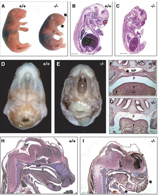
Dlgh-12/2embryos were viable at all embryonic stages examined. However, at the gross level Dlgh-12/2 embryos exhibited an abnormal head shape that included a shortened snout (Figs. 2A, H–I).Dlgh-12/2mice were smaller than theirDlgh-1+/+
counter-parts. This reduction in size was apparent overtly (Fig. 2A) and in histological section by E14.5 (Figs. 2B, C). At the gross anatomic level, Dlgh-12/2 mice also exhibited a secondary cleft-palate (Figs. 2D, E) that was consistent with the lack of a nasopharynx in these mutant animals observed in histological sections (Figs. 2F–I). All of these phenotypes of theDlgh-12/2mice were observed with 100% penetrance on both the FVB/NJ and C57BL/6J genetic backgrounds and were similar to those observed in theDlgh-1gt/gt
mice [34]. Additionally, the kidneys of theDlgh-12/2 mice were smaller in size and exhibited a reduced tubule to mesenchyme ratio (data not shown), consistent with a defect in branching morphogenesis, which was reported for the previously described
Dlgh-1mutant mice [35,38]. Finally, defects in the ocular lens were also noted in the Dlgh-12/2 mice (data not shown) as reported previously for theDlgh-1gt/gtmice [36] and mice in whichDlgh-1
was specifically deleted in the lens [40]. Together, these data show thatDlgh-1is required for mouse survival, likely in part through its contribution to palate formation, and for multiple aspects of mouse development.
Effect of Loss ofDlgh-1on Eyelid and Neural Tube Closure
C57BL/6JDlgh-12/2 mice at E18.5 (Fig. 3). The severity of this phenotype varied. In some cases the epidermis had extended and fused; however, the dermis had not filled in (Fig. 3E). In other cases, the epidermis had failed to extend, leading to a complete failure of eyelid closure (Fig. 3F). To determine if the effect of Dlgh-1 deficiency on eyelid closure may be a direct effect, head sections from Dlgh-1+/+ and Dlgh-12/2 mice were subjected to
double immunofluorescence experiments using anti-Dlgh-1 and anti-Vangl2 antibodies (Fig. 4). Staining for Dlgh-1 was observed in theDlgh-1+/+eyelid epithelium (Fig. 4A, B) but was absent in
theDlgh-12/2eyelid (Fig. 4D, E), suggesting that the open eyelid defect may be a direct result of loss of Dlgh-1 function.
Interestingly, the failure of eyelid closure is also noted in the
circletail(Crc/Crc) mouse, in which a defective Scrib protein lacking the C-terminal two PDZ domains is expressed [42], and in the
looptail (Vangl2Lp/Lp) mouse, in which a defective Vangl2 protein carrying an amino acid substitution mutation (S464N) is expressed [43,44]. The Vangl2 protein is normally expressed in the eyelid
epithelium [43,44,45] and is primarily localized to the plasma membrane [45]. However, the mutant Vangl2 protein inVangl2Lp/
Lp
mice fails to localize to the membrane; rather it is cytoplasmic [45]. Immunofluorescent staining of eyelids fromDlgh-1+/+mice
showed that Vangl2 was tightly localized to the membranes in the eyelid epithelium (arrow, Fig. 4A, C) where there was overlap with Dlgh-1 staining. In contrast, Vangl2 was cytoplasmic in the eyelid of theDlgh-12/2mice (Fig. 4D, F). Thus, the correct subcellular localization of Vangl2 in the eyelid epithelium depends onDlgh-1.
Vangl2Lp/Lp and Crc/Crc mice also exhibit craniorachischisis
[42,44,46], a severe neural tube closure defect in which the neural tube is open from the midbrain-hindbrain boundary to the tail. This same neural tube defect was observed in C57BL/6JDlgh-12/
2mice at both E16.5 (Figs. 5C, D) and E18.5 (not shown). Unlike
the open eyelid phenotype, however, craniorachischisis occurred with low penetrance; only 6.7% (2 out of 30) of the C57BL/6J
Dlgh-1 null mice displayed this defect. Dlgh-12/2 mice that exhibited craniorachischisis also exhibited gastroschisis, an
exter-Figure 1. Generation ofDlgh-1null mice.(A) Schematic representation of the targeting of theDlgh-1allele. WT, wild-type allele; C, construct, plasmid containing loxP sites flanking exon 8 ofDlgh-1and neo cassette, and the TK cassette; F, the floxed allele allele, containing lox P sites flanking
Dlgh-19s exon 8 and the neo cassette; R, recombined allele, desired recombination event lacking exon 8 and neo. Abbreviations: 7, exon 7; 8, exon 8; 9, exon 9; P,PstI; triangles, lox P sequences; neo, neomycin; TK, thymidine kinase. 39probe for Southern blot analysis is denoted by black bars. Fragments expected fromPstI digestion and 39probe hybridization are depicted by double-arrowed lines. (B) Southern blot analysis ofPstI digested genomic DNA isolated from mice that were WT (+/+), heterozygote (+/2), or null (2/2) for theDlgh-1allele. Sizes of hybridizing bands are shown in kb. (C) Western blot analysis of Dlgh-1 protein levels. Protein lysates of brain, heart, lung, kidney, skin, eye, liver, and mouse embryo fibroblasts (MEF) fromDlgh-1wild type (+/+), heterozygote (+/2), or null (2/2) mice were resolved by SDS-PAGE and immunoblotted with an anti-Dlgh-1 antibody, as described in Materials and Methods. Membranes were reprobed with an anti-b-actin antibody as a loading control.
nalization of the gut (Fig. 5A, C). Closure of the eyelid and neural tube involves a process of polarized cell movement known as convergent extension [3,47]. Thus, the defects in eyelid and neural
tube closure exhibited in theDlgh-12/2mice suggest a new role for
Dlgh-1in the mouse in the regulation of convergent extension.
Figure 2. Phenotype ofDlgh-12/2mice.(A) NewbornDlgh-12/2mice are smaller then theirDlgh-1+/+
counterparts and have a shortened snout and mandible (arrows). (B–C) Hematoxylin and eosin stained paraffin sections of FVB/NJ E14.5 Dlgh-1+/+ (B) andDlgh-12/2 (C) mice. At this
developmental stage, the reduced size of theDlgh-12/2is noticeable. (D–E). Dissected heads ofDlgh-1+/+
andDlgh-12/2C57BL/6J E18.5 embryos
showing the cleft palate (asterisk*) inDlgh-12/2mice. (F–G) Frontal sections ofDlgh-1+/+(F) andDlgh-12/2(G) E18.5 embryos on the C57BL/6J
background showing the absence of the nasopharyngeal passage (NP, arrow in F). (H–I) Sagittal H&E stained sections of E18.5Dlgh-1+/+
Effect of the Loss ofDlgh-1on Cochlear Hair Cell Stereociliary Bundle Orientation
Recent studies in vertebrates have shown that convergent extension is regulated, at least in part, by a group of proteins that are involved in planar cell polarity (PCP) inDrosophila [3]. PCP refers to the polarization of cells within the plane of a cell sheet. In
Drosophila, PCP regulates wing hair, body bristle, and eye ommitidial cluster orientation. In mammals, the uniform orienta-tion of stereociliary bundles of the hair cells within the cochlea represents a distinctive example of PCP. InVangl2Lp/Lpmice, the stereociliary bundles in the inner (IHC) and outer (OHC) hair cell layers are misoriented [4]. Similarly, in Crc/Crc mice the stereociliay hair bundles in the second and third row of OHCs are misoriented [4], Together, these data identify a role forVangl2
andScribin the regulation of the PCP pathway in vertebrates. Becausedlgandscribare known to interact inDrosophila[2] and because the open eyelid, neural tube, and gut phenotypes of the
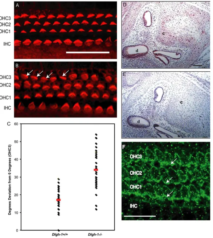
Dlgh-12/2embryos (Fig. 4) resemble that ofCrc/Crcmutant mice [42], we asked if stereociliary bundle orientation was affected in theDlgh-1null mice. Cochlear sensory epithelia (organs of Corti) from E18.5Dlgh-1+/+
and Dlgh-12/2 embryos were stained with phalloidin to label hair cell sterocilia, a structure rich in filamentous actin, and the specimens analyzed by confocal microscopy. Normally, the stereociliary bundles are arranged uniformly around the primary cilia and oriented toward the outer edge of the organ. The IHC bundles are arranged in a curved shape while the OHC bundles are arranged in a v-shape [48]. The stereociliary bundles in the IHC row of theDlgh-12/2mice were uniformly polarized, as in theDlgh-1+/+
mice (Fig. 6A, B). The stereociliary bundles in all three rows of OHCs of theDlgh-1+/+
mice were uniformly oriented (Fig. 6A). However, many stereo-ciliary bundles in the third row of OHCs in the Dlgh-12/2
cochleae were misoriented (Fig. 6B, arrows). Occasionally, stereociliary bundles in the second OHC layer were also misoriented (Fig. 6B). The angle of orientation of individual
Figure 3.Dlgh-12/2mice display defects in eyelid closure.E18.5Dlgh-+/+2andDlgh-12/2mice on the C57BL/6 background were collected and
analyzed for the presence of closed eyelids.Dlgh-1+/+mice (A) form closed eyelids whileDlgh-12/2(B, C) littermates show defects in eyelid closure.
(D–F) Hematoxylin and eosin staining of histological sections collected from E18.5 mice show completely closed eyelids inDlgh-1+/+
mice (D) whereas eyelids inDlgh-12/2mice were only partially closed (E) or completely open (F). Insets show a higher magnification of the eyelids. Arrows show the position of the tips of the dermis inDlgh-1+/+
(D) andDlgh-12/2(E) embryos, which failed to extend fully in theDlgh-12/2eye. Scale bar = 250mm and
150mm for insets. c, cornea; e, eyelids; l, lens.
doi:10.1371/journal.pone.0054410.g003
Figure 4. Expression of Dlgh-1 and Vangl2 proteins in the eyelid ofDlgh-1+/+and
Dlgh-12/2mice.Sections of sagittally oriented, paraffin embedded heads from newborn Dlgh-1+/+
(A–C) and Dlgh-12/2 (D–F) mice on the C57BL/6J background were subjected to double immunofluorescence analysis with anti-Dlgh-1 (green) and anti-Vangl2 (red) antibodies. Shown are representative merged and unmerged images of the staining in the eyelid ofDlgh-1+/+
(A–C) andDlgh-12/2(D–F) eyelids. Dlgh-1 (A, B) and Vangl2 (A, C) localized to the membranes of the
epithelium (arrow in A) including the site of eyelid fusion, inDlgh-1+/+mice. Staining for Dlgh-1 was absent in eyelid of theDlgh-12/2mice (E) and
stereociliary bundles in the OHC3 layer relative to the neural-abneural axis inDlgh-1+/+
andDlgh-12/2cochlea were measured and the degrees of deviation from this axis calculated (Fig. 6C). The average degrees of deviation for bundles in the Dlgh-12/2
mice (33.7+/21.87 degrees) was significantly higher (p = 1.561029) than in theDlgh-1+/+
mice (17.3+/20.87 degrees). To determine if the effect of Dlgh-1 deficiency on the cochlea might be a direct effect, head sections from day E15.5Dlgh-1+/+
embryos were immunostained with an anti-Dlg-1 antibody (Fig. 6D). Immunoreactivity was observed in the epithelial cells of the duct, which give rise to the sensory cells, and cartilage within the cochlea, indicating the presence of Dlgh-1 protein in these structures. To further determine if the effect of Dlgh-1 deficiency on hair cell orientation might be a direct effect, cochlear explants from P2 control mice were subjected to immunofluorescence for Dlgh-1. Immunoreactivity was observed in the basal-lateral membrane domain of the outer hair cells (Fig. 6F, arrows). The expression of Dlgh-1 in tissues such as the eyelid and hair cells, which show defects in the Dlgh-12/2 mouse, together with the
open eyelid, mislocalization of PCP protein Vangl2 in the open eyelid, craniorachischisis, and misorientation of cochlear stereo-ciliary in theDlgh-12/2 mice (Figs. 3, 4, 5, 6) suggest a role for
Dlgh-1in the regulation of PCP in the mouse.
Effect of the Loss ofDlgh-1in Skeletogenesis
TheDlgh-1gt/gtmice exhibited a cleft palate and a hypotrophic mandible [34], suggesting that Dlgh-1 may be required for the formation of at least certain craniofacial bones. To test this hypothesis, the skeletal frames of E18.5Dlgh-1+/+, Dlgh-1+/2,and
Dlgh-12/2mice on both the FVB/NJ and C57BL/6J background were stained with alizarin red and alcian blue to visualize bone and cartilage, respectively.Dlgh-12/2mice on both the FVB/NJ (Fig. 7) and C57BL/6J (not shown) genetic backgrounds had shorter mandibles (MA) and maxillas (MX), which was consistent with the short snout phenotype. However, uniquely on the FVB/ NJ background, Dlgh-12/2 mice also showed additional skeletal defects. Dlgh-12/2 embryos exhibited reduced ossification of multiple craniofacial structures including the nasal bone (N), the retrotympanic process (RTP), and the tympanic bone (T) (Figs. 7A, C). Interestingly, the developing calvarium was also affected as the ossification of the frontal bone (F) and the parietal bone (P) were reduced or absent in theDlgh-12/2mice (Fig. 7C). Furthermore, the parietal bone was absent in one Dlgh-1+/2 specimen on the
FVB/NJ background, suggesting a dosage-dependent effect of Dlgh-1 on calvarium development (Fig. 7B).
The skeletal defects in the Dlgh-12/2 mice on the FVB/NJ background extended to a reduction in the ossification of the ribs, as well as the second, third, and fourth sternebrae (S) (Fig. 7E). The long bones of the fore- and hindlimbs were also shortened by approximately 20% and 18%, respectively, in theDlgh-12/2mice when compared to their Dlgh-1+/+ counterparts (Figs. 7F, G).
Calcification in the phalanges, while apparent in the limbs of the
Dlgh-1+/+
mice, was absent in the limbs of the Dlgh-12/2 mice (Figs. 6F, G, arrow). Taken together, these results suggest that
Dlgh-1is required for proper skeletogenesis of craniofacial, axial and long bones. Furthermore, as the calvarium forms through intramembranous bone formation whereas the other craniofacial bones and the axial bones form through endochondral ossification [49], these data show that both forms of bone development are affected whenDlgh-1is ablated in the mouse.
Discussion
The MAGUK protein Dlgh-1, which is highly conserved cross-species, has emerged as an important factor in the regulation of cell-cell adhesion, apical-basal polarity, and cell proliferation in
Drosophila[1,2]. The regulation of these cellular aspects is critical for the establishment of specialized organs during embryogenesis. In the current study, we assess the role of Dlgh-1 in mouse embryogenesis (see Table 1 for a summary of phenotypes). We provide evidence that loss of Dlgh-1 affects PCP during mouse embryogenesis. We further provide evidence that loss ofDlgh-1
causes defects in skeletal structures arising through both endochondral and intramembranous ossification. These findings demonstrate newly discovered roles for Dlgh-1 in vertebrate development and suggest that the Dlgh-12/2 mouse may serve as an animal model to study major congenital birth defects in humans such as defects in neural tube closure and skeletal malformations.
Role ofDlgh-1in Mouse Organogenesis
The results presented in this study suggest that Dlgh-1 plays a vital role during mouse organogenesis. Defects in the mandible,
Figure 5. A fraction ofDlgh-12/2mice display defects in neural
tube closure.E16.5Dlgh-1+/+andDlgh-12/2embryos on the C57BL/6J
background were collected, examined for overt defects, and photo-graphed under a dissecting microscope. Profile (A) and dorsal (B) views of an E16.5Dlgh-1+/+
embryo. Profile (C) and dorsal (D) views of an E16.5
Dlgh-12/2embryo. In addition to craniorachischisis (C, D arrows), the
Dlgh-12/2embryo exhibits gastroschisis (C, arrowhead) and a rightward
kink to the body axis (D).
Figure 6.Dlgh-12/2mice display defects in outer cell hair polarity.(A, B) Representative images of the OHC and IHC layers in the cochlea of
Dlgh-1+/+
andDlgh-12/2mice. The organ of Corti was isolated from the cochleae of E18.5Dlgh-1+/+
(A) andDlgh-12/2(B) embryos, stained with phalloidin to visualize the actin bundles of the inner and outer hair cells, and viewed by confocal microscopy. Arrows indicate cells with misoriented stereociliary bundles in OHC3 row of theDlgh-12/2mice. (C) Measurement of misorientation of sterociliary bundles in OHC3 layer ofDgh-1+/+
and
Dlgh-12/2mice. The angle of sterociliary bundles in OHC3 ofDlgh-1+/+andDlgh-12/2mice was measured in reference to a line parallel to the
neural-abneural axis and perpendicular to the plane of the pillar cells. Plotted is the deviation in degrees from the neural-neural-abneural axis for each OHC3 bundle measured in (4 cochleae from 3 mice of each genotype, 36 cells forDlgh-1+/+and 33 cells forDlgh-12/2mice). No deviation from the neural-abneural
axis was assigned a value of 0u. The average angle deviation from 0ufor OHC3 bundles inDlgh-12/2cochlea (33.7u+/21.869, red bar) was significantly
higher (p = 1.561029) than that of theDlgh-1+/+(17.3
u+/20.869, red bar). (D, E) Immunohistochemical detection of Dlgh-1 protein in the cochlea of E15.5 mouse embryos. Transverse sections of paraffin embedded heads from controlDlgh-1+/+
mice were subjected to immunohistochemistry using (D) an anti-Dlgh-1 specific antibody and alkaline phosphatase detection or (E) no primary antibody. The pink color in the epithelium of the cochlear duct and cartilage in (D) is indicative of the presence of Dlgh-1 protein in these structures. (F) Immunofluorescent detection of Dlgh-1 protein in the cochlear hair cells of P2 mice control. Fixed cochlear explants fromDlgh-1+/+
mice were subjected to immunofluorescence using an anti-Dlgh-1 (green) antibody. Arrows show concentration of Dlgh-1 on the basal-lateral membranes of the OHC cells. IHC, inner hair cells, OHC1, 2, 3, outer hair cell rows 1, 2, and 3. c, cartilage in cochlea, d, cochlear duct. Scale bar = 50mm.
the kidneys, and the lens have been previously described in the
Dlgh-1gt/gtmice [34,35,36]. The formation of these structures and organs was likewise affected in our Dlgh-1null mice (Fig. 2 and data not shown). More recently, novel defects in the ureteric smooth muscle and the urogenital tract have been observed in
Dlgh-12/2mice [38,39]. In the present study, we provide evidence for Dlgh-1playing a role in the skeletogenesis of trunk and limb structures, in neural tube and eyelid closure, and in the
organization of the stereociliary bundles in the cochlea. These latter phenotypes were not reported previously in mice carrying the gene trap insertion inDlgh-1[34] and we have not observed open eyelids or open neural tubes in C57BL/6Dlgh-1gt/gtmice in our laboratory (M.M. Nguyen, C. Rivera, and A.E. Griep, unpublished observations). As the N-terminal portion of Dlgh-1 including the L27 and three PDZ domains are intact in the Dlgh-1gtfusion protein [34,37], it is likely that the function of one or more of these protein-protein interaction domains is required for Dlgh-19s role in the novel phenotypes we have observed.
Role ofDlgh-1in Planar Cell Polarity
Planar cell polarity is the mechanism through which the orientation of cells is coordinated within the plane of a cell sheet. In vertebrates, PCP has been suggested to be required for several developmental processes that involve convergent extension, such as lengthening of the body axis, eyelid closure, neural tube closure, branching morphogenesis in the kidney and lung, and the sterociliary cell hair bundle orientation in cochlea [3].Dlgh-12/2
mice, with variable penetrance, exhibited a reduced body length to width ratio, open eyelids (Fig. 3), craniorachischisis (Fig. 5), and misorientation of stereocilia in the third row of cochlear OHCs (Fig. 6). Moreover, Dlgh-1 is expressed in the eyelid epithelium (Fig. 4A, B) and cochlear hair cells (Fig. 6F) andDlgh-1deficiency led to mislocalization of the core PCP protein, Vangl2, in the mutant eyelid (Fig. 4D, F). Thus, this study provides the first evidence in support of a role forDlgh-1in the regulation of PCP in the mousein vivo.
Although much remains to be learned about the molecular and genetic mechanisms regulating PCP, from studies in theDrosophila
eye and wing, factors including Frizzled (Fz), Dishevelled (Dsh),
Figure 7.Dlgh-12/2mice display defects in skeletogenesis.Eviscerated carcasses of E18.5Dlgh-1+/+
,Dlgh-1+/2, andDlgh-12/2mice on the
FVB/NJ background were prepared and stained for cartilage and bone with alcian blue and alizarin red, respectively. Craniofacial skeleton in (A) Dlgh-1+/+,
(B)Dlgh-1+/2, and (C)Dlgh-12/2mice. Arrows show loss of bone formation inDlgh-1+/2andDlgh-12/2mice. F, frontal bone; IP, intraparietal
bone; P, parietal bone; N, nasal; Ro, rostral bone; RTP, retrotemporal process; T, temporal bone. (D–G) Axial structures ofDlgh-1+/+(D) andDlgh-12/2
mice (E). Forelimb (F) and hindlimb (G) structures. Arrows show the absence of bone formation inDlgh-12/2mice. S, sternebrae; H, humerus; R, radius;
U, ulna; F, femur; Fi, fibula; T, tibia. Scale bar in A-E = 1000mm, in F = 650mm and in G = 500mm. doi:10.1371/journal.pone.0054410.g007
Table 1.Summary of Phenotypes inDlgh-1Null Mice.
Defect Mouse Strain Penetrance
Reported inDlggt
mice
Perinatal lethality Both 100% Yes
Cleft palate Both 100% Yes
Shortened snout Both 100% Yes
Reduced size Both 100% Yes
Eyelid closure C57BL/6J 100%a No Neural tube
closure
C57BL/6J 6.7% No
Cochlear hair cells C57BL/6J 100% No
Skeletalb FVB/NJ 100% No
aEyelid closure defects were observed in 100% ofDlgh-12/2mice on the C57BL/
6J background. However, the severity of the phenotype varied from animal to animal.
bDefects in bone formation were observed throughout the head, trunk and limbs of theDlgh-12/2embryos on the FVB/NJ background.
Strabismus/Van Gogh (stbm/Vang), and Starry night/Flamingo (Stan/Fmi) are thought to mediate PCP through non-canonical Wnt signaling [3]. This pathway appears to be highly conserved in vertebrates. Wnt signaling has been shown to be required for mediating the organization of cochlear OHCs into their distinctive unidirectional orientation during development [48,50]. Fz32/
2;Fz62/2,Dvl12/2;Dvl22/2, andLp/Lpmice show reduced body
length to width ratio, craniorachischisis, failure of eyelid closure, and misorientation of stereociliary bundles at the apices of sensory hair cells [4,42,51,52,53] Additionally, this small group of factors in vertebrates includes Celsr1 [54], a protocadherin and homolog ofStan/Fmi, and PTK 7, a protein tyrosine kinase [55].
Interestingly, Fz3, Fz6, and Vangl2 contain C-terminal PDZ binding motifs whereas Dvl1 and Dvl2 are PDZ domain proteins, suggesting that PDZ domain proteins and their ligands interact to play a major role in PCP. Dvl2genetically interacts with Vangl2
and Vangl2 is required for the proper membrane localization of Dvl2 in the cochlear hair cells [52]. Vangl2 interacts with Fz3 and is required for targeting of Fz3 to the membrane of the cochlear hair cells [56]. It has recently been shown that these core PCP proteins also interact with other PDZ proteins in vertebrate systems. Fz4 and Fz7 have been shown to interact with the MAGUK protein, MAGI-3, through the PDZ binding motif of Fz and the first PDZ domain of MAGI-3in vitroand in epithelial cells [57] and Dlgh-1 has been shown to interact through its PDZ domain with the C-terminal PDZ binding motif in Fz1, Fz4, and Fz7 at least in yeast two hybrid experiments [58,59]. Mice defective for the PDZ protein Scrib (Crc/crcmice) exhibit the same phenotypes as mouse Fz, Dvl, and Vangl2 mutants. Scrib been shown to genetically interact withVangl2to modulate PCP in the cochlea [4] and Scrib binds to Vangl2 [56]. In this study, we have shown that the phenotype of Dlgh-1 null mice bears many similarities to the phenotypes of PCP mutant mice. Furthermore, we have shown thatDlgh-1is expressed in the eyelid epithelium (Fig. 4A, B) and the outer hair cells of the cochlea (Fig. 6F) and is required for correct subcellular localization of Vangl2 in the eyelid (Fig. 4D, F). Therefore, we suggest that Dlgh-1 also is a member of this group of factors involved in the regulation of PCP in the mouse, which is has very recently been shown to include another mouse Dlg, Dlg-3 [60]. The role for Dlgh-1 in PCP in properly localizing Vangl2 to the membrane may be a critical function for PCP as Vangl2 has been shown to be required for proper localization of Fz3 [56] and Dvl2 [61,62].
We observed differences in the phenotypes of Dlgh-12/2
embryos depending on the genetic background. On the C57BL/ 6J background, eyelid closure defects were present in 100% and craniorachischisis was present in 6.7% of Dlgh-12/2 embryos (Figs. 3, 5). Neither of these phenotypes was observed inDlgh-12/2
embryos on the FVB/NJ background. The low penetrance of craniorachischisis in Dlgh-12/2 embryos on the C57BL/6J background is similar to observations of low penetrance of craniorachischisis in Dvl12/2;Dvl22/2 mice on the C57BL/6J background. By contrast the penetrance of craniorachischisis is high on the 129 background [52]. The observation that the penetrance of some phenotypes associated with PCP is dependent on mouse genetic background suggests either that there are additional genetic modulators of PCP that have yet to be identified or that polymorphisms in one or more of the already identified genes on these different genetic backgrounds influences PCP.
Role ofDlgh-1in Skeletogenesis
The cleft palate and hypotrophic mandible in the Dlgh-1gt/gt
mice indicated that this PDZ domain protein might be important in the development of craniofacial skeletal structures [34]. The
Dlgh-12/2 mice not only recapitulated the craniofacial defects observed in the Dlgh-1gt/gt mice, but also displayed reduced ossification of the maxilla, middle ear, frontal, and parietal bones and overall reduced size of the skeleton (Fig. 7).
One mechanism through which loss of Dlgh-1 may affect craniofacial development is through disruption of WNT/PCP signaling [63]. Recently, it has been shown that certain core Wnt/ PCP proteins are expressed in cranial neural crest (CNC) cells [64,65], which give rise to most of the skeletal structures of the head and neck, and these core PCP proteins are required for CNC migration [66]. As noted above, Dlgh-1 has been shown to bind to several Fz receptors, including those commonly associated with Wnt/PCP signaling [58,59]. Likewise, Wnt5a, which has been suggested to be a regulator of Wnt/PCP signaling in the mouse [50], is expressed in outgrowing regions of the facial primordia and Wnt5a mutant mice display abnormally shaped head and shortened snout and mandible [67]. Finally,Wnt5adeficiency in mice results in cleft palate formation due to inhibition of the directional migration of cells that is required for palate formation [68]. Thus, the similarity of the craniofacial defects inWnt5a2/2,
Vangl2Lp/Lp, and Dgh-12/2 mice suggests that Dlgh-1 may be a regulator of craniofacial skeletogenesis through a mechanism that involves modulating Wnt/PCP.
The Dlgh-12/2 mice exhibited defects not only in skeletal structures of the head, but also of the trunk and limbs and the skeletons were reduced in size overall. In particular, the forelimbs and hindlimbs ofDlgh-12/2mice were approximately 20% shorter than those ofDlgh-1+/+
mice and the humerus was clearly wider in the mutants as compared to controls (Fig. 6). One possibility is that the reduced size is due to an inhibition of proliferation, as Dlgh-1 has been associated with regulating proliferation [22]. However, Wnt/PCP also has been shown to play a role in limb development [69,70].Wnt5amutant mice exhibit truncated limbs [67] and the directional outgrowth of the limb has been shown to require Wnt5a/JNK signaling in the distal mesenchyme [71,72]. Similar-ly,Vangl2Lp/Lpmice display shorter and wider limbs [73]. Recent studies have demonstrated a genetic and mechanistic link between Wnt5a and Vangl2 through the frizzled-like receptor, Ror2 [73], which binds Wnt5a [74], clearly establishing a connection between Wnt5a and a core PCP protein. Thus, the similarities in the limb phenotypes of these mouse mutants and the Dlgh-12/2 mice combined with the known mechanistic link between Dlgh-1 and Vangl2 suggest that the mechanism through whichDlgh-1exerts its effects may be through modulating Wnt/PCP.
Although, the most likely explanation for the defects in skeletogenesis in theDlgh-1mutant mice is thatDlgh-1is required the Wnt/PCP-dependent development of these skeletal structures, analysis of mouse mutants has demonstrated that numerous other signaling pathways play a role in skeletogenesis. These include FGFR [75,76], TGFbII [75,77], BMP [78], and Wnt/b-catenin [79,80,81,82]. Further studies will be required to determine if the mechanism through which Dlgh-1 affects bone formation is through an effect on Wnt/PCP and how Wnt/PCP signaling coordinates with these other signaling pathways to direct the proper formation of the skeleton.
In summary, we provide evidence of new roles forDlgh-1in the regulation of PCP and both endochondral and intramembranous bone formation in the mouse.
Materials and Methods
Ethics Statement
Health and the ARVO Statement for the Use of Animals in Ophthalmic and Vision Research. The protocol covering these studies (protocol #M00712) was approved by the Institutional Animal Care and Use Committee of the University of Wisconsin School of Medicine and Public Health (Animal Welfare Assurance
#A3368-01).
Generation of theDlgh-1Null Mouse
The generation of the mice carrying the gene targetedDlgh-1
conditional allele (Fig. 1A) has been described in detail previously (Rivera et al., 2009). To generate mice carrying a germline null mutation inDlgh-1, female Dlgh-1fn/+
mice were mated to male
EIIA-Cretransgenic mice [41] (Fig. 1). Removal of exon 8, which encodes a portion of the first PDZ domain of the Dlgh-1 protein, results in a frameshift mutation that generates a termination codon 3 codons downstream of the deletion. Male F1 progeny were mated to stock C57BL/6J or FVB/NJ females. Tail DNAs from the F2 progeny were screened by PCR for both the WT allele and the null allele using the Dlgh-1 59 primer CATCATGGTT-GAAGTGCTCTGGGC paired to the Dlgh-1 39 primer GGAAGGAAACTCACGGATGGTCC (Fig. 1).
Gross and Microscopic Analyses
For examination of morphological defects at the gross level,
newborn mice were examined for overt defects and photographed under a dissecting microscope. To examine palate formation, E18.5 mice were collected from pregnant dams and an incision made through the temporal-mandibular joint to expose the palate surface. Embryos were staged by designating the morning of the vaginal plug as day E0.5.
For histological analysis, E14.5, E16.5, and E18.5 embryos and neonates fromDlgh-1+/+, Dlgh-1+/2, andDlgh-12/2animals were
fixed in 4% paraformaldehyde at 4uC for 2–3 days, dehydrated through graded alcohols and xylenes, and embedded in paraffin for transverse, coronal, and sagittal sectioning. E18.5 day embryos were decalcified prior to dehydration. Serial 5mm paraffin sections were stained with hematoxylin and eosin and viewed by light microscopy. For detection of Dlgh-1 protein by immunohis-tochemistry, transverse oriented sections from E15.5 embryos were rehydrated and stained with an anti-SAP97 antibody (1:500 dilution, obtained from J. Hell, University of Iowa), followed by Vectastain ABC-AP and Vector Red (Vector Labs) according to manufacturer’s instructions. For detection of Dlgh-1 and Vangl2 proteins by immunofluorescence, sagittal sections from paraffin embedded newborn Dlgh-1+/+ and Dlgh-12/2 mice were
rehy-drated, boiled in a rice cooker for 30 minutes in a solution of 10 mM Tris/1 mM EDTA/0.05% Tween-20 pH 9.0, blocked in 5% donkey serum diluted in 1X PBS, incubated with an anti-Dlgh-1 antibody (1:1000 dilution, catalog number NBP1-48054, Novus Biologicals) and an anti-Vangl2 antibody (1:100 dilution, catalog number sc-46560, Santa Cruz Biotechnology) overnight at 4uC. The next day, sections were washed and incubated with fluorescein conjugated horse anti-mouse and Alexa Fluor 568 conjugated goat anti-rabbit secondary antibodies for 1 hour at room temperature. Stained sections were viewed on a Zeiss Axioimager M2 microscope and images captured using Axiovision 4.8.2 software.
Cochlear Hair Cell Stereociliary Bundle Staining
The intact organ of Corti from E18.5Dlgh-1+/+, Dlgh-1+/2, and
Dlgh-12/2embryos was isolated in Hank’s Balanced Salt Solution (HBSS, Gibco, Invitrogen) and fixed in 4% paraformaldehyde for 3 hours at 4uC. After initial fixation, the stria vascularis and tectorial membrane were removed and then the explants were
fixed in 4% paraformaldehyde for overnight at 4uC. The fixed explants were permeabilized with 0.5% triton-X100 in HBSS for 30 minutes at room temperature, then stained with Alexa Fluor 568-phalloidin (1mg/ml, Molecular Probes) for 1 hour, and finally mounted as a surface preparation on a glass slide. Stained sections were viewed on a Nikon Diaphot 200 confocal microscope and images were captured using BioRad 1024 software. The orienta-tion of individual stereociliary bundles was determined relative to a line parallel to the neural-abneural axis and perpendicular to the row of pillar cells, as described by Montcouquiol et al. [4]. The deviation from the neural-abneural axis was measured in degrees with a protractor. A cell with the vertex of the sterociliary bundle parallel to the neural-abneural axis was assigned 00
deviation. A total of 4 cochleae from 3 different mice of each genotype were analyzed. The data were subjected to statistical analysis using the two-sided Wilcoxon Rank Sums test and p,.05 was considered statistically significant. For detection of Dlgh-1 in the hair cells by immunofluorescence, fixed explants were immersed in 150 mM Tris-HCl, pH 9.0 at 65uC for 15 minutes followed by acetone treatment at220uC for 20 minutes. After washing, explants were blocked in 10% serum diluted in 0.5% Triton X-100 in PBS for 2 hours at room temperature. Explants then were incubated with anti-Dlgh-1 antibody (1:500 dilution, Novus Biologicals) overnight at 4uC followed by incubation with fluorescein conjugated horse anti-mouse secondary antibody for 2 hours at room temperature. Immunostained explants were viewed and photographed as described above.
Immunoblot Analysis
Whole tissues and MEFs were lysed in RIPA Buffer [1% NP-40, 0.1% sodium dodecyl sulfate (SDS), 0.5% deoxycholatate sodium (DOC), 150 mM NaCl, 50 mM Tris, pH 8.0] and centrifuged at 13,200 rpm for 15 minutes at 4uC. Protein concentrations in cell extracts were determined by the bicinchoninic acid (BCA) assay. Fiftymg of RIPA-soluble lysate from E18.5Dlgh-1+/+
, Dlgh-1+/2,
andDlgh-12/2animals were run on a 7.5% acrylamide gel and transferred to a PVDF membrane. The blots were blocked in 5% nonfat dry milk 1X PBS containing 0.1% Tween-20 and then incubated with mouse anti-Dlgh-1 (catalog number 610874, BD Transduction Laboratories), which was raised against amino acids 5–213 of human Dlgh-1 (amino acids upstream of the first PDZ domain), for 3 hours at room temperature in block solution. After washing and incubation with HRP-coupled goat-mouse (Pierce), detection was performed with ECL plus (Amersham). Blots were reprobed with mouse anti-b-actin (Sigma) as loading control.
Skeletal Staining
Eviscerated carcasses from E18.5 Dlgh-1+/+
, Dlgh-1+/2, and
Dlgh-12/2animals were fixed in 95% ethanol for 5 days at room temperature. After fixation, samples were cleared in 1% KOH for 2 days at RT. Samples were stained in 0.5% alcian blue 8GS (in 70% ethanol, 5% glacial acetic acid) and 0.2% alizarin red S solution (95% ethanol, 5% glacial acetic acid) for 5 days at room temperature. Samples were destained in 1% KOH for 2 days at room temperature and in graded glycerol/1% KOH at room temperature.
Acknowledgments
Author Contributions
Conceived and designed the experiments: CR SJSS IFY SS MMN MB PFL AEG. Performed the experiments: CR SJSS IFY SS MMN MB.
Analyzed the data: CR SJSS SS MB PFL AEG. Wrote the paper: CR SJSS IFY SS MMN MB PFL AEG.
References
1. Woods DF, Hough C, Peel D, Callaini G, Bryant PJ (1996) Dlg protein is required for junction structure, cell polarity, and proliferation control in Drosophila epithelia. J Cell Biol 134: 1469–1482.
2. Bilder D, Li M, Perrimon N (2000) Cooperative regulation of cell polarity and growth by Drosophila tumor suppressors. Science 289: 113–116.
3. Wang Y, Nathans J (2007) Tissue/planar cell polarity in vertebrates: new insights and new questions. Development 134: 647–658.
4. Montcouquiol M, Rachel RA, Lanford PJ, Copeland NG, Jenkins NA, et al. (2003) Identification of Vangl2 and Scrb1 as planar polarity genes in mammals. Nature 423: 173–177.
5. Bossinger O, Klebes A, Segbert C, Theres C, Knust E (2001) Zonula adherens formation in Caenorhabditis elegans requires dlg-1, the homologue of the Drosophila gene discs large. Dev Biol 230: 29–42.
6. Firestein BL, Rongo C (2001) DLG-1 is a MAGUK similar to SAP97 and is required for adherens junction formation. Mol Biol Cell 12: 3465–3475. 7. Koppen M, Simske JS, Sims PA, Firestein BL, Hall DH, et al. (2001)
Cooperative regulation of AJM-1 controls junctional integrity in Caenorhabditis elegans epithelia. Nat Cell Biol 3: 983–991.
8. McMahon L, Legouis R, Vonesch JL, Labouesse M (2001) Assembly of C. elegans apical junctions involves positioning and compaction by LET-413 and protein aggregation by the MAGUK protein DLG-1. J Cell Sci 114: 2265–2277. 9. Segbert C, Johnson K, Theres C, van Furden D, Bossinger O (2004) Molecular and functional analysis of apical junction formation in the gut epithelium of Caenorhabditis elegans. Dev Biol 266: 17–26.
10. Bilder D (2001) PDZ proteins and polarity: functions from the fly. Trends Genet 17: 511–519.
11. Bellaiche Y, Radovic A, Woods DF, Hough CD, Parmentier M-L, et al. (2001) The Partner of Inscuteable/Discs-large complex is required to establish planar polarity during asymmetric cell division in Drosophila. Cell 106: 355–366. 12. Caruana G (2002) Genetic studies define MAGUK proteins as regulators of
epithelial cell polarity. Int J Dev Biol 46: 511–518.
13. Lin L, Sahr KE, Chishti AH (1997) Identification of the mouse homologue of human discs large and rat SAP97 genes. Biochim Biophys Acta 1362: 1–5. 14. Lue RA, Marfatia SM, Branton D, Chishti AH (1994) Cloning and
characterization of hdlg: the human homologue of the Drosophila discs large tumor suppressor binds to protein 4.1. Proc Natl Acad Sci U S A 91: 9818– 9822.
15. Muller BM, Kistner U, Veh RW, Cases-Langhoff C, Becker B, et al. (1995) Molecular characterization and spatial distribution of SAP97, a novel pre-synaptic protein homologous to SAP90 and the Drosophila discs-large tumor suppressor protein. J Neurosci 15: 2354–2366.
16. McLaughlin M, Hale R, Ellston D, Gaudet S, Lue RA, et al. (2002) The distribution and function of alternatively spliced insertions in hD1g. J Biol Chem 277: 6406–6412.
17. Nguyen MM, Rivera C, Griep AE (2005) Localization of PDZ domain containing proteins Discs Large-1 and Scribble in the mouse eye. Mol Vis 11: 1183–1199.
18. Thomas U, Phannavong B, Muller B, Garner CC, Gundelfinger ED (1997) Functional Expression of Rat Synapse-Associated Proteins Sap97 and Sap102 in Drosophila Dlg-1 Mutants - Effects On Tumor Suppression and Synaptic Bouton Structure. Mech Dev 62: 161–174.
19. Rafael JA, Hutchinson TL, Lumeng CN, Marfatia SM, Chishti AH, et al. (1998) Localization of Dlg at the mammalian neuromuscular junction. Neuroreport 9: 2121–2125.
20. Laprise P, Viel A, Rivard N (2004) Human homolog of disc-large is required for adherens junction assembly and differentiation of human intestinal epithelial cells. J Biol Chem 279: 10157–10166.
21. Dow LE, Brumby AM, Muratore R, Coombe ML, Sedelies KA, et al. (2003) hScrib is a functional homologue of the Drosophila tumour suppressor Scribble. Oncogene 22: 9225–9230.
22. Ishidate T, Matsumine A, Toyoshima K, Akiyama T (2000) The APC-hDLG complex negatively regulates cell cycle progression from the G0/G1 to S phase. Oncogene 19: 365–372.
23. Valiente M, Andres-Pons A, Gomar B, Torres J, Gil A, et al. (2005) Binding of PTEN to specific PDZ domains contributes to PTEN protein stability and phosphorylation by microtubule-associated serine/threonine kinases. J Biol Chem 280: 28936–28943.
24. Kim E, Niethammer M, Rothschild A, Jan YN, Sheng M (1995) Clustering of Shaker-type K+channels by interaction with a family of membrane-associated guanylate kinases. Nature 378: 85–88.
25. Yamamoto Y, Irie K, Asada M, Mino A, Mandai K, et al. (2004) Direct binding of the human homologue of the Drosophila disc large tumor suppressor gene to seven-pass transmembrane proteins, tumor endothelial marker 5 (TEM5), and a novel TEM5-like protein. Oncogene 23: 3889–3897.
26. Lee S, Fan S, Makarova O, Straight S, Margolis B (2002) A novel and conserved protein-protein interaction domain of mammalian Lin-2/CASK binds and recruits SAP97 to the lateral surface of epithelia. Mol Cell Biol 22: 1778–1791. 27. Kiyono T, Hiraiwa A, Fujita M, Hayashi Y, Akiyama T, et al. (1997) Binding of high-risk human papillomavirus E6 oncoproteins to the human homologue of the Drosophila discs large tumor suppressor protein. Proc Natl Acad Sci U S A 94: 11612–11616.
28. Lee SS, Weiss RS, Javier RT (1997) Binding of human virus oncoproteins to hDlg/SAP97, a mammalian homolog of the Drosophila discs large tumor suppressor protein. Proc Natl Acad Sci U S A 94: 6670–6675.
29. Suzuki T, Ohsugi Y, Uchida-Toita M, Akiyama T, Yoshida M (1999) Tax oncoprotein of HTLV-1 binds to the human homologue of Drosophila discs large tumor suppressor protein, hDLG, and perturbs its function in cell growth control. Oncogene 18: 5967–5972.
30. Shai A, Brake T, Somoza C, Lambert PF (2007) The human papillomavirus E6 oncogene dysregulates the cell cycle and contributes to cervical carcinogenesis through two independent activities. Cancer Res 67: 1626–1635.
31. Frese KK, Latorre IJ, Chung SH, Caruana G, Bernstein A, et al. (2006) Oncogenic function for the Dlg1 mammalian homolog of the Drosophila discs-large tumor suppressor. Embo J 25: 1406–1417.
32. Simonson SJ, Difilippantonio MJ, Lambert PF (2005) Two distinct activities contribute to human papillomavirus 16 E6’s oncogenic potential. Cancer Res 65: 8266–8273.
33. Hirata A, Higuchi M, Niinuma A, Ohashi M, Fukushi M, et al. (2004) PDZ domain-binding motif of human T-cell leukemia virus type 1 Tax oncoprotein augments the transforming activity in a rat fibroblast cell line. Virology 318: 327–336.
34. Caruana G, Bernstein A (2001) Craniofacial dysmorphogenesis including cleft palate in mice with an insertional mutation in the discs large gene. Mol Cell Biol 21: 1475–1483.
35. Naim E, Bernstein A, Bertram JF, Caruana G (2005) Mutagenesis of the epithelial polarity gene, discs large 1, perturbs nephrogenesis in the developing mouse kidney. Kidney Int 68: 955–965.
36. Nguyen MM, Nguyen ML, Caruana G, Bernstein A, Lambert PF, et al. (2003) Requirement of PDZ-containing proteins for cell cycle regulation and differentiation in the mouse lens epithelium. Mol Cell Biol 23: 8970–8981. 37. Feng W, Long JF, Fan JS, Suetake T, Zhang M (2004) The tetrameric L27
domain complex as an organization platform for supramolecular assemblies. Nat Struct Mol Biol 11: 475–480.
38. Mahoney ZX, Sammut B, Xavier RJ, Cunningham J, Go G, et al. (2006) Discs-large homolog 1 regulates smooth muscle orientation in the mouse ureter. Proc Natl Acad Sci U S A 103: 19872–19877.
39. Iizuka-Kogo A, Ishidao T, Akiyama T, Senda T (2007) Abnormal development of urogenital organs in Dlgh1-deficient mice. Development 134: 1799–1807. 40. Rivera C, Yamben IF, Shatadal S, Waldof M, Robinson ML, et al. (2009)
Cell-autonomous requirements for Dlg-1 for lens epithelial cell structure and fiber cell morphogensis. Dev Dyn 238: 2292–2308.
41. Lakso M, Pichel JG, Gorman JR, Sauer B, Okamoto Y, et al. (1996) Efficient in vivo manipulation of mouse genomic sequences at the zygote stage. Proc Natl Acad Sci U S A 93: 5860–5865.
42. Murdoch JN, Henderson DJ, Doudney K, Gaston-Massuet C, Phillips HM, et al. (2003) Disruption of scribble (Scrb1) causes severe neural tube defects in the circletail mouse. Hum Mol Genet 12: 87–98.
43. Rachel RA, Murdoch JN, Beermann F, Copp AJ, Mason CA (2000) Retinal axon misrouting at the optic chiasm in mice with neural tube closure defects. Genesis 27: 32–47.
44. Murdoch JN, Doudney K, Paternotte C, Copp AJ, Stanier P (2001) Severe neural tube defects in the loop-tail mouse result from mutation of Lpp1, a novel gene involved in floor plate specification. Hum Mol Genet 10: 2593–2601. 45. Torban E, Wang HJ, Patenaude AM, Riccomagno M, Daniels E, et al. (2007)
Tissue, cellular and sub-cellular localization of the Vangl2 protein during embryonic development: effect of the Lp mutation. Gene Expr Patterns 7: 346– 354.
46. Murdoch JN, Rachel RA, Shah S, Beermann F, Stanier P, et al. (2001) Circletail, a new mouse mutant with severe neural tube defects: chromosomal localization and interaction with the loop-tail mutation. Genomics 78: 55–63. 47. Keller R (2002) Shaping the vertebrate body plan by polarized embryonic cell
movements. Science 298: 1950–1954.
48. Dabdoub A, Donohue MJ, Brennan A, Wolf V, Montcouquiol M, et al. (2003) Wnt signaling mediates reorientation of outer hair cell stereociliary bundles in the mammalian cochlea. Development 130: 2375–2384.
49. Hartmann C (2006) A Wnt canon orchestrating osteoblastogenesis. Trends Cell Biol 16: 151–158.
51. Kibar Z, Vogan KJ, Groulx N, Justice MJ, Underhill DA, et al. (2001) Ltap, a mammalian homolog of Drosophila Strabismus/Van Gogh, is altered in the mouse neural tube mutant Loop-tail. Nat Genet 28: 251–255.
52. Wang J, Hamblet NS, Mark S, Dickinson ME, Brinkman BC, et al. (2006) Dishevelled genes mediate a conserved mammalian PCP pathway to regulate convergent extension during neurulation. Development 133: 1767–1778. 53. Wang Y, Guo N, Nathans J (2006) The role of Frizzled3 and Frizzled6 in neural
tube closure and in the planar polarity of inner-ear sensory hair cells. J Neurosci 26: 2147–2156.
54. Curtin JA, Quint E, Tsipouri V, Arkell RM, Cattanach B, et al. (2003) Mutation of Celsr1 disrupts planar polarity of inner ear hair cells and causes severe neural tube defects in the mouse. Curr Biol 13: 1129–1133.
55. Lu X, Borchers AG, Jolicoeur C, Rayburn H, Baker JC, et al. (2004) PTK7/ CCK-4 is a novel regulator of planar cell polarity in vertebrates. Nature 430: 93– 98.
56. Montcouquiol M, Sans N, Huss D, Kach J, Dickman JD, et al. (2006) Asymmetric localization of Vangl2 and Fz3 indicate novel mechanisms for planar cell polarity in mammals. J Neurosci 26: 5265–5275.
57. Yao R, Natsume Y, Noda T (2004) MAGI-3 is involved in the regulation of the JNK signaling pathway as a scaffold protein for frizzled and Ltap. Oncogene 23: 6023–6030.
58. Hering H, Sheng M (2002) Direct interaction of Frizzled-1, -2, -4, and -7 with PDZ domains of PSD-95. FEBS Lett 521: 185–189.
59. Wawrzak D, Luyten A, Lambaerts K, Zimmermann P (2009) Frizzled-PDZ scaffold interactions in the control of Wnt signaling. Adv Enzyme Regul 49: 98– 106.
60. Van Campenhout CA, Eitelhuber A, Gloeckner CJ, Giallonardo P, Gegg M, et al. (2011) Dlg3 trafficking and apical tight junction formation is regulated by Nedd4 and Nedd4–2 E3 ubiquitin ligases. Dev Cell 21: 479–491.
61. Bastock R, Strutt H, Strutt D (2003) Strabismus is asymmetrically localised and binds to Prickle and Dishevelled during Drosophila planar polarity patterning. Development 130: 3007–3014.
62. Park M, Moon RT (2002) The planar cell-polarity gene stbm regulates cell behaviour and cell fate in vertebrate embryos. Nat Cell Biol 4: 20–25. 63. Topczewski J, Dale RM, Sisson BE (2011) Planar cell polarity signaling in
craniofacial development. Organogenesis 7: 255–259.
64. Bekman E, Henrique D (2002) Embryonic expression of three mouse genes with homology to the Drosophila melanogaster prickle gene. Mech Dev 119 Suppl 1: S77–81.
65. Darken RS, Scola AM, Rakeman AS, Das G, Mlodzik M, et al. (2002) The planar polarity gene strabismus regulates convergent extension movements in Xenopus. EMBO J 21: 976–985.
66. De Calisto J, Araya C, Marchant L, Riaz CF, Mayor R (2005) Essential role of non-canonical Wnt signalling in neural crest migration. Development 132: 2587–2597.
67. Yamaguchi TP, Bradley A, McMahon AP, Jones S (1999) A Wnt5a pathway underlies outgrowth of multiple structures in the vertebrate embryo. De-velopment 126: 1211–1223.
68. He F, Xiong W, Yu X, Espinoza-Lewis R, Liu C, et al. (2008) Wnt5a regulates directional cell migration and cell proliferation via Ror2-mediated noncanonical pathway in mammalian palate development. Development 135: 3871–3879. 69. Barrow J (2011) Wnt/planar cell polarity signaling: an important mechanism to
coordinate growth and patterning in the limb. Organogenesis 7: 260–266. 70. Romereim SM, Dudley AT (2011) Cell polarity: The missing link in skeletal
morphogenesis? Organogenesis 7: 217–228.
71. Wyngaarden LA, Vogeli KM, Ciruna BG, Wells M, Hadjantonakis AK, et al. (2010) Oriented cell motility and division underlie early limb bud morphogen-esis. Development 137: 2551–2558.
72. Gros J, Hu JK, Vinegoni C, Feruglio PF, Weissleder R, et al. (2010) WNT5A/ JNK and FGF/MAPK pathways regulate the cellular events shaping the vertebrate limb bud. Curr Biol 20: 1993–2002.
73. Wang B, Sinha T, Jiao K, Serra R, Wang J (2011) Disruption of PCP signaling causes limb morphogenesis and skeletal defects and may underlie Robinow syndrome and brachydactyly type B. Hum Mol Genet 20: 271–285. 74. Gao B, Song H, Bishop K, Elliot G, Garrett L, et al. (2011) Wnt signaling
gradients establish planar cell polarity by inducing Vangl2 phosphorylation through Ror2. Dev Cell 20: 163–176.
75. Sasaki T, Ito Y, Bringas P, Jr., Chou S, Urata MM, et al. (2006) TGFbeta-mediated FGF signaling is crucial for regulating cranial neural crest cell proliferation during frontal bone development. Development 133: 371–381. 76. Trokovic N, Trokovic R, Mai P, Partanen J (2003) Fgfr1 regulates patterning of
the pharyngeal region. Genes Dev 17: 141–153.
77. Ito Y, Yeo JY, Chytil A, Han J, Bringas P, Jr., et al. (2003) Conditional inactivation of Tgfbr2 in cranial neural crest causes cleft palate and calvaria defects. Development 130: 5269–5280.
78. Dudas M, Sridurongrit S, Nagy A, Okazaki K, Kaartinen V (2004) Craniofacial defects in mice lacking BMP type I receptor Alk2 in neural crest cells. Mech Dev 121: 173–182.
79. Brault V, Moore R, Kutsch S, Ishibashi M, Rowitch DH, et al. (2001) Inactivation of the beta-catenin gene by Wnt1-Cre-mediated deletion results in dramatic brain malformation and failure of craniofacial development. De-velopment 128: 1253–1264.
80. Day TF, Guo X, Garrett-Beal L, Yang Y (2005) Wnt/beta-catenin signaling in mesenchymal progenitors controls osteoblast and chondrocyte differentiation during vertebrate skeletogenesis. Dev Cell 8: 739–750.
81. Hill TP, Spater D, Taketo MM, Birchmeier W, Hartmann C (2005) Canonical Wnt/beta-catenin signaling prevents osteoblasts from differentiating into chondrocytes. Dev Cell 8: 727–738.