doi: 10.15171/jcvtr.2015.22 http://journals.tbzmed.ac.ir/jcvtr
Biosynthetic Versus Polytetrafluoroethylene Graft in Extra-anatomical
Bypass Surgery of Takayasu Arteritis Patients With Supra-aortic
Disease
Berkan Ozpak1*, Gokhan Ilhan2
1Department of Cardiovascular Surgery, Tekirdag State Hospital, Tekirdag, Turkey
2Department of Cardiovascular Surgery, Faculty of Medicine, Recep Tayyip Erdogan University, Tekirdag, Turkey
Introduction
Takayasu arteritis (TA) is a rarely seen non-specific chronic vasculitis, which generally involves aorta, and its main branches. Clinical manifestations differ according to the severity of occlusive vascular lesion.1 The involved
arteries can progress to critical stenosis or occlusion not reversible by medical treatment. The clinical symptoms may differ considerably from one patient to the next; such as decreased or disappeared pulse, bruits, hypertension, renal artery stenosis, aortic regurgitation and pulmonary artery involvement.2
TA treatment strategies include immunosuppressive agents or medication with steroids and revascularization procedures.3 In TA, the primary treatment is
immunosup-pression with corticosteroids which results remission in 40%-60% of the patients.3,4 Approximately 40% of patients
with steroid resistance response to cytotoxic agents4 and
the percentage of patients who required surgical bypass has been reported to reach 16%-28%.5 Biosynthetic grafts,
which are more frequently used in recent years, can be an
alternative to artery or vein conduits in cases where native arteries are damaged because of inflammation especially in autoimmune diseases.
The aim of this study was to evaluate the mid-term results of biosynthetic grafts, comparing treatment outcomes of 24 patients, who underwent supra-aortic extra-anatomi-cal bypass using biosynthetic and polytetrafluoroethylene (PTFE) grafts.
Material and Methods
Study Design
This retrospective multicentre study included TA patients considered indicated for surgical revascularization be-tween January 2005 and May 2011from 2 vascular surgi-cal units in Turkey. A total of 24 patients were evaluated in 2 groups depending on the graft used: Biosynthetic group (n = 12) and PTFE group (n = 12). As the disease most fre-quently affects young Asian women of childbearing age there was no significant difference between basic demo-graphic characteristics of two groups.
*Corresponding author: Berkan Ozpak, Email: drberkanozpak@mynet.com
© 2015 The Author(s). This is an open access article distributed under the terms of the Creative Commons Attribution License (http://creativecommons.
Original Article
Publishing Group TUOMS
Article info
Article History: Received: 30 March 2015 Accepted: 9 July 2015
Keywords: Takayasu Arteritis Vascular Surgical Procedures Tissue Engineered Vascular Grat
Abstract
Introduction: To evaluate treatment outcomes of patients diagnosed with Takayasu arteritis (TA), who underwent extra-anatomical bypass surgery using biosynthetic grafts.
Methods: This retrospective study included 12 TA patients considered eligible for surgical revascularization between January 2005 and May 2011 from two vascular surgical units in Turkey. Control group consisted of 12 peripheral arterial disease patients who underwent supra-aortic extra-anatomical bypass surgery using polytetrafluoroethylene (PTFE) graft. Preoperatively, all patients underwent Doppler ultrasound and arteriography. Patients were examined every 3 months for clinical findings after monthly follow-up during the first 6 months, first, second and third year controls. Graft patencies were evaluated by Doppler ultrasound at each visit.
Results: The mean age was 38.6 ± 4.2 years and the mean follow-up time was 37.9 ± 6.9 months for the study group. In Biosynthetic Group, subclavian-subclavian (n = 2), axillo-axillary (n =9) and carotico-subclavian (n = 1) bypass operations were performed. In PTFE group, subclavian-subclavian (n = 3), axillo-axillary (n = 7), subclavian-subclavian-left ulnar (n = 1), subclavian-subclavian-distal brachial (n = 1) bypass operations were performed. Graft occlusion occurred in four patients in PTFE Group during follow-up period. These occlusive lesions were treated successfully according to the routine of each vascular unit.
Graft Choice
The selection of the graft type was based on the surgeons’ preference at first. However, biosynthetic graft became the first choice of surgeons after availability and excellent short-term results.
Preoperatively, all patients underwent Doppler ultraso-nography and arteriography, with recording of anatomical details of inflow and outflow arteries. Patient characteris-tics, risk factors, indications for surgery and arteriograph-ic and postoperative findings are shown in Table 1 and Ta-ble 2. There were no significant differences between basic demographic characteristics (age, smoking, dyslipidemia, hypertension) of 2 groups. TA was diagnosed according to the American College of Rheumatology Criteria for the Classification of TA.6 At least 3 of the following 6 criteria
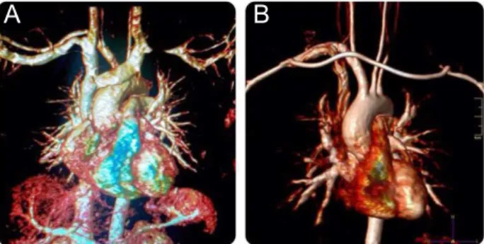
were enough to diagnose our TA patients: Claudication of upper extremities, brachial artery pulselessness or de-creased brachial artery pulse, onset age under 40 years, systolic arterial pressure gradient higher than 10 mm Hg between upper extremities, arteriography showing steno-sis or occlusion of aorta and its primary branches includ-ing large arteries in the proximal upper and lower extrem-ities (Figure 1).
Surgery was performed using end-to-side anastomosis in all cases both proximally and distally.
The patients who failed medical therapy were considered for operation. This included the patients with acute oc-clusive or claudication symptoms and chronic symptoms with under maximum medical therapy. All revascular-izations were performed by open surgical technique us-ing biosynthetic or PTFE graft. No balloon or stentus-ing was applied. Non-inflamed arterial districts were chosen
for both anastomotic sites. Regional inflow and outflow of bypass anastomotic sites were evaluated peropertively to maintain maximum graft patency. Perioperative anti-thrombotic therapy and antibiotics were administered ac-cording to the routine of the individual vascular unit. All patients were administered 100 mg acetylsalicylic acid at the first postoperative day of operation.
The biosynthetic graft used in this study; Omniflow II (Bio Nova International Pty Ltd, North Melbourne, VIC, Australia) is a collagen-polyester composite that has been successfully used for peripheral vascular replacement.7
Outcome Parameters
Patients were examined for clinical findings monthly during the first 6 months. All patients were invited for the follow up visit 10 days after the discharge, and their out-comes and complaints were recorded. Graft patency was evaluated with arterial ultrasonography at sixth month, first, secondand third year controls.
Results
A total of 24 patients met the eligibility criteria for the study. Of the 24 patients (17 females, seven males), the mean age was 38.6 ± 4.2 (range 26 to 47) years. The mean duration of follow-up for the study group was 37.9 ± 6.9 months. Both biosynthetic and PTFE groups included 12 patients. The mean age was 37.6 ± 4.81 (range 26 to 44) years for Biosynthetic group, and 39.5 ± 3.5 (range 34 to 47) years for PTFE group. The mean follow-up time was 37.6 ± 4.8 months for Biosynthetic group, and 39.5 ± 3.5 months for PTFE group.
The patients presented with various complaints as
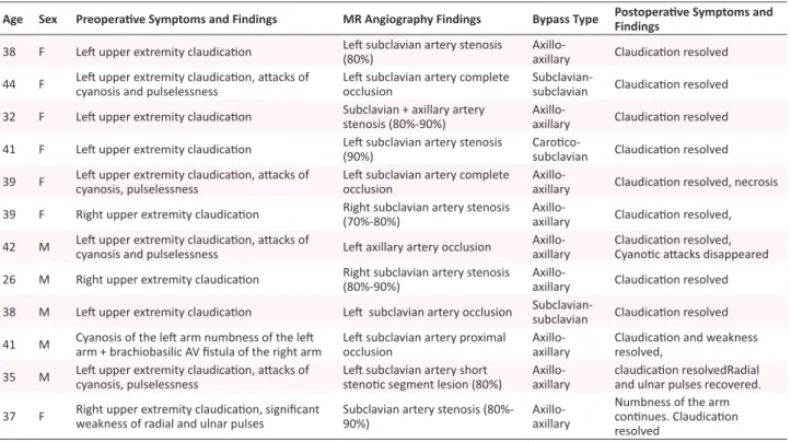
im-Table 1. Clinical Manifestations and Postoperative Findings of Patients Treated With Biosynthetic Grafts
Age Sex Preoperaive Symptoms and Findings MR Angiography Findings Bypass Type Postoperaive Symptoms and Findings
38 F Let upper extremity claudicaion Let subclavian artery stenosis (80%)
Axillo-axillary Claudicaion resolved
44 F Let upper extremity claudicaion, atacks of cyanosis and pulselessness Let subclavian artery complete occlusion Subclavian- subclavian Claudicaion resolved
32 F Let upper extremity claudicaion Subclavian + axillary artery
stenosis (80%-90%) Axillo-axillary Claudicaion resolved
41 F Let upper extremity claudicaion Let subclavian artery stenosis
(90%) Caroico-subclavian Claudicaion resolved 39 F Let upper extremity claudicaion, atacks of cyanosis, pulselessness Let subclavian artery complete occlusion Axillo-axillary Claudicaion resolved, necrosis
39 F Right upper extremity claudicaion Right subclavian artery stenosis (70%-80%) Axillo-axillary Claudicaion resolved,
42 M Let upper extremity claudicaion, atacks of cyanosis and pulselessness Let axillary artery occlusion Axillo-axillary Claudicaion resolved, Cyanoic atacks disappeared
26 M Right upper extremity claudicaion Right subclavian artery stenosis
(80%-90%) Axillo-axillary Claudicaion resolved
38 M Let upper extremity claudicaion Let subclavian artery occlusion Subclavian- subclavian Claudicaion resolved
41 M Cyanosis of the let arm numbness of the let arm + brachiobasilic AV istula of the right arm Let subclavian artery proximal occlusion Axillo-axillary Claudicaion and weakness resolved,
35 M Let upper extremity claudicaion, atacks of cyanosis, pulselessness Let subclavian artery short stenoic segment lesion (80%) Axillo-axillary claudicaion resolvedRadial and ulnar pulses recovered.
37 F Right upper extremity claudicaion, signiicant weakness of radial and ulnar pulses Subclavian artery stenosis (80%-90%) Axillo-axillary
Numbness of the arm coninues. Claudicaion resolved
paired vision, fainting, weakness of arm muscles, and numbness. Physical examination revealed hypertension, and differences in blood pressures, and pulse rates be-tween both extremities.
In Biosynthetic group, subclavian-subclavian (n = 2), ax-illo-axillary (n = 9) and carotico-subclavian (n=1) bypass operations were performed. In PTFE group, an-subclavian (n = 3), axillo-axillary (n = 7), subclavi-an-left ulnar (n = 1), subclavian-distal brachial (n = 1) bypass operations were performed. In PTFE group, 4 pa-tients developed recurrent claudication. Three papa-tients who underwent subclavian-subclavian bypass surgery developed graft occlusion at 6th, 23th and 24th months and 2 patients who had axillo-axillary bypass surgery
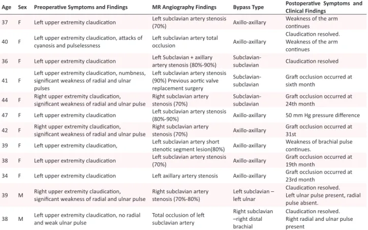
de-Table 2. Clinical Manifestations and Follow-up Findings of Patients Treated With Polytetraluoroethylene (PTFE) Grafts
Age Sex Preoperaive Symptoms and Findings MR Angiography Findings Bypass Type Postoperaive Symptoms and
Clinical Findings
37 F Let upper extremity claudicaion Let subclavian artery stenosis
(70%) Axillo-axillary
Weakness of the arm coninues
40 F Let upper extremity claudicaion, atacks of
cyanosis and pulselessness Let subclavian artery total occlusion Axillo-axillary
Claudicaion resolved. Weakness of the arm coninues
36 F Let upper extremity claudicaion Let Subclavian + axillary
artery stenosis (80%-90%) Subclavian- subclavian Claudicaion resolved
41 F
Let upper extremity claudicaion, numbness, signiicant weakness of radial and ulnar pulses
Let subclavian artery stenosis (90%) Previous aoric valve replacement surgery
Subclavian-subclavian Grat occlusion occurred at sixth month
44 F Right upper extremity claudicaion,
signiicant weakness of radial and ulnar pulse Right subclavian artery stenosis (70%) Subclavian- subclavian Grat occlusion occurred at 24th month
47 F Let upper extremity claudicaion Let subclavian artery stenosis
(80%-90%) Axillo-axillary 50 mm Hg pressure diference
42 F Right upper extremity claudicaion,
signiicant weakness of radial and ulnar pulse Right subclavian artery stenosis (70%) Axillo-axillary Grat occlusion occurred at 31st 39 F Let upper extremity claudicaion, Let subclavian artery short
stenoic segment lesion(80%) Axillo-axillary Weakness of brachial pulse coninues.
38 F Let upper extremity claudicaion Let subclavian artery stenosis
(70%) Axillo-axillary
Grat occlusion occurred at 19th month
34 F Let upper extremity claudicaion Let axillary artery stenosis Axillo-axillary Grat occlusion occurred at
23rd month
39 M Right upper extremity claudicaion,
signiicant weakness of radial and ulnar pulse Right subclavian artery stenosis (70%-80%) Let subclavian – let ulnar
Claudicaion resolved. Let ulnar pulse present, radial pulse absent.
38 M Let upper extremity claudicaion, no radial
and weak ulnar pulse Total occlusion of let subclavian artery
Right subclavian –right distal brachial
Claudicaion resolved. Right radial and ulnar pulse present
Abbreviations: F, female; M, male.
Figure 1. MR angiography image of a 42 year old male TA patient. Both left subclavian and carotid arteries are occluded.
veloped graft occlusion at 19th and 31st months. These occlusive lesions in 5 patients were treated according to the routine of each vascular unit . Vascular revasculariza-tion was successfully achieved in all cases. No early or late infection, major amputation, or perioperative death was observed in both groups.
Discussion
TA is a chronic large vessel vasculitis of unknown etiology. The histopathological appearance, involving all the arteri-al warteri-alls, is characterized by granulomatous inflammation and “panarteritis” with developed intimal fibrous thick-ness.8 This inflammation leads to arterial stenosis,
throm-bosis and aneurysm. Prognosis changes depending upon the vascular involvement and arterial inflammation. Ex-tensive stenosis/occlusion depending on the involvement of extremity arteries and associated claudication were present in our cases. Upper extremity pulses in involved area were poor or not palpated, and upper extremity isch-emia symptoms like cyanosis, numbness including clau-dication were dominant symptom, dominant symptoms. Arteriography is considered as the gold standard in clini-cal diagnosis and classification of TA.9 Contrast-enhanced
computed tomography angiography and particularly magnetic resonance angiography can demonstrate arte-rial anatomy, wall enhancement, edema, and thickening which might enable early disease detection where lumi-nal diameter is still preserved.10 We preferred to use both
common involvement of the upper and lower extremity. To avoid complications, the surgical or endovascular procedure is recommended when the disease is dormant or inactive.1 In a comprehensive study conducted by
Saadoun et al, surgical revascularization conducted when the inflammation markers are at their highest level have more complications compared to multivariate analysis results.11 Some have claimed that surgical or
endovascu-lar interventions should be avoided during the acute in-flammatory stage of TA to avoid anastomotic dehiscence or restenosis. According to Fields et al,12 performing
sur-gery in patients with active-stage TA increases the risk of early graft revision and the progression of symptomatic disease in other arterial beds. In this study to assess TA disease activity, C-reactive protein (CRP) level was used as biologic marker of inflammation along magnetic reso-nance imaging (MRI). Active-stage TA was defined as one or more of the following: CRP (>0.9 mg/dL), arterial wall thickening on MRI or the presence of systemic symptoms. We used C-reactive protein levels for both treatment, timing of surgery. It appears that patients with CRP lev-els below 1 mg/dL had an excellent long-term outcome, but 4 patients with 1mg or higher had undergone surgery because of emergency circumstances and close long-term follow-up was required in these patients. However, in se-verely symptomatic TA patientswith SAA lesions, it is not always possible to postpone SAA reconstruction until bio-logic markers return to normal. On the other hand, these four patients had biosynthetic graft and when assesed by MR angiography grafts’ anastomotic sites were not affect-ed and the disease in the extremities did not progress. Postoperative anti-inflammatory therapy was performed for these patients.
Pharmacologic therapy is the primary therapy for TA, but surgical or endovascular treatment may be required to treat organ ischemia, renovascular hypertension, or aneurysmal lesions in late-stage TA.12 However, an
evi-dence-based consensus regarding indications for surgi-cal or endovascular intervention and optimal treatment options for patients with TA involving SAA lesions are lacking.
The multiplicity of progressive lesions is challenging in the management of TA, with endovascular treatment tak-ing priority over surgery. In TA, unlike atherosclerotic le-sions, the vessels are firm and fibrotic.Thus, arch vessels require higher balloon inflation pressure. However, this challenging anatomical structure of the lesion in TA be-comes the main reason for technical failure in subclavian artery stenosis. On the other hand, although endovascular treatment can be attempted as a less-invasive treatment for short stenotic subclavian artery lesions this treatment is associated with a higher risk of restenosis or occlusion than bypass surgery, even after postoperative anti-inflam-matory treatment.13 In another recent study the results for
endovascular intervention (angioplasty and stenting) are less encouraging in comparison and surgical treatment had a better outcome compared to endovascular interven-tion in terms of postoperative complicainterven-tions (37.5% and
50%, respectively).11 Long-term survival rates following
surgical bypass procedures are good in recent studies.12,14
Cong et al5 echo this, where restenosis occurred in 34.7%
of surgical bypasses and 77.3% of angioplasty procedures. In the light of previous studies it is quite difficult to com-pare percutaneous transluminal angioplasty (PTA) and extra-anatomic bypass procedures to each other since the subclavian artery stenosis requiring treatment is seldom observed. Aburahma et al15 compared 51 patients who had
carotico-subclavian bypass (CSB) to 12 patients who had PTA in 2007. One, 3 and 5 years patency was 100%, 98% and 96% for the CSB group and 93%, 78% and 70% for PTA group (P < .0001).On the other hand, Sharma et al16
reported a technical success of 89% in 66 TA patients who had undergone endovascular treatment. Restenosis rate was 16% at 22 months.16 Also a cohort study conducted
recently resulted in disappointment from an endovascular procedures point of view. Patients who had both endo-vascular and surgical reendo-vascularizations had good initial results, while 78% of patients on whom endovascular re-vascularization is applied and 36% of patients who under-went surgical revascularization developed restenosis.17 In
another study by Liang et al18 a high failure rate for
en-dovascular revascularization procedures to treat various arterial lesions in TA patients was reported.Surgical re-construction is mainly performed to improve brain isch-emic symptoms or to prevent stroke in patients with SAAs caused by TA.19 Generally, indications for surgery of TA
include hypertension with critical renal artery stenosis, extremity claudication limiting activities of daily living, cerebrovascular ischemia or critical stenosis of three or more cerebral vessels, moderate aortic regurgitation, and cardiac ischemia with confirmed coronary artery involve-ment.3 Extremity claudication was the most common
symptom found in our patients. In the present study ar-terial occlusion was most common in the left subclavian artery.
In anatomical locations where bypass grafting is not possi-ble or hazardous, extraanatomical bypass methods can be preferred to ensure blood supply to the ischemic region.20
TA patients included in this study have been operated using various extraanatomical bypass procedures in con-sideration of the location of the lesion and general health state of the patients.
For upper extremities, extra-anatomic bypass methods are utilized for revascularization since innominate and subclavian arteries are restricting, and bypass operations conducted through an anatomic path within thorax are associated with a high mortality and morbidity rate.21,22
Possible paths of extra anatomical bypass graft are CSB, carotid-subclavian transposition, axillo-axillary bypass and subclavian-subclavian bypass. Not many detailed studies regarding these procedures are present and in the existing studies primary and secondary patency rates are varying between 82% to 100%.23-25 Kieffer et al26 reported
re-quired secondary revascularization procedures.
Axillo-axillary bypass, one of our techniques of choice (Figure 2 A,B), is an advantageous surgical technique, be-cause it is simple and supraclavicular incision and tempo-rary carotid occlusion are not needed. In a previous study, in which long-term follow-up of axillo-axillary bypass ap-plied in 32 cases, 3- and 10-year graft patency rates have been reported as 95% and 73%, respectively.27 Thus, we
used axillo-axillary bypass in nine of 12 TA patients due to the compatibility of lesion areas. Comparative studies between CSB and axillo-axillary bypass revealed contra-dictory data about long-term patency rates. However, mortality and morbidity rates of both operations are close to each other.28 Linni et al29 compared PTA and CSB and
reported that vessels with PTA were occluded in 48% of the patients with subclavian artery occlusion, while all CSB grafts maintained their patency. We preferred CSB procedure in one TA patient with a brachiobasilic fistu-la in one arm without contrafistu-lateral carotid stenosis and achieved total graft patency for one year (Figure 3). Graft material chosen in graft interpositioning is an im-portant factor that affects patency rate. The biosynthetic vascular graft used in this study (Omniflow II®
) is com-posed of bovine collagen stabilized with glutaraldehyde, and has high biocompatibility due to biosynthetic mate-rial in its content. Polyester mesh also provides graft wall microvascularization.23 On the other hand; biosynthetic
material is also an attractive long-term option due to its bio-compatibility allowing the integration of the graft into host tissues as well as true micro-vascularisation of the graft vessel wall.Immunohistological studies of
bio-Figure 2. Preoperative and postoperative(axillo-axillary bypass) MR angiography image of a 39-year-old female.
A
B
Figure 3. Postoperative (carotico-subclavian bypass) MR angiography image of a 41-year-old female patient.
synthetic grafts show that original bovine collagen is still present in the graft and it is strengthened with the host-re-lated connective tissue even after 4 years following the transplantation of graft.30
Surgical outcomes have improved over time, aided by ad-vances in surgical techniques as well as improvements in graft technology. Our study about the use of biosynthetic grafts for TA patients has some limitations. Although our study is limited with small number of subjects, we assessed the short and midterm results of biosynthetic graft for su-pra-aortic arterial lesions in patients with TA. However, it was reassuring to achieve complete patency in biosyn-thetic graft group. A recent study by Ziomek et al31
docu-mented that prosthetic grafts had a better patency than sa-phenous vein grafts at thia arterial district. Furthermore, it was reported that ringed PTFE grafts were more durable than Dacron grafts by previous studies.32,33 However, we
could not find a study comparing biosyhnthetic and PTFE graft at this location.
In conclusion, we assessed encouraging results in man-agement of arterial lesions with biosyhnthetic grafts at su-pra-aortic location of this special patient group progress-ing with inflammation attacks, in that they have satisfac-tory short and midterm graft patencies, and lower rates of complications (ie, restenosis) requiring re-intervention.
Ethical issues
None to be declared.
Competing Interests
Authors declare no conlict of interest in this study.
References
1. Ogino H, Matsuda H, Minatoya K, Sasaki H, Tanaka H, Matsumura Y et al. Overview of late outcome of medical and surgical treatment for Takayasu arteritis.
Circulation 2008;118:2738-2747. doi: 10.1161/ CIRCULATIONAHA.107.759589
2. Johnston SL, Lock RJ, Gompels MM. Takayasu arteritis: a review. J Clin Pathol 2002; 55:481-486. 3. Kerr GS, Hallahan CW, Giordano J, Leavitt RY, Fauci
AS, Rottem M, et al. Takayasu arteritis. Ann Intern Med 1994;120:919-929.
4. Hoffman GS, Leavitt RY, Kerr GS, Rottem M, Sneller MC, Fauci AS. Treatment of glucocorticoid-resistant or relapsing Takayasu arteritis with methotrexate.
Arthritis Rheum 1994;37:578-582.
5. Cong XL, Dai SM, Feng X, Wang ZW, Lu QS, Yuan LX, et al. Takayasu’s arteritis: clinical features and outcomes of 125 patients in China. Clin Rheumatol
2010;29:973-981. doi: 10.1007/s10067-010-1496-1 6. Arend WP, Michel BA, Bloch DA. The American
College of Rheumatology 1990 criteria for the classification of Takayasu arteritis. Arthritis Rheum
1990;33:1129-1134.
1997;67:637-39.
8. Numano F, Okawara M, Inomata H, Kobayashi Y. Takayasu’s arteritis. Lancet 2000;356:1023-1025. 9. Hata A, Noda M, Moriwaki R, Numano F.
Angiographic findings of Takayasu arteritis: new classification. Int J Cardiol 1996; 54 Suppl:S155-63. 10. Mason JC. Takayasu arteritis-advances in diagnosis
and management. Nat Rev Rheumatol 2010;6:406-415. doi: 10.1038/nrrheum.2010.82
11. Saadoun D, Lambert M, Mirault T, Resche-Rigon M, Koskas F, Cluzel P, et al. Retrospective analysis of surgery versus endovascular intervention in Takayasu arteritis: a multicenter experience.
Circulation 2012;125:813-819. doi: 10.1161/ CIRCULATIONAHA.111.058032
12. Fields CE, Bower TC, Cooper LT. Takayasu’s arteritis: operative results and influence of disease activity. J Vasc Surg 2006;43(1)64–71.
13. Kim YW, Kim DI, Park YJ, Yang SS, Lee GY, Kim DK, et al. Surgical bypass vs endovascular treatment for patients with supra-aortic arterial occlusive disease due to Takayasu arteritis. J Vasc Surg 2012;55(3):693-700.
14. Miyata T, Sato O, Koyama H, Shigematsu H, Tada Y. Long-term survival after surgical treatment of patients with Takayasu’s arteritis. Circulation
2003;108(12):1474–1480.
15. AbuRahma AF, Bates MC, Stone PA, Dyer B, Armistead L, Scott Dean L, et al. Angioplasty and stenting versus carotid-subclavian bypass for the treatment of isolated subclavian artery disease. J Endovasc Ther 2007; 14:698-704.
16. Sharma S, Gupta H, Saxena A. Results of renal angioplasty in nonspecific aortoarteritis (Takayasu disease). J Vasc Interv Radiol 1998;9:429-435. 17. Maksimowicz-McKinnon K, Clark TM, Hoffman
GS. Limitations of therapy and a guarded prognosis in an American cohort of Takayasu arteritis patients.
Arthritis Rheum 2007;56:1000-1009.
18. Liang P, Tan-Ong M, Hoffman GS. Takayasu’s arteritis: vascular interventions and outcomes. J Rheumatol 2004;31:102-106.
19. Giordano JM. Surgical treatment of Takayasu’s arteritis. Int J Cardiol 2000;75(Suppl 1):S123-8. 20. Rutherford RB, Patt A, Pearce WH. Extra-anatomic
bypass: A closer view. J Vasc Surg 1987;6:437-446. 21. Erentug V, Bozbuga UN, Omeroglu NS.
Ekstra-anatomik bypass girişimleri [Article in Turkish].
Turkish J Thorac Cardiovasc Surg 2003;11:42-45.
22. Goksin I, Onem G, Baltalarli A. Ekstremite revaskülarizasyonu için alternatif yaklaşım: Ekstraanatomik bypass greftleme [Article in Turkish]
Turkish J Thorac Cardiovasc Surg 2004; 12:40-46. 23. Werkmeister JA, White JF, Ramshaw JA. Evaluation of
the Omniflow collagen-polymer vascular prosthesis.
Med Prog Technol 1994;20:231-242.
24. Ballotta E, Da Giau G, Abbruzzese E, Mion E, Manara R, Baracchini C. Subclaviancarotid transposition for symptomatic subclavian artery stenosis or occlusion. A comparison with the endovascular procedure. Int Angiol 2002;21:138-44.
25. Mingoli A, Sapienza P, Feldhaus RJ, Bartoli S, Palombi M, di Marzo L, et al. Long-term results and outcomes of crossover axilloaxillary bypass grafting: a 24-years experience. J Vasc Surg 1999;29:894-901.
26. Kieffer E, Piquois A, Bertal A, Bletry O, Godeau P. Reconstructive surgery of the renal arteries in Takayasu’s disease. Ann Vasc Surg 1990;4:156-165. 27. Rosenthal D, Ellison RG Jr, Clark MD, Lamis PA,
Stanton PE Jr, Codner MA, et al. Axilloaxillary bypass: Is it worthwhile? J Cardiovasc Surg (Torino)
1988; 29:191-195.
28. Mingoli A, Feldhaus RJ, Farina C, Schultz RD, Cavallaro A. Comparative results of carotid-subclavian bypass and axillo-axillary bypass in patients with symptomatic subclavian disease. Eur J Vasc Surg 1992;6:26-30.
29. Linni K, Ugurluoglu A, Mader N, Hitzl W, Magometschnigg H, Hölzenbein TJ. Endovascular management versus surgery for proximal subclavian artery lesions. Ann Vasc Surg 2008;22:769-775. doi: 10.1016/j.avsg.2008.08.001
30. Edwards GA, Roberts G. Development of an ovine collagen-based composite biosynthetic vascular prosthesis. Clin Mater 1992;9:211-223.
31. Ziomek S, Quinones-Baldrich WJ, Busuttil RW, Baker JD, Machleder HI, Moore WS. The superiority of synthetic arterial grafts over autogenous veins in carotid-subclavian bypass. J Vasc Surg 1986;3:140-145.
32. Law MM, Colburn MD, Moore WS, Quinones-Baldrich WJ, Machleder HI, Gelabert HA. Carotid-subclavian bypass for brachiocephalic occlusive disease. Stroke 1995;26:1565-71.
33. Owens LV, Tinsley EA, Criado E, Burnham SJ, Keagy BA. Extrathoracic reconstruction of arterial occlusive disease involving the supraaortic trunks. J Vasc Surg