CASE REPORT
Subclavian vein angioplasty during arteriovenous fistula
surgery: case report and literature review
Angioplastia de veia subclávia no intraoperatório de fístula arteriovenosa: relato de caso e
revisão de literatura
Marcus Vinícius Martins Cury1, Marcelo Fernando Matielo1, Ana Carolina Calixtro2, Giuliano de Almeida Sandri1,
Marcos Roberto Godoy1, Roberto Sacilotto3
Introduction
Hemodialysis is the main treatment in end-stage renal disease (ESRD). When possible, this treatment should be administered via an arteriovenous istula (AVF), which of-fers the best vascular access1. Compared with other options
of kidney replacement therapy, this modality of treatment is associated with reduction in infection complications and less morbidity and mortality2.
In the United States, approximately 300,000 patients are treated by hemodialysis, and vascular access is the leading
Abstract
Patients with chronic kidney disease stage 5 are generally treated by hemodialysis, preferentially performed via an arteriovenous istula (AVF). We report the case of a 58-year-old male patient with diabetes mellitus, hypertension and end-stage renal disease in whom hemodialysis was conducted via a long-term catheter. His medical record described numerous central venous cannulations and several AVF creations. he patient developed subclinical subclavian stenosis that required creation of a new vascular access route. he purpose of this case report is to describe treatment of subclavian vein stenosis during AVF creation.
Keywords: renal insuiciency, chronic; arteriovenous istula; angioplasty.
Resumo
Pacientes portadores de Insuiciência Renal Crônica (IRC) estágio V são geralmente tratados por hemodiálise (HD), preferencialmente por fístula arteriovenosa (FAV). Descrevemos um relato de caso de um paciente de 58 anos, masculino, portador de diabetes mellitus, hipertensão arterial sistêmica e IRC terminal. Seus antecedentes demonstram múltiplos acessos para implante de cateter de hemodiálise, assim como tentativas prévias de realização de FAV. Esse paciente desenvolveu estenose subclínica da veia subclávia, limitando a HD pelo membro superior. O propósito deste relato foi descrever o tratamento endovascular de estenose de veia subclávia, concomitante à realização de uma nova FAV.
Palavras-chave: insuiciência renal crônica; fístula arteriovenosa; angioplastia.
Study carried out at Vascular Surgery Department at the Hospital do Servidor Público Estadual São Paulo (HSPE) – São Paulo (SP), Brasil. 1 Assistant doctor of Vascular and Endovascular Surgery Service at the HSPE – São Paulo (SP) Brasil.
2 Resident doctor of Vascular and Endovascular Surgery Service at the HSPE – São Paulo (SP) Brasil. 3 Head of Vascular and Endovascular Surgery Service at the HSPE – São Paulo (SP) Brasil. Financial support: none.
Conlict of interest: nothing to declare. Submitted on: 28.07.11. Accepted on: 02.01.12. J Vasc Bras. 2012;11(2):154-157.
cause of hospitalization3.In Brazil, approximately 80,000
patients use hemodialysis treatment4.
Nearly 40% of patients who have previously received a subclavian vein hemodialysis catheter develop stenoses5.
Unless these lesions are corrected, creation of an upper ex-tremity AVF is limited6. Endovascular treatment has a high
initial technical success, but it is associated with low pri-mary patency and high failure rate with its consequences (e.g. development of upper limb edema)7,8.
Brief title: Subclavian vein PTA during AVF surgery - Cury MVM et al. J Vasc Bras 2012, Vol. 11, Nº 2 155
Case report
A 58-year-old patient with diabetes mellitus, hyperten-sion and chronic kidney disease (stage 5) had been treated by hemodialysis via a long-term catheter inserted in the right femoral vein. His past medical history included many central venous cannulations (right internal jugular, let in-ternal jugular, right subclavian, let subclavian, right femo-ral and let femofemo-ral veins). he patient had had a previous let femoral deep venous thrombosis (DVT), and creation of an upper extremity AVF was attempted three times (ra-diocephalic and brachiocephalic in the right arm, and bra-chiobasilic in the let arm) without success.
A color Doppler study performed to investigate the cervical and upper extremity veins revealed the absence of arm veins suitable for new surgery and detected signs of previous axillary vein thrombosis. Upper extremity phle-bography performed prior to attempting to create a new AVF showed let axillary vein occlusion and stenosis of the proximal right subclavian vein.
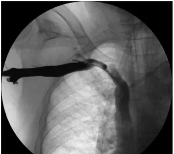
Surgery was then performed, by right axillary vein and ipsilateral brachial artery dissection. A loop subcutaneous tunnel was created in the anterior arm and a terminal-lateral nonringed polytetraluoroethylene (PTFE) 6.0 mm anasto-mosis was performed with the vein. he prosthesis was then punctured and a 10 F sheath was introduced (Figure 1). Intraoperative phlebography was obtained (Figure 2) and a hydrophilic 0.035” guide wire (Roadrunner® 260 cm;
Cook) was passed beyond the subclavian lesion to reach the inferior vena cava. he diameter of the vein was esti-mated as 12 mm; ater infusion of 5000 UI of intravenous heparin, subclavian vein angioplasty was performed using a 14×40 mm vascular balloon (XXL®; Boston Scientiic)
in-lated to 10 atm for 3 minutes. Recoil was noted following a
second angiography, and a self-expanding stent (16x40 mm Wallstent®; Boston Scientiic) was satisfactorily deployed,
preserving the right internal jugular ostium (Figure 3). he PTFE grat was passed through the tunnel, and brachial anastomosis was performed. Lastly, the sheath was removed and the prosthesis closed with Prolene sutures. A thrill was identiied at the axillary vein, and evolution of AVF and the patient was satisfactory. Clopidogrel and Aspirin were initiated on the irst day ater surgery.
Color Doppler study surveillance performed four weeks ater surgery showed AVF patency, signs of PTFE integra-tion and a low rate of 640 mL/minute. Hemodialysis via the AVF was satisfactorily initiated six weeks ater surgery.
Figure 3. Final image.
Figure 1. PTFE puncture with 10 F sheath.
Brief title: Subclavian vein PTA during AVF surgery - Cury MVM et al. J Vasc Bras 2012, Vol. 11, Nº 2
156
Discussion
Many guidelines recommend that kidney replacement treatment should preferably be performed by an autogenous AVF3,5. Until 2002 in the United States, access was created
using prosthesis in approximately 80% of cases for reasons of accessibility, early cannulation and thrombectomy treat-ment in cases of occlusion9.Cumulative patency, infection
rate and survival analysis have shown better results with autogenous conduits; however, these indings were derived from low-evidence reports10.
Central venous stenoses are common in central vein cannulation, especially in subclavian vein hemodialysis catheters. Almost 50% of patients with these catheters have stenoses or occlusions11, which are a major impediment to
upper extremity access8. Ater obtaining arteriovenous
ac-cess, central venous stenoses sometimes lead to swelling of the arm; ater obtaining prosthetic vascular access, attempts to treat these lesions can result in an arm hematoma and related complications. We could ind no previous reports regarding central venous angioplasty during AVF creation.
Central venous lesions are short and have ibrotic fea-tures12. his condition makes endovascular treatment a
favorable approach that has high initial technical success (approximately 90%)13. he 1-year primary patency rate for
balloon angioplasty is approximately 30%, but the feasibil-ity of performing repeat angioplasty makes this treatment a feasible option8. Treatment by primary stenting has been
studied previously, but is not associated with an increase in primary patency, and thus it should be used selectively8,14.
Stent placement is recommended in situations of recoil or failure, which is identiied when a symptomatic patient re-turns for treatment (e.g. development of arm swelling, un-successful hemodialysis)14. he primary assisted program
may require multiple angioplasties to achieve a 1-year cu-mulative patency (CP) result of 70%8,13,15.
Preservation of the ostium of the internal jugular vein is important during stent placement to ensure that the pos-sibility of future central venous cannulation is retained. Similarly, preservation of the ostium is also beneicial in stenting of the contralateral innominate vein16.
Wallstent® self-expanding stents are generally used in
treatment of central venous stenosis7,8,13-16; the use of Nitinol
stents is also reported for this purpose17. Both types
pro-duced similar results regarding primary patency and free-dom from symptoms, but, in the United States, Nitinol stent placement is an of-label procedure for central veins. Considerations in stent selection are that elgiloy alloy (Wallstent®) has better resistance to external compression,
but that Nitinol has greater radial strength and conforms better to the wall of the vein.
Recently, Haskal et al. conducted a multi center study,
in which dysfunctional access were randomized to receive balloon angioplasty or stent grat (Bard Flair stent®). At
six months follow-up, stent grat group had better results of freedom from reintervention and patency (51x23%; p<0.001)18.
Creation of lower extremity arteriovenous access is an alternative treatment in patients with upper extremity vein outlow obstruction. Saphenous vein tight transposition has 1-year cumulative patency of 93%, but sometimes the use of this conduit is not possible because of previous usage or the presence of peripheral arterial disease. Otherwise, there is the possibility of prosthetic vascular access, which has a rate of complications from infection of 20%19.
Surveillance of arteriovenous hemodialysis access re-mains controversial20. Some types of surveillance are
poten-tially beneicial: in particular, measurement of volume low rate can detect early dysfunction of vascular access, and detection should be encouraged as a method of increasing vascular patency. his approach is reported as a method of reducing costs, hospitalization, morbidity and mortality. A flow rate of less than 600 mL/minute or a 20% reduction in low rate over 1 month are predictive signs of arterio-venous occlusion5,21.
Conclusion
he present case report describes an alternative treat-ment of subclavian stenosis that was performed via the axil-lary vein. his approach enabled treatment without another vein puncture and with the advantage of performing the an-gioplasty under favorable conditions (absence of arm swell-ing and hematoma development ater prosthesis puncture). In the present patient, previous deep venous thrombosis of the lower extremity limited access through the inferior vena cava. he present procedure was successful. Previous reports regarding angioplasty and stenting favor endovas-cular treatment.
References
1. Ravani P, Marcelli D, Malberti F. Vascular acess surgery managed by renal physicians: the choice of native arteriovenous istulas for hemodialysis. Am J Kidney Dis. 2002;40(6):1264-76.
Brief title: Subclavian vein PTA during AVF surgery - Cury MVM et al. J Vasc Bras 2012, Vol. 11, Nº 2 157
3. National Kidney Foundation. KDOQI Clinical Practice Guidelines and Clinical Practice Recommendations for 2006 Updates: Hemodialysis Adequacy, Peritoneal Dialysis Adequacy and Vascular Access.Am J Kidney Dis. 2006;48(Suppl):1-322.
4. Tordoir J, Canaud B, Haage P, et al. European best practice guide-lines on vascular access. Nephrol Dial Transplant 2007;22(Suppl 2): ii88-117.
5. Padberg Jr FT, Calligaro KD, Sidawy AN. Complications of arterio-venous hemodialysis access: Recognition and management. J Vasc Surg 2008;48(Suppl):S55-80
6. Kim YC, Won JY, Choi SY, et al.Percutaneous treatment of central venous stenosis in hemodialysis patients: long-term outcomes. Cardiovasc Intervent Radiol. 2009;32(2):271-8
7. Bakken AM, Protack CD, Saad WE, Lee DE, Waldman DL, Davies MG. Long-term outcomes of primary angioplasty and primary stenting of central venous stenosis in hemodialysis patients. J Vasc Surg. 2007;45(4):776-83.
8. Huber TS, Carter JW, Carter RL, Seeger JM. Patency of autogenous and polytetraluoroethylene upper extremity arteriovenous he-modialysis accesses: a systematic review. J Vasc Surg. 2003;38(5): 1005-11.
9. Murad MH, Elamin MB, Sidawy AN, et al. Autogenous versus pros-thetic vascular access for hemodialysis: a systematic review and meta-analysis. J Vasc Surg. 2008;48(5 Suppl):34-47.
10. Schwab SJ, Quarles LD, Middleton JP, Cohan RH, Saeed M, Dennis VW. Hemodialysis-associated subclavian vein stenosis. Kidney Int. 1988;33(6):1156-9.
11. Begin V, Ethier J, Dumont M, Leblanc M. Prospective evaluation of the intra-access low of recently created native arteriovenous istulae. Am J Kidney Dis. 2002;40(6):1277-82.
12. Nael K, Kee ST, Solomon H, Katz SG. Endovascular management of central thoracic veno-occlusive diseases in hemodialysis patients: a single institutional experience in 69 consecutive patients. J Vasc Interv Radiol. 2009;20(1):46-51.
13. Maya ID, Saddekni S, Allon M. Treatment of refractory central vein stenosis in hemodialysis patients with stents. Semin Dial. 2007;20(1):78-82.
14. Oderich GS, Treiman GS, Schneider P, Bhirangi K. Stent placement for treatment of central and peripheral venous obstruction: a
long-term multi-institutional experience. J Vasc Surg. 2000;32(4): 760-9.
15. Haage P, Vorwerk D, Piroth W, Schürmann K, Günther RW. Treatment of hemodialysis-related central venous stenosis or oc-clusion: results of primary Wallstent placement and follow-up in 50 patients. Radiology. 1999;212(1):175-80.
16. Rajan DK, Saluja JS. Use of nitinol stents following recanalization of central venous occlusions in hemodialysis patients. Cardiovasc Intervent Radiol. 2007;30(4):662-7.
17. Antoniou GA, Lazarides MK, Georgiadis GS, Sfyroeras GS, Nikolopoulos ES, Giannoukas AD. Lower-extremity arteriovenous access for haemodialysis: a systematic review. Eur J Vasc Endovasc Surg. 2009;38(3):365-72.
18. Haskal ZJ, Trerotola S, Dolmatch B, et al. Stent graft versus bal-loon angioplasty for failing dialysis-access grafts. N Engl J Med. 2010;362(6):494-503.
19. Casey ET, Murad MH, Rizvi AZ, et al. Surveillance of arteriovenous hemodialysis access: a systematic review and meta-analysis. J Vasc Surg. 2008;48(5 Suppl):S48-54.
20. Malik J, Slavikova M, Svobodova J, Tuka V. Regular ultrasonograph-ic screening signiultrasonograph-icantly prolongs patency of PTFE grafts. Kidney Int. 2005;67(4):1554-8.
21. Kim YO, Yang CW, Yoon SA, et al. Access blood low as a predic-tor of early failures of native arteriovenous istulas in hemodialysis patients. Am J Nephrol. 2001;21(3):221-5.
Correspondence
Marcus Vinícius Martins Cury Serviço de Cirurgia Vascular Av. Pedro de Toledo, 1800, 14º andar/ala ímpar - Vila Clementino CEP 04039-901 – São Paulo (SP), Brazil E-mail: [email protected]
Author’s contributions