9
Prevalence of some mastitis causes in dromedary camels in Abu Dhabi,
United Arab Emirates
A.A. Al-Juboori
1, N.K. Kamat
2and J.I. Sindhu
21 Development Sector, Research and Development Division, 2 Animal Wealth Sector, Animal Health Division, ADFCA, UAE
(Received August 6, 2012; Accepted January 20, 2013)
Abstract
The present study was designed to determine the prevalence of different types of mastitis in camels in U.A.E. and to identify the causative microorganisms and their sensitivity to different antimicrobial agents. From 162 lactating she-camels, 630 milk samples were collected from different cities in Abu Dhabi Emirate/UAE. The overall prevalence of mastitis was 18.52% (7.94% on quarter basis), the prevalence of clinical and sub clinical mastitis was found to be 24.70% and 11.67% on animal basis, respectively; it being 9.70% and 5.86% on quarter basis, respectively. The hind quarters were more frequently affected than the fore quarters. Bacteriological examination of milk samples revealed that Staphylococcus was the chief
etiological agents both in clinical and sub clinical mastitis (41.67%) in camels, followed by Streptococcus spp. (21.67%), Enterobacter spp. (15.00%), C. pyogenes (10.00%), Micrococcus spp. (5.00%), Pasteurells spp. (5.00%) and Pseudomonas aeruginosa (1.66%). Most of the Staphylococcus spp., Streptococcus spp. and C. pyogenes strains were sensitive to
carbenicillin, gentamycin, kanamycin, and erythromycin, but resistant to colistin and sulphamethoxazole. Other pathogens like
Enterobacter, Micrococcus, Pasteurella spp. and Ps. aeuroginosa isolates showed variable sensitivities to the antimicrobials.
Keywords: Camelus dromedaries; Clinical mastitis; Incidence; Etiological agents; Antimicrobial agents.
Available online at http://www.vetmedmosul.org/ijvs
! " #
$
# % &
' ! (
)!
*
+
,+ - + . /0 1
2 3
4
! " # $ %&
'
156
( ) *++
, -+
.
/
0 1 %
23 )4 564
7 0+ %8 ( .9 :)3
3 ; < 7 .+
= >+
? @ !,
-!,
! " #
.
A ,
*
B
)
BCD
E 9 F 4
(
! " #
3 23 GH F3 )4 23
.
. # I9
/
0 1 %
* J K
L,M
) %
NOPQ
%
R 2
(
I9
+J
* J S
. * 8 S
.
0 1 %
@
Q,ND
) %
P,ND
%
R 2
(
,BN
) %
M,LB
%
R 2
(
!
T 4
.
J
U V W8 JR
0 != > E <
S
. * 8 S
.
0 1 %
)
BP,BP
%
NB,QN
%
,
!
T 4
(
!3 3R E <
W3
)
CD,CD
%
COMC
%
!
T 4
(
.
* (
X
WF- 7
K+
)
Q
,BN
(%
S
. * 8 S
.
0 1 % 7Y 9 23 JR )W
% 8
.+ 7
K+ Z ; &
)
,BN
(%
W 7 A4 Z ; & Z;
)
M,DD
(%
; &
Z
W 7 )8
)
D,DD
(%
Z ; &
W ) 7
K+
)
M,DD
(%
(
Z ; &
V
)
M,DD
(%
Z ; &
[' 6
W
)
,BB
%
(
.
( .
\ ]'
* ^ #
Z_-3 ` Z ; < [ >+
7Y64
WF- 7
K+ Z ; &
W 7 )8
Z ; &
.+ 7
K+
)
J#
23
LD
(%
* J
S
0+ ( .9
2 . 3 F< 2 .F K
2 . 3 FK
2 . 3
R
5 $ .J 3 = .
2 . K 3 W3 %FK
.
10
6-+
^ + Z ; < ! 7 %"#
Z ; & ? 3
7 -+
W ) 7
K+
W [' 6
V
(
#
+
>3 H
=
( .
7 0+ a <8
.
Introduction
The dromedary camel (Camelus dromedarius) is the most important livestock animal in the desert and semi-desert areas of Northern and Eastern Africa as well as in the deserts of the Arabian Peninsula. Dromedary camels rearing in the UAE are mainly for milk, meat production and for racing purposes (1). In UAE, the population of dromedary camels is around 459,242 (2). All are one-humped camels and are commonly found in certain parts of UAE, especially Abu Dhabi and Al-Ain cities and Western region. Peak milk yield of 20-40 liter per day has been recorded (3-5). Mastitis is a complex disease occurring world-wide among dairy animals, with heavy economic losses (6-9) due to of reduced milk yield, degradation of milk quality and additional cost in the care and treatment of mastitis (10). Bacterial infections are considered the primary cause of mastitis in domestic animals (10). Reports of inflammation of the camel udder have appeared from various countries, such as Egypt (11,12), India (13), Saudi Arabia (14,15), Somalia (16,17), Sudan (18), UAE (19). Few available literatures indicate that Staphylococcus aureus, Streptococcus spp. (10,14,20-25), Micrococcus spp. (12,20,26), Streptococcus agalactiae (19,27,28), coagulase negative staphylococci (20), Pasteurella haemolytica (21), Escherichia coli (20,21) and Corynebacterium spp. (14,24) have been implicated as causes of mastitis in camels. Accordingly, the present study was taken up with a view to determine the prevalence of different types of mastitis in camels in U.A.E., to identify the causative microorganisms and to determining the sensitivity of mastitis pathogens to different antimicrobial agents. Accordingly, the present study was taken up with a view to determine the prevalence of different types of mastitis in camels in U.A.E., to identify the causative microorganisms and to determining the sensitivity of mastitis pathogens to different antimicrobial agents.
Materials and methods
For assessing the prevalence of different types of clinical mastitis in camels, a systemic survey was conducted; this was done by visiting different camel herds around Abu Dhabi Emirate in the time period between September 2009 to December 2011. The camels were allowed to graze freely in the desert, but were also supplemented with concentrate feed and green grass. The information pertaining to the camels examined during this study was conducted. This included age, lactation number, stage of lactation, pregnancy, previous mastitis history, etc.
Clinical mastitis was recognized by abnormal milk secretion, signs of udder inflammation and detection of mastitis pathogens by bacteriological culture, whereas subclinical mastitis was recognized by apparently normal milk, total count of somatic cells and the presence of pathogenic microorganisms.
Collection of milk samples
A total of 630 milk samples from 162 lactating she-camels,were collected from the individual quarters infected with different types of subclinical and clinical mastitis separately in the sterilized test tubes under aseptic precautions, in accordance with International Dairy Federation standards (29). The milk samples immediately after collection were taken to the central laboratory at Al Wathba Veterinary Hospital, Baniyas, Abu Dhabi for bacteriological examinations.
Cultural isolation
Using standard microbiology techniques including: Culture on general, selective and special indicators media with the special supplement and different atmospheres necessary for different bacteria. Whether they are fast or slow growing and fastidious or regular in their growth needs (aerobic, 10% Co2 and anaerobic conditions), (30). Primary incubation may take from 24-72 hrs according to the microorganism species. Temperature of incubation is 37 °C for most of cases although some cases need to be incubated on another range (25 -45°C) (31,32). Identification using commercially available sets of biochemical and enzymatic testing for the identification of the isolates (API 20 A, NE, E and API 20 Coryn) and rapid microbial detection systems like Vitek 2 (Biomerioux France).
Antimicrobial sensitivity test
The antimicrobial sensitivity test was conducted using Bauer–Kirby technique, as described by (33). The antimicrobial agents used in this study were carbenicillin, gentamycin, kanamycin, erythromycin, ampicillin, cephalothin, tetracycline, penicillin G, colistin, sulphomethoxazole and streptomycin.
Statistical Analysis
11
ResultsPrevalence of mastitis
In order to determine the prevalence of subclinical and clinical mastitis in camels, a total of 162 lactating she-camels (630 quarters) were examined for the presence of mastitis. The results are presented in Table 1. Out of 85 camels (340 quarters) examined for the presence of clinical mastitis, 21 camels (33 quarters) were found to have clinical mastitis, the incidence being 24.70% (9.70% on quarter basis). A total of 77 camels were examined for estimating the prevalence of subclinical mastitis, 30 (11.67%) camels were found to have subclinical mastitis; these animals were carrying mastitis pathogens in their udder secretion. Subclinical infection of mastitis was detected in 17 (5.86%) of the 290 quarters examined. Taking into consideration all the 162 camels examined during this study, the prevalence of mastitis on animal basis was found to be 18.52%, while on quarter basis 7.94%. It
was further observed that hind quarters were affected with clinical and subclinical infections (69.69% and 76.47%, respectively) more frequently than fore quarters (30.30% and 23.53 %, respectively) (Table 2).
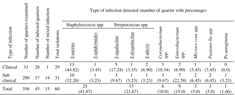
Frequency distribution of mastitis pathogens
The data available on the relative frequency of different types of microorganisms encountered in udder infection (Table 3) revealed that Staphylococcus was the most
important organism involved in the causation of clinical and subclinical mastitis in camels (41.67 %). Streptococcus spp. came next in importance (21.67%), followed by Enterobacterium spp. (15.00%), C. pyogenes (10.00%),
while the prevalence of miscellaneous organisms was low. Among the staphylococcal infections, S.aureus was the
predominant in the infections of clinical and subclinical mastitis (38.33%). while among the streptococcal infection, Str.agalactiae was predominant (13.33 %).
Table 1: Incidence of clinical and subclinical mastitis in camels. Type of
infection camels examined Number of quarters examined Number of Number showing mastitis Animals Quarters On animal basis Prevalence rate On quarter basis
Clinical 85 340 21 33 24.70% 9.70%
Sub clinical 77 290 9 17 11.67% 5.86%
Total 162 630 30 50 18.52% 7.94%
Table 2: Distribution of clinical and subclinical infections in camels according to the position of quarters involved.
Type of infection Number of infected quarters Left-hind Left-fore Distribution Right-hind Right-fore
Clinical 33 (33.33%) 11 (9.10%) 3 (36.36%) 12 (21.21%) 7
Subclinical 17 (23.53%) 4 (5.88%) 1 (52.94%) 9 (17.65%) 3
Total 50 (30.00 %) 15 (10.00 %) 4 (42.00 %) 21 (20.00 %) 10
Sensitivity of mastitis pathogens
As many as 60 isolates obtained from the cases of clinical and subclinical mastitis, during the present study, were subjected to sensitivity test, using 11 different antimicrobial agents as listed in Table 4. It is evident from the table that most of the Staphylococcus spp., Streptococcus spp. and C. pyogenes strains (> 80%) were
sensitive to carbenicillin, gentamycin, kanamycin and erythromycin. All these isolates were however found resist
to colistin and sulphamethoxazole. The Enterobacter
isolates showed moderate sensitive to carbenicillin and sulphamethoxazole, while less sensitive or even resist to the other antimicrobial agents. All the Micrococcus isolates
were found highly sensitive to carbenicillin, gentamycin, cephalothin and sulphamethoxazole. While the Pasteurella spp. showed highly sensitive only to carbenicillin and
cephalothin. Ps. aeruginosa were found resist to all
12
Table 3: Relative frequency of different type of mastitis pathogens in clinical and sub clinical infections.
T yp e of in fe ct io n N um be r of q ua rt er s ex am in ed N um be r of in fe ct ed q ua rt er s N um be r of m ix ed in fe ct io n T ot al is ol at io ns
Type of infection detected (number of quarter with percentage)
Staphyloccoccu spp. Streptococcus spp.
C o ry n eb a ct e ri u m sp p . E n te ro b a ct e ri u m sp p . M ic ro c o c cu s sp p . P a st eu re l la s p p . Ps .a er ug in os a S . a u re u s S . e p id e rm id e s S . a g a la ct ia e S . d y sg a la c ti a e S . u b e ri s
Clinical 33 28 1 29 (44.82) 13 (3.45) 1 (17.24) 5 (3.35) 1 (6.90) 2 (10.34) 3 (6.90) 2 (3.45) 1 (3.45) 1 (0.0) 0 Sub
clinical 290 17 14 31 (32.26) 10 (3.23) 1 (9.67) 3 (3.23) 1 (3.23) 1 (9.67) 3 (22.58) 7 (6.45) 2 (6.45) 2 (3.23) 1 Total 356 45 15 60 (41.67) 25 (21.67) 13 (10.0) 6 (15.0) 9 (5.0) 3 (5.0) 3 (1.66) 1
Table 4: In vitro sensitivity of mastitis pathogens isolates.
Antimicrobial agent C on ce nt ra tio n pe r di sc
Sensitivity in percentage
S ta p h y lo c o cc u s sp p . ( 25 ) * S tr e p to c o c cu s sp p . ( 13 ) * C . p y o g e n e s ( 6 )* E n te ro b a ct e ri a l sp p . ( 9 )* M ic ro c o cc u s sp p . ( 3 )* P a st eu re ll a sp p . ( 3 )* P s. a e ru g in o sa ( 1 )*
Carbenicillin mcg 100 S: 96.00 I: 4.00 100.00 0.0 100.00 0.0 88.89 11.11 100.00 0.0 100.00 0.0 100.00 0.0
Gentamycin mcg 10 S: 92.00 I: 8.00 100.00 0.0 100.00 0.0 77.78 22.22 100.00 0.0 66.67 33.33 100.00 0.0
Kanamycin mcg 30 S: 88.00 I: 12.00 84.62 15.38 16.67 83.33 55.56 44.44 66.67 33.33 66.67 33.33 100.00 0.0
Erythromycin mcg 15 S: 80.00 I: 20.00 100.00 0.0 16.67 83.33 55.56 44.44 66.67 33.33 66.67 33.33 100.00 0.0
Ampicillin mcg 10 S: 76.00 I: 24.00 76.92 23.08 33.33 66.67 55.56 44.44 66.67 33.33 66.67 33.33 100.00 0.0
Cephalothin mcg 30 S: 60.00 I: 40.00 61.54 38.46 50.00 50.00 77.78 22.22 100.00 0.0 100.00 0.0 100.00 0.0
Tetracycline mcg 30 S: 68.00 I: 32.00 76.92 23.08 33.33 66.67 33.33 66.67 100.00 0.0 100.00 0.0 100.00 0.0
Penicillin G units 10 S: 64.00 I: 36.00 61.54 38.46 33.33 66.67 11.11 88.89 33.33 66.67 100.00 0.0 100.00 0.0
Colistin mcg 10 S: 44.00 I: 56.00 30.77 69.23 83.33 16.67 77.78 22.22 66.67 33.33 66.67 33.33 100.00 0.0
Sulphamethoxazole mcg 50 S: 36.00 I: 64.00 23.08 76.92 66.67 33.33 88.89 11.11 100.00 0.0 66.67 33.33 100.00 0.0
13
DiscussionThe prevalence of camel mastitis as revealed during the present study is considered low, especially when compared to the report of (10,19,21,25,34-37), which revealed the prevalence rate ranging from 38%-83%. There is no definite explanation for the relative low prevalence of mastitis in camels in this study, but the possible factors contributing to it may be the hygienic milking procedures followed by the camel owners and the good hygienic condition of the milking area. It was also noticed during the present study that the prevalence of clinical mastitis was higher than subclinical infections. However, (12-14,19,21,36,38-41) who found subclinical mastitis seems to be more common. The majority of the clinical cases in this study were sub acute infections, with signs of swelling of the udder and teats, heat, congestion and pain of the mammary gland as well as abnormal milk secretion. However, (42) who reported chronic mastitis (57.15%) was the commonest clinical mastitis. Peracute, chronic and gangrenous mastitis have been described in camels (13,14,18,21,25,41-43). It was further observed that hind quarters were affected with clinical and subclinical infections more frequently than fore quarters. A higher risk of infections in hind quarters compared to the front ones which could be due to the unfavorable hygienic condition; greater exposure to dung and urine. In addition, due to the shorter length of the hind teats with a corresponding shorter teat canal, the defence potential in the hind quarter could be decreased. However, (26) found that subclinical mastitis was higher in fore quarters than hind quarters. Bacteriological examination of milk samples revealed that Staphylococci was the chief etiological agents both in
clinical and sub clinical mastitis (41.67%) in camels, followed by Streptococcus spp. (21.67%), Enterobacter spp. (15.00%), and C. pyogenes (10.00%), while the
incidence of miscellaneous organisms was low. These findings, in general, agree with those of other workers investigating in camels in Iraq (21,42), in Saudi Arabia (14,25,43,45), in Egypt (21,34,39) in U.A.E. (19,46,47), in Sudan (18), In India (13,36), in Nigeria (48), and in Kingdom of Bahrain (50). However, (10) from Ethiopia found the predominant etiological agents of camel mastitis in the study area were found to be coagulase negative staphylococci. (24) reported that the most predominant
bacterial isolates were Micrococcus spp., Staphylococcus aureus, Streptococcus spp. and Corynebacterium spp. The
isolation of genera of pathogenic bacteria from the camel milk samples suggests the need for strict hygienic measures during the production and handling of camel milk to reduce public health hazards. Education of the camel owners about the importance of hygienic milking practices would minimize the adverse effect of mastitis on the yield and quality of camel milk. A study to determine the sensitivity
of mastitis pathogens, isolated during the present investigations, revealed that Most of the Staphylococcus spp., Streptococcus spp. and C. pyogenes strains were
sensitive to carbenicillin, gentamycin, kanamycin, and erythromycin, but resistant to colistin and sulphamethoxazole. The other mastitis pathogens like
Enterobacter, Micrococcus, Pasteurella spp. and Ps. aeruginosa isolates were showed variable pattern of
sensitivity to the antimicrobial agents. This suggested that these antimicrobial agents could be used for treatment of mastitis in camels in U.A.E. More or less similar pattern of sensitivity of the bacterial isolates in the present study to some of the above mentioned antimicrobial agents have been reported by (24,26,44,48,50).
References
1. Abdulwahhab Y. Lameness of camels in United Arab Emirates. Proceeding of the 16th Symposium and 8th Conference of Lameness in
Ruminants 2011, 28 February-3rd March, Rotorua, New Zealand.
2. Statistics Center- Abu Dhabi (SCAD). 2010. P: 227-228.
3. Knoess KH. Milk production in the dromedary, in IFS Int. Symp. Camels, Sudan: 1979. 201-214.
4. Qureshi MH. The camel. Seminar on the Camel. FAO. Oct 1986. Lahore, Pakistan.
5. Khanna ND, Rai AK. Milk production potential of Indian camel. Asian Livestock (FAO). 1993;18 (2): 19-21.
6. AL-Ani FK. Camel Management and Diseases. /Animal Diseases/Camel/ Infection Diseases.first Edition AL-Sharq Printing Press, Jordan. 2004.;pp: 331-335.
7. Sudhan NA, Neelesh Sharma. Mastitis- An Important Production Disease of Dairy Animals. SMVS‘DAIRY YEAR BOOK. 2010. 8. Maichomo MW, Kibugu J, Kurgat R, Malonza VM. Importance of
Sub-Clinical Mastitis Due To Streptococcus Agalactiae in Camels. Kenya Agricultural Research Institute, Trypanosomiasis Research Centre, Wednesday, 2011, 03 August.
9. Bhikane AV, Kawitkar SB. Hand book for Veterinary Clincian.Venkatesh Books. Udgir, India. 2000.
10. Eyassu S, Bekele T. Prevalence and etiology of mastitis in traditionally managed camels (Camelus dromedarius) in selected pastoral areas in eastern Ethiopia. Ethiop Vet J. 2010;14 (2):103-113. 11. Hassanien A, Soliman AS, Ismail MA. Clinical case of mastitis in
she-camel (Camelus dromedarius) caused by Corynebacterium pyogenes. Assiut Vet Med. J. 1984;12: 239-241.
12. Moustafa AS, Ragale AM, Safwat EE, Sayed Z, Mervat j. El-Rehaman A, El-Danaf NA, Shouman MT. Examination of raw she-camel milk for detection of subclinical mastitis. J. Egypt Vet Med Ass. 1987;47:117-128.
13. Kapur MP, Khanna BM, Singh RP. A peracute case of mastitis associated with Klebsiella pneumoniae and Escherchia coli. Indian Vet J. 1982.59: 650-651.
14. Barbour EK, Nabbut NH, Frerichs WM, Al-Nakhli HM. Al-Mukayel AA. Mastitis in Camelus dromedarius in Saudi Arabia. Trop Anim Hlth Prod. 1995;17: 173-179.
15. Hafez AM, Razing SA, Al-Amrousi S, Ramadan RO. 1987. Studies on mastitis in farm animals in Al-Hassa. 1. Analytical studies. Assiut Vet J. 19:139-145.
16. Abdurahman O, Bornstein S, Osman K, Abdi AM, Zakrisson G. Prevalence of mastitis among camels in southern Somalia: a pilot study. Camel forum, working paper, 1991;pp: 37:1-9.
14
Somalia. Bullettino Scientifica della Facolta di Zootecniue Veterinaria, Universita Nazionale Somalia. 1984;4:99.
18. Obeid AI. Field investigation, clinical and laboratory findings of camel mastitis. MVSc. Thesis, University of Khartoum. 1983. 19. Quandil SS, Quadar J. Bacteriological study of some cases of mastitis
in the dromedary (Camelus dromedarius) in the United Arab Emirates. Review Med. Vet J. 1984;135:705-707.
20. Abdurahman OA, Agab H, Abbas B, Åström G. Relations between udder infection and somatic cells in camel (Camelus dromedarius) milk. Acta Vet Scand. 1995; 36: 423-431.
21. Al-Ani FK, Al-Shareefi MR. Studies on mastitis in lactating one-humped camels (Camelus dromedarius) in Iraq. J Camel Pract Res. 1997; 4: 47-49.
22. Younan M, Ali Z, Bornstein S, Mülle W. Application of the California mastitis test in the intramammary Streptococcus agalactiae and Staphylococcus aureus infections of camels (Camelus dromedarius) in Kenya. Prev Vet Med. 2001;51: 307-316.
23. Abdurahman O. Udder health and milk quality among camels in the Errer valley of eastern Ethiopia. Livestock Research for Rural Development. 2006; 18 (8).
24. Hawari AD, Hassawi D. Mastitis in One Humped She-Camels (Camelus dromedarius) in Jordan. J Biol Sci. 2008; 8 (5): 958. 25. Bakhsh AS, Fouda AT, Al-Juboori AA. Clinical and epidemiological
aspects of mastitis in Camelus dromedarius in Saudi Arabia. Proceedings of the XXV11 world Buiatrics congress, Lisbon Portugal, 2012;pp:12.
26. Saleh SK, Faye B. Detection of subclinical mastitis in dromedary camels (Camelus dromedaries) using somatic cell counts, California
mastitis test and udder pathogen. Emir J Food Agric. 2011; 23 (1): 48-58.
27. Khanna ND, Rai AK. Milk production potential of Indian camel. Asian Livestock (FAO). 1993;18 (2):19-21.
28. AL-Ani FK. Camel Management and Diseases. /Animal Diseases/Camel/ Infection Diseases.first Edition AL-Sharq Printing Press, Jordan. 2004;pp: 331-335.
29. International Dairy Federation: Laboratory methods for use in mastitis work. Brussels: IDF, Document 132; 1981.
30. Quinn PJ, Carter M., Bryan Markey. Clinical Veterinary Microbiology. 5th Press Mosby. 2002.
31. Betty AF, Daniel FS, Alice SW, Bailey and Scott’s Diagnostic Microbiology. 1998.
32. Dwight CH, Yuang C. Veterinary Microbiology. 1st ed. Press
Blackwell Science. 1999.
33. Bauer AW, Kirby WM, Sherris JC, Truck M. Antibiotic susceptibility testing by a standardized single disc method. Am J Clin Path. 1966; 45: 493.
34. Karmy SA. Bacteriological studies on mastitis in small ruminants and she camel in upper Egypt. J Egypt Vet Med Assoc. 1990;50:69-79. 35. Obied AI. Bagadi O, Mukhtar MM. Mastitis in Camelus dromedarius
and the somatic cell content of camel’s milk. Res Vet Sci. 1996;61:55-58.
36. Lenin Bhatt AC, Tuteja FC, Deepak V. Prevalence, etiology and antibiogram of subclinical mastitis isolates from camel. Vet Pract. 2004;5(1): 61-65.
37. Aljumaah RS, Almutairi FF, Ayadi M, Alshaikh MA, Aljumaah AM, Hussein MF. Factors influencing the prevalence of subclinical mastitis in lactating dromedary camels in Riyadh Region, Saudi Arabia. Trop Anim Health Prod. 2011; 43(8):1605-10.
38. Manefield GW, Tinson AH. Camels, A Compendium. University of Sydney Post Graduate Foundation in Veterinary Science. 1997. 39. El-Jakee J. Microbiological studies on mammary glands of one –
humped she-camels in Egypt. J Camel Pract Res. 1998;5:243-246. 40. Hegazy AA, El Dughaym A, Alaknah M, HousawiFMT. and Hatem
ME. Studies on Mastitis in Female Camel with Special Reference to Brucellosis. J Camel Sci. 2004;1:96-102.
41. Fiaz QM, Chaudhary H, Junaid A, Waqar A, Tariq J, Abdul Wahab, Farhan HM, Adeel S, Zeeshan F. Prevalence of sub-clinical mastitis in one humped camel of lesser cholistan, Pakistan. International Workshop on Dairy Science Park, Peshawar, Pakistan. 2011. 42. Al-Tofaily YI, Al-Rodhan MA. Study on Clinical Mastitis
(Bacteriological) in She-Camels (Camelus dromedarius) in Some Areas of Middle Euphrates in Iraq. Al-Qadisiya JVet Med Sci. 2011;10(2).
43. Ramadan RO, Hassan AM, Abdin-Rey R, Al-Gansnawi YA, Ardalla AC, Fayed AA. Chronic obstructive mastitis in the camels: clinico-pathological study. Cornell Vet. 1987;77: 132-150.
44. Bakeer AM, Afify M, El Jakee, Hemeda M. Pathological and bacteriological studies on mammary gland affections in one humped she camel. Vet Med J. Giza, 1994; (B):321-326.
45. Alhendi AA. Clinical aspects of she camel mastitis (Camelus dromedarius) in Saudi Arabia. Assiut Vet Med J.
2000;42(84):112-119.
46. Moustafa T, Omer EA, Basyouni SM and Ali N. Camel mastitis in the eastern region of the United Arab Emirates. Proceeding of the third annual meeting for animal production under arid conditions, Al – Ain, U.A.E. 1997;pp:138.
47. Abdulwahhab, Yas. Camels: Diseases & Treatment. First Edition. Amrit Advertising, UAE, ISBN – 9948 – 03 – 059 – 1. 2003. 48. Kalla DJU, Butswat ISR, Mbap ST, Abdussamad AM, Ahmed MS
and konkwo IO. Microbiological examination of camel (Camelus dromedarius) milk samples and sensitivity of milk microflora to commonly-available antibiotics in Kano, Nigeria. Savanna J Agricul. 2008;3:1-8
49. Abubakr MI, Abdelrahman AO. Mirghani E.F. Bacterial Camel Mastitis in the Kingdom of Bahrain. The Third conference of ISOCARD. Muscat, Oman, 2012, 105-107.
50. Muhammad BF, Alkali HA, Kalla DJ. Incidence of Mastitis in One-Humped Camels (Camelus dromedarius) Under Pastoral Management