K. Somasundaram
1and T. Genish
21Image Processing Lab, Department of Computer Science and Applications, Gandhigram
Rural Institute, Gandhigram, Tamil Nadu, India.
2
Image Processing Lab, Department of Computer Science and Applications, Gandhigram Rural Institute, Gandhigram, Tamil Nadu, India.
A
BSTRACTMedical image segmentation plays a crucial role in identifying the shape and structure of human anatomy. The most widely used image segmentation algorithms are edge-based and typically rely on the intensity inhomogeneity of the image at the edges, which often fail to provide accurate segmentation results. This paper proposes a boundary detection technique for segmenting the hippocampus (the subcortical structure in medial temporal lobe) from MRI with intensity inhomogeneity without ruining its boundary and structure. The image is pre-processed using a noise filter and morphology based operations. An optimal intensity threshold is then computed using K-means clustering technique. Our method has been validated on human brain axial MRI and found to give satisfactory performance in the presence of intensity inhomogeneity. The proposed method works well even for weak edge. Our method can be used to detect boundary for accurate segmentation of hippocampus.
K
EYWORDSSegmentation, intensity inhomogeneity, hippocampus, morphological operations, k-means, thresholding, MRI
1.
I
NTRODUCTIONIntensity inhomogeneity [1] often occurs in real-world images due to various factors, such as spatial variations in illumination and imperfections in imaging devices, which complicates many problems in image processing and computer vision. Image segmentation may become considerably difficult for images with intensity inhomogeneity overlapping with the ranges of the intensities in the regions to be segmented. This makes it impossible to identify these regions based on the pixel intensity. The widely used image segmentation algorithms [2] [3] usually rely on intensity homogeneity, and therefore are not applicable to images with intensity inhomogeneity. In general, intensity inhomogeneity has been a challenging task in image segmentation.
12
thresholding [6] [7], annealing-based optimal threshold determining method [8] [9], image intensity standardization for correcting acquisition-to-acquisition signal intensity variations [10] [11] [12], homomorphic methods [13], curvelet and wavelet based methods [14] [15] [16] [17]. However such methods sometimes are sensitive to small data variations and it is difficult to find the optimum threshold value for segmenting the hippocampus from brain regions. In order to overcome this problem, the image is first pre-processed by mean filter followed by grey-scale morphological operations to remove or subdue the background noise. Since mean filter is consistently more effective than median-based algorithms [11] for removing intensity inhomogeneity in MR images, it is used in our proposed scheme. The filtered image is then binarized using the threshold value computed by K-means clustering technique [18] [19]. Application of our method on few MRI of human head scans show that our approach gives improved results. The remaining paper is organized as follows. In Section 2, the proposed scheme is explained. In section 3, the details of materials used are given. In section 4, we present the results and discussions. In Section 5, the conclusions are given.
2.
PROPOSED METHODThe first step in the proposed method is to remove the noise. We apply mean filter to the input image to remove noise. We consider a small mask of size 3x3 pixels and the computation is made from top-left to bottom-right of an image to remove the background noise in the MRI. Then, the grey-scale morphology based top-hat and bottom-hat filtering are used to smooth the pixel intensities of the image to correct uneven background. Generally morphological operations are used for binary images to dilate or to shrink. But, here we perform morphological operations on grey-scale images to make the darker regions as lighter and vice-versa. After performing morphological top-hat and bottom-hat filtering, the region of interest (hippocampus) appeared as a darker part. In this operation, the boundary of the region is not affected. The flow chart of the proposed scheme is shown in Figure 1.
The top-hat filtering is given as:
Itop = f - (A ° B) (1)
and the bottom-hat filtering is given as:
Ibot = f - ( A • B ) (2)
where, f is the original image, A ° B and A • B are the morphological opening and morphological closing of an image A by a structuring element B. Since the appearance of the hippocampus is small, structuring element B of size 3x3 is used. The filtered image is then obtained by:
IF = ( f + Itop ) - Ibot (3)
2.1. Generating binary image
Figure 1. Flow cha
The criteria we use to assess the the separation of grey matter from above, the KM meets the demand method. The Figure 2. shows th and KM.
(a) (b
Figure 2. (a) Original image. Bin KM.
From the above figure, we can satisfactory result when compare
2.2. Finding Threshold using
K-Means algorithm is an unsupe into multiple classes based on the
In Apply Morphol and botto Binary obtaine Compute using k-me
chart of the proposed method.
e binary images is focused on the hippocampus region om the white matter. Among the thresholding techniqu nd and hence it is used to compute threshold value in the results of binary images generated by Otsu`s, R
(b) (c) (d)
inary images obtained by(b) Otsu`s (c) Ridler`s Calva
can observe that the binary image generated by K red to other two methods.
ng KM Clustering
pervised clustering algorithm that classifies the inpu their inherent distance from each other. The algorithm
Stop Start
Input slice
ply 3X3 mean
ological top-hat ttom-hat filtering
ary image ned usingT
te threshold T means clustering
ion which aims iques described in the proposed Ridler Calvard
lvard and (d)
KM produces
14
consists of the following steps to classify a data set xi, i=1, 2, 3, .. n into k clusters.
Step 1: Initialize the centroids with k random intensities.
Step 2: Assign each data point xi to the group that has the closest centroid.
Step 3: When all the data points are assigned to any of the cluster, calculate the positions of k centroids.
Step 4: Repeat steps 2 and 3 until the cluster labels of the image does not change anymore.
This algorithm aims at minimizing an objective function given by:
F = ∥ ( )− ∥ (4)
where, ∥ ( )− ∥ is a measure of intensity distance between a data point xi and the cluster
center Cj. In the proposed method, the value of k is assumed as 3. The total mean value of the
image IF and its doubled, tripled products are assigned as an initial centroids. The final clusters
obtained by the k- means algorithm , and are used to compute the threshold value T given by:
T = ( + + ) / 2 (5)
The given input image IF (x ,y) is converted to a binary image g as:
g ( x , y) = 1 if IF ( x , y ) > = T
= 0 otherwise (6)
3.
MATERIALS USEDThe materials used for our experiment are obtained from Neuroimaging Informatics Tools and Resources Clearinghouse (NITRC) [20] that facilitates neuroimaging resources for functional and structural neuroimaging analyses. In NITRC, Paul Yushkevich posted list of high-resolution postmortem MRI of the human hippocampus entitled Penn Hippocampus Atlas (PHA). The T2-weighted (T2w) MRI data was acquired from 9.4T scanner. It consists of 130 axial 0.2 mm slices of dimension 300 X 280 pixels. Among them, the slices numbered from Penn002L_01078 to Penn002L_01092 are considered for experiments because the hippocampus is not present in other slices.
4.
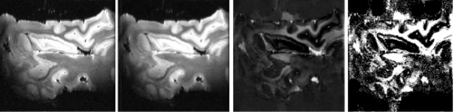
RESULT AND DISCUSSIONS(
Figure3. The binarized image (a) hat and bottom-hat filtered image The 3 x 3 mask is considered in resembles an oval structure. Th hippocampus region does not me
(a) (b)
a) binarization of original image using T (b) binarizati ge using T.
in the morphological filtering since the shape of the The binarized image produces satisfactory result and
erge or join with the adjacent regions in most of the c
ation of top-
Figure 4. Image binarization usi after mean filtering (c) Image ob means clustering.
5.
CONCLUSIONIn this paper, we have proposed a MR images to remove the artifa filtering operations and k-means identified by our method can be used to develop a generalized me brain MRI.
A
CKNOWLEDGMENTSThis work is supported by a rese Grant No: M.R.P, F.No-37- 154/2
R
EFERENCES[1] Chumming Li, Huang, R. Din for Image Segmentation in th IEEE Transactions on Image P [2] Chan, T. & Vese, L, (2001 Processing,Vol. 10, No. 2, pp2 [3] Ronfard R, (1994)
”Region-Computer Visio,. Vol. 13, No. [4] Shen, D. Susan, M.S. Resnic
Hippocampus in MR Images 422-434.
[5] Ghanei, A. Zadeh, H.S. & Win using deformable contours”, C 216.
[6] Freeborough, P.A. Fox, N.C. & quantitation of 3-D MRI brain No. 1, pp15-25.
[7] Boyes, B.J. Lewis, R.G. Sch calculation of hippocampal a integral”, Neurobiology of Agi [8] Jorge Haddock & John Mitte Computers & Industrial Engine
sing the proposed scheme (a) Original image (b) Im obtained after morphological filtering (d) Binarized im
d a binarization technique for correcting intensity inho ifacts. The combination of morphological top-hat an ns clustering technique are used to find edges sharpl e used as a pre-processing technique to avoid artifact method to segment the hippocampus automatically fro
search grant by University Grants Commission, New 4/2009(SR).
ing, Z. Chris Gatenby, J. & Metaxas, D.N, (2011) “A Lev the Presence of Intensity In homogeneities With Applica Processing, Vol. 20, No. 7, pp2007-2016.
01) “Active contours without edges”, IEEE Transactio p266-277.
-based strategies for active contour models”,Internation o. 2, pp229-251.
nick, M. & Davatzikos, C, (2002) “Measuring Size and s Using a Deformable Shape model”, NeuroImage, Vol.
indham, J.P, (1998) “Segmentation of the hippocampus fr , Computerized Medical Imaging and Graphics, Vol. 22, N
. & Kitney, R.I, (1997) “Interactive algorithms for the seg ain scans”, Computer Methods and Programs in Biomed
chott, E.B. Frost, J.M.C. Scahill, R.I& Fox, N.C, (2007 atrophy rates using a hippocampal template and the b
ging28 (2007), Vol. 28, No. 11, pp1657-1663.
ittenthal, (1992) “Simulation optimization using simulate ineering, Vol. 22, No. 4, pp387-385.
Image obtained image using
k-homogeneity in and bottom-hat rply. The edges acts and can be rom the human
w Delhi, India.
evel Set Method ication to MRI”,
tions on Image
ional Journal of
nd Shape of the l. 15, No. 2, pp
from brain MRI No. 3, pp
203-egmentation and edicine, Vol. 53,
07) “Automatic boundary shift
18 thresholds”, Image and Vision Computing, Vol. 23, No. 1, pp 69-85.
[10] Somasundaram, K. & Kalaiselvi, T, (2010) “Fully automatic brain extraction algorithm for axial T2-weighted magnetic resonance images”, Computers in Biology and Medicine. Vol. 40, No. 10,pp811-822.
[11] Heckemann, R.A. Hajnal, J.V. Aljabar, P. Rueckert, D. & Hammer, A, (2006) “Automatic anatomical brain MRI segmentation combining label propagation and decision fusion”, NeuroImage, Vol. 33, No. 1, pp115-126.
[12] Thillou, C. &Gosselin, B. Robust, (2004)“Thresholding Based on Wavelets and ThinningAlgorithms for Degraded Camera Images”, Proceedings of ACIVS, tcts.fpms.ac.be.
[
13] Sonka, M. Hlavac & Boyle, (2007), “Digital Image Processing and Computer Vision”, Thomson Learning Inc., Second Edition.
[14] Starck, J.L. Candes, E.J. & Donoho, D.L, (2002) “The curvelet transform for image denoising”, IEEE Transactions on Image Processing, Vol. 11, No. 6, pp 670-684.
[15] Portilla, J. Strela, V. Wainwright, M.J.&Simoncelli, E.P, (2003), “ Image denoising using scale mixtures of Gaussians in the wavelet domain” , IEEE Transactions on Image Processing,Vol. 12, No. 11, pp1338-1351.
[16] Saeedi, J. Moradi, M.H. & Faez, K, (2010) “ A new wavelet-based fuzzy single and multi-channel image denoising”, Image and Vision Computing, Vol. 28, No. 12, pp1611-1623.
[17] Huang, K. Wu, Z.Y. Fung, S.K. and Chan, H.Y, (2005) “Color image denoising with wavelet thresholding based on human visual system model”, Signal Processing: Image Communication, Vol. 20, No. 2, pp115-127.
[18] Li-Hong Juang & Ming-Ni Wu, (2010) “MRI brain lesion image detection based on color-converted K-means clustering segmentation”, Measurement, Vol. 43, No. 7, pp941–949.
[19] D.A. Clausi, (2002)“K-means Iterative Fisher (KIF) unsupervised clustering algorithm applied to image texture segmentation”,Pattern Recognition, Vol. 35, No. 9, pp1959-1972.
Authors
K. Somasundaram was born in the year 1953. He received the M.Sc degree in Physics from University of Madras, Chennai, India in 1976, the Post Graduate Diploma in Computer Methods from Madurai Kamaraj University, Madurai, India in 1989 and the Ph.D degree in theoretical Physics from Indian Institute of Science, Bangalore, India in 1984. He is presently the Professor and Head of the Department of Computer Science and Applications, and Head, Computer Centre at Gandhigram Rural Institute, Gandhigram, India. From 1976 to 1989, he was a Professor with the Department of Physics at the same Institute. He was previously a Researcher at an International Centre for Theoretical Physics, Trieste, Italy and a Development Fellow of Commonwealth Universities at the school of Multimedia, Edith Cowan University, Australia. His research interests are in image processing, image compression and medical imaging. He is a Life member of Indian Society for Technical Education and Telemedicine Society of India. He is also an annual member in ACM, USA and IEEE Computer Society, USA.