Induced Liver Fibrosis
Jie Liang
1, Bei Zhang
1*, Ruo-wu Shen
2*, Jia-Bao Liu
3, Mei-hua Gao
1, Ying Li
1, Yuan-Yuan Li
1, Wen Zhang
11 Department of Immunology, Medical College of Qingdao University, Qingdao, China, 2 Department of Anatomy, Medical College of Qingdao University, Qingdao, China, 3 Department of Radiology, Affiliated Hospital of Qingdao University Medical College, Qingdao, China
Abstract
Halofuginone (HF) is an active component of extracts derived from the plant alkaloid febrifugine and has shown therapeutic promise in animal models of fibrotic disease. Our main objectives were to clarify the suppressive effect of HF on concanavalin A (ConA)-induced liver fibrosis. ConA injection into the tail vein caused a great increase in the serum aspartate aminotransferase (AST) and alanine aminotransferase (ALT) levels, while orally administration of HF significantly decreased the levels of the transaminases. In addition, the levels of hyaluronic acid (HA), procollagen III (PCIII) and TGF- 1 in the serum and collagen I, α-SMA, tissue inhibitors of metalloproteinase β (TIMPβ) and Smadγ in the liver tissue were significantly lowered with the treatment of HF. Histological examination also demonstrated that HF significantly reduced the severity of liver fibrosis. Since ConA-induced liver fibrosis is caused by the repeated activation of T cells, immunomodulatory substances might be responsible for the suppressive effect of HF. We found that the production of nuclear factor (NF)-kB in the serum was increased in ConA-treated group, while decreased significantly with the treatment of HF. The changes of inflammatory cytokines tumor necrosis factor (TNF-α), IL-6 and IL-1 in the serum followed the same rhythm. All together, our findings indicate that orally administration HF (10ppm) would attenuate the liver fibrosis by suppressing the synthesis of collagen I and inflammation-mediated liver injury.
Citation: Liang J, Zhang B, Shen R-w, Liu J-B, Gao M-h, et al. (β01γ) Preventive Effect of Halofuginone on Concanavalin A-Induced Liver Fibrosis. PLoS ONE 8(1β): e8ββγβ. doi:10.1γ71/journal.pone.008ββγβ
Editor: Bernhard Ryffel, French National Centre for Scientific Research, France
Received July γ0, β01γ; Accepted October ββ, β01γ; Published December 16, β01γ
Copyright: © β01γ Liang et al. This is an open-access article distributed under the terms of the Creative Commons Attribution License, which permits unrestricted use, distribution, and reproduction in any medium, provided the original author and source are credited.
Funding: This study was supported by grant-in-aids for The National Natural Science Foundation of China (8107βγ98). The funders had no role in study design, data collection and analysis, decision to publish, or preparation of the manuscript.
Competing interests: The authors have declared that no competing interests exist. * E-mail: zhangbei1β[email protected] (BZ); [email protected] (RWS)
Introduction
In any chronic liver disease (CLDs), whatever the aetiology, reiteration of liver injury results in persisting inflammation and progressive fibrogenesis. The normal liver structure may ultimately develop into overt cirrhosis after distorted by scar tissue. In these progress, tissue injury recruits inflammatory cells and activates hepatic stellate cells (HSC) which is the major source of ECM proteins in the injured liver[1] and of many of the metalloproteinases (MMPs) and their inhibitors (TIMPs)[β]. MMPs are a family of highly homologous metal-dependent endopeptidases that can cleave the majority of constituents of the extracellular matrix such as collagen, fibronectin, laminin and elastin[γ]. MMPs are inhibited by endogenous tissue inhibitor of metalloproteinases (TIMPs)[4]. Chronic liver injury and activation of HSCs lead to the upregulation of TIMPs and growth factor -1 (TGF- 1) with the inhibition of MMP activity. The TIMPs activation thus stimulates collagen I synthesis and matrix proteins accumulation in the extracellular space[5]. At cellular levels, the perisinusoidal HSC
has been extensively reported as a key effector of fibrogenesis[5,6]. Following hepatocyte injury, HSC differentiates into an “activated” myofibroblast-like phenotype[7]and contributes to fibrillar collagen formation, which plays an important role in controlling liver fibrosis[8,9]. Moreover, it increases until vascular structures are linked and the architecture of the liver is disrupted significantly [10,11]. Although numerous agents have been tried, the lack of specific inhibitors of ECM components in general and the lack of specific inhibitors of collagen type I in particular, limits the progress in the treatment of hepatic fibrosis.
fibrogenesis and EMT[1β,1γ]. IL-1 and TNF-α play similarity effects on fibroblasts[14]. IL-1, a ubiquitous and pleiotropic cytokine, particularly IL-1 , inhibits collagen production but it enhances fibroblast proliferation. Similarly, TNF-α, a primary immune and inflammatory regulator, stimulates fibroblast chemotaxis and proliferation meanwhile it inhibits collagen production[15,16]. IL-6, another inflammatory cytokine, which may affect differentiation of fibroblast to myofibroblast, plays an important role in fibrosis diseases[17,18].
NF-KB (nuclear factor kappa-light-chain-enhancer of activated B cells) is a family of transcription factors which plays a critical role in regulation of immunity and inflammation by stimulating the transcription of a wide range of cytokine-encoding genes, including TNF-α and IFN- . This family is composed of five related transcription factors (p50, p5β, p65, c-Rel, and RelB), and they can form homo- and heterodimers[19]. The most important NF-kB dimmers are formed by p65 and p50 in NF-kB signaling pathway[β0]. NF-kB mediated transcriptional activation plays a critical role in the HSC activation [β1]. NF-kB activity can induce the expression and secretion of the various inflammatory cytokines and adhesion molecules, which play a major role in hepatic fibrosis [ββ,βγ]. Upon stimulation by inflammatory cytokines such as TNF-α, IkB is phosphorylated by IKK and degraded. NF-kB is then released and translocates to the nucleus from cytoplasm, and activates the transcription of its target genes[ββ]. Therefore, inhibition of NF-kB activity is considered as an underlying mechanism for anti-fibrosis[β4,β5].
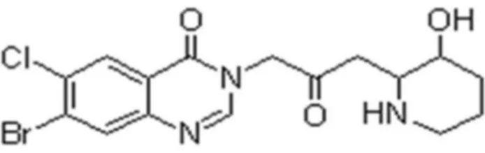
For centuries, the roots of Dichroa Febrifuga, a saxifragaceous plant, have been used in china in the treatment of malarial fever. Febrifugine and its stereoisomere, isofebrifugine, were identified as the active antimalarial components[β6]. Halofuginone (HF) [7-bromo-6-chloro-γ-(γ-hydroxy-β-piperidine)-β-oxopropyl-4(γH)-quinazoline] is one of the febrifugine analogous used world-wide for almost β0 years in commercial poultry production to prevent coccidosis[β7,β8]. In addition, halofuginone has been found to attenuate collagen α 1 (I) gene expression and collagen production by murine ,avian and human skin fibroblasts derived from either scleroderma or chronic graft-versus-host disease(cGvHD) patients[β9,γ0]. It can inhibit collagen α 1(I) gene expression in many models of fibrosis involving skin, liver, urethra, heart, surgical and traumatic adhesions[γ1,γβ]. This inhibitory effect was probably mediated by blocking TGF- induced Smadγ activation [γγ]. TGF- is a central regulator in chronic liver disease, being involved in all stages of the disease progression, from initial liver injury, inflammation, fibrosis, to cirrhosis and hepatocellular carcinoma at the end[β7]. In addition, M. Leiba showed that halofuginone inhibited DTH response in mice, indicating suppression of T cell-mediated inflammation and pro-inflammatory cytokine production in vivo[γ4].
Concanavalin A (ConA)-induced liver injury is well accepted as a rat model of immune-mediated liver injury that resembles viral and autoimmune hepatitis in humans. The intravenous injection of ConA into rat can increases transaminase and activate T cells infiltrate in liver[γ5]. The activation of T cells by ConA results in increased levels of inflammatory cytokines,
including TNF-α, IFN- and IL-6 [γ6]. In the present study, we assessed the preventive effect of HF on ConA-induced chronic liver fibrosis.
Materials and Methods
Ethics Statement
All the procedures and care administered to the animals have been approved by the Institutional Ethics Committee of Qingdao University Medical College, under a permit of animal use (SCXK40090007) in the Center of Experimental Animal and Animal Experiment at Qingdao, compliance with the Experimental Animal Regulations by the National Science and Technology Commission, China.
Animals
Male Wistar rats (weighing β10-βγ0 g) were supplied by the Experimental Animal and Animal Experiment Center of Qingdao, Shandong, China. They were housed in the animal facility with a 1β h light/dark cycle, the temperature was maintained at ββ-βγ°C and relative humidity was 60%.
Materials
ConA and HF were purchased from Sigma-Aldrich Co. (St Louis, MO, USA) and Jilan Technology Development Co. (Shanghai, China), respectively. NF-kB, TGF- 1, TNF-á, IL-6, IL-1 , HA and PC-III enzyme-linked immune sorbent assay(ELISA) kits and α-SMA, Collagen1, TIMPβ, Smadγ polyclonal antibodies were from Solarbio Science &Technology Co. (Beijing , China) and Biosynthsis Biotechnology Co. (Beijing, China).
Experimental design
HF (Figure 1), an alkaloid originally extracted from the plant Dichroa febrifuga was given in the diet at concentrations of 10 ppm. The animals were randomly distributed into three groups: group 1 (G1), rats received a weekly iv injection of γ00ul PBS for 8 weeks (n =15); group β (Gβ), rats received a weekly iv injection of ConA (17.5mg/kg, in γ00ul PBS) for 8 weeks (n =15); group γ (Gγ), weekly iv injection of ConA (17.5mg/kg, in γ00ul PBS) with 10ppm HF in diet for 8 weeks (n =15). During all experiments rats were maintained in individually ventilated cages under specific pathogen-free conditions. Animals were sacrificed on d57, blood was collected via cardiac puncture and serum was prepared by centrifugation at β000g for 10 min and stored at -β0°C. Livers were taken after perfusion with 4% paraformaldehyde.
Figure 1. The chemical structure of halofuginone (HF).
Liver function and fibrosis index
The serum levels of alanine aminotransferase (ALT), aspartate aminotransferase (AST), total protein(TP) and the albumin (ALB) were measured with an auto-biochemical analyzer (Roche P800, Basle Switzerland). HA, PCIII were measured using ELISA micro-titer plates pre-coated with antibodies specific to HA or PCIII according to the instructions of the manufacturer. Each experiment was done in quadruplicate, and the results are expressed as the mean ±SEM.
Cytokine Secretion
Sera aliquots collected from all rats were assayed for transforming growth factor-beta (TGF- 1) as a fibrogenesis-driving cytokine, the nuclear transcription factor NF-kB and the pro-inflammatory cytokine
Interleukin TNF-α, IL-6 and IL-1 as important signals in liver injury. All of them were assayed by enzyme-linked immune sorbent assay using ELISA micro-titer plates pre-coated with antibodies specific to NF-kB, TNF-α, IL-6 or IL-1 according to the instructions of the Manufacturer. Each experiment was done in quadruplicate, and the results are expressed as the mean±SEM.
Haematoxylin-eosin (HE) Staining
After rats were sacrificed, vessels were perfused with PBS, followed by 4% paraformaldehyde. Paraformaldehyde-fixed liver specimens were dehydrated in a graded alcohol series. Following xylene treatment, the specimens were embedded in paraffin blocks, cut into 4-μm thick sections, and placed on glass slides. The sections were then stained with hematoxylin and eosin (HE) according to standard procedures.
Histopathological evaluation of liver
Liver fibrosis was assessed with Masson trichrome (MT) stain according to standard procedures. To describe and evaluate liver pathological changes, a pathologist who was blinded to the research design examined 10 different low-power fields of MT-stained sections (selected fields were in almost the same location) for each rat. In addition, the percentage of collagen calculated by a multimedia color image analysis system (Image-Pro Plus 6.0) was measured as a relative objective index (because a histological/fibrosis score that is evaluated by pathologists is susceptible to the ability and subjective judgment of the pathologist) to evaluate the degree of liver fibrosis. Each MT-stained section was examined at high-power fields (HPFs) (magnification ×400). Every field analyzed contained a granuloma, portal area, or a centrilobular vein. Fibrotic areas were scanned and summed by the software. The level of fibrosis was scored according to liver fibrosis semi-quantitative scoring system (SSS) method[γ7].
Immunohistochemistry
Paraffin blocks were cut into 4-µm sections, deparaffinized in xylene, and rehydrated in graded ethanol solutions. Immunohistochemical SP method was used to detect the expressions of collagen I, α-SMA and Smadγ. The tissue
sections were first incubated a specific antibody (anti-α-SMA, anti-collagen I and anti-Smadγ) overnight at 4 °C, washed in Phosphate Buffer Solution (PBS) for three times, and was then incubated with biotinylated secondary antibody (anti-immunoglobulin from the animal species from which the specific antibody was obtained) for β0 minutes at γ7°C. After washed in PBS, the sections were incubated with avidin conjugated to HRP for β0min at γ7°C, then colored with γ,γ-diaminobenzidin (DAB). After counterstained with haematoxylin for 5 mins, tissue sections were washed and then dehydrated with ethanol from 70% to 80%, then 90%, 95%, 100%, followed by xylene (γX). Finally, the sections were covered with cover slips by neutral gums and observed by microscope. PV-6000 ploymer detection system was used to detect the expressions of TIMPβ in rat liver tissue according to the manufacturer’s instructions.
Image Pro plus 6.0 (Media Cybernetics, Bethesda, MD, USA) was used to quantify immune-staining results. This software was designed to select stained nuclei or cells based on color intensity and nuclear shape. Brown staining was considered positive. The chromatic area and the strength (light density values) of positive cells were calculated and represented as a percentage of total positively stained cells with integral light density values (integral optical density).
Statistics
All statistical analyses in this study were carried out using Sigma-Plot 10 and SPSS 19.0, and results are expressed as the mean±SEM. A one-way analysis of variance (ANOVA) followed by the post-hoc Dunnett’s test was used to analyze multiple groups for evaluating statistical significance. A value of P<0.05 was considered significant.
Results
Treatment with HF decrease LBWR in ConA-treated rats There are 15 rats in G1, 10 and 1γ left in Gβ and Gγ respectively when the animals were sacrificed after 8 weeks treatment. The body and liver weights were significant different among three groups. The liver-to-bodyweight ratio (LBWR) was significantly higher in Gβ compared with that in G1, and simultaneous administration of halofuginone (8 weeks, 10 ppm) decreased the LBWR in Gγ by 10.4% (Table 1), which was consistent with the macroscopic change of liver. In contrast to the livers in control group, a typical fibrosis appearance with increased volume and extensive nodular formation was present in the livers of ConA- treatment group, while the hepatic lesion were hardly observed under microscopy in ConA+HF group (Figure β). Those results indicated that HF can decrease LBWR in ConA-induced liver fibrosis rats.
HF attenuates the impairment in liver function
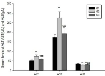
time-dependent manner, reaching a maximum and minimum at eight weeks (data not shown). ConA stimulation significantly increased plasma ALT(7γ.γ7±8.4βU/L), AST(β74.64±γβ.10U/L) and decreased ALB(β7.58±1.87g/L) levels in Gβ compared to G1, but simultaneous treatment with HF significantly lowered the level of ALT(59.β9±5.6γU/L), AST(19γ.0β±γ0.8γU/L) and increased the level of ALB(γ1.17±β.51 g/L) (Figure γ). Those results thus indicated that HF can attenuate the impairment of liver function induced by ConA injection.
HF reduce fibrosis index
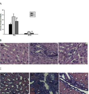
The levels of HA and PCIII, which have been shown as two liver fibrosis indexes, were detected by ELISA kits. Their production were significantly higher in Gβ after ConA treatment for eight weeks. However, simultaneous administration of halofuginone (8 weeks, 10 ppm) decreased HA and PCIII production by β6% and 47% respectively, suggesting that HF can reduce the degree of liver fibrosis (Figure 4A).
HF alleviates liver fibrosis level in ConA-treated rats Weekly ConA injection for eight weeks resulted in advanced liver fibrosis, characterized by distorted tissue architecture with collagen bundles surrounding the lobules and large fibrous septa. Histopathologic findings of the rats liver tissues were shown in Figure 4B. In contrast to the normal foliages and cell structures seen in G1 liver tissue, HE staining displayed that the tissue architecture in Gβ was distorted, with collagen bundles surrounding the lobules and a large number of inflammatory cell infiltrations together with hepatocellular necrosis. When compared with the Gβ, fibrous strips and Table 1. Comparison of LBWR in three groups.
Group n LBWR
G1 15 4.5γ±0.00β4
Gβ 10 5.4β±0.0056**
Gγ 1γ 4.86±0.00γ1*##
There are significant difference in LBWR among this three groups. Data are presented as the mean ±SEM. Note: *p<0.05,**P<0.01 compared with G1, # p<0.05, ## p<0.01 compared with Gβ.
doi: 10.1γ71/journal.pone.008ββγβ.t001
Figure 2. Macroscopic view of the liver from different groups. Macroscopic view of the liver from ConA-treated rats, showing a bigger volume and a rough surface with many nodules that developed on a liver with fibrosis, while it was similar to the normal and no obvious damage to the naked eye from ConA+HF treated rats.
doi: 10.1γ71/journal.pone.008ββγβ.g00β inflammatory cell infiltration were markedly attenuated in theGγ. The semi-quantitative scoring system (SSS) value for Gγ
was significantly lower than Gβ (Table β, P<0.05).
The degree of fibrosis determined by MT staining of the liver sections from all the treated groups was shown in Figure 4C. Liver sections from normal rats appeared normal foliages without signs of collagen deposition. In contrast, liver sections from the Gβ rats displayed increased deposition of collagen fibers surrounding the congested central vein, and a large number of inflammatory cell infiltration along with hepatocellular necrosis. Liver tissues from rats treated with HF showed mild collagen deposition and mild congestion around the central vein. The mean score of fibrosis in the Gγ group was significantly lower than that of Gβ (P<0.05). Measurement of the degree of fibrosis thus confirmed the previous findings that treatment with HF might protect the animals’ liver from development of fibrosis.
HF supress the synthesis of Collagen 1, α-SMA and TIMP2
In our present study, a remarkable collagen a 1(I) accumulation was observed in the rats from Gβ group which received ConA treatment for eight weeks. In addition, TIMP-β and stellate cells positive for α-SMA were also noted Gβ group rats. HF-treated rats presented minimal levels of collagen1, α-SMA and TIMPβ expression throughout the experiment (Figure 5A-C). Neither α-SMA positive cells nor collagen and less TIMP-β staining cells were observed in control animals. .
HF down-regulates TGF-β1/Smad3 signaling pathway Since the TGF- 1/Smadγ signaling pathway has been shown to be involved in the regulation of fibrosis, and the expression of TGF- 1/Smadγ signaling pathway related protein Figure 3. Halofuginone suppress liver injury induced by ConA. Blood was collected to determine the serum levels of AST, ALT and ALB. HF significantly decreased ALT, AST and increased ALB levels in serum. The values are the means ±SEM. *p<0.05, **p< 0.01 as compared to G1, # p<0.05, ##p<0.01 as compared to Gβ.
collagen a 1(I), a-SMA and TIMP-β have been measured by immunohistochemical methods. We further tested the expression of Smadγ in the liver tissue and the expression of TGF- 1 in the serum, in order to explore whether TGF- 1/ Smadγ pathway was also affected with the treatment of HF. As shown Figure 6A, the expression of Smadγ was significantly increased in Gβ rats treated with ConA for eight weeks, while Figure 4. Halofuginone alleviate liver fibrosis level induced by ConA. (A)Treatment with halofuginone can markedly decreased the raised levels of HA and PCIII in serum induced by ConA. (B) H&E staining show enhanced collagenous fiber deposition expands in the portal area as well as in the bile duct in ConA-treated rats and the collagenous fiber attenuate obviously when there are halofuginone was added in diet. (C) MT staining shows collagen accumulation were the highest in ConA-treated rats, while it was decreased significantly with the treatment of halofuginone. Magnification for all photographs, ×400, the values are the means ±SEM. *p<0.05, **p< 0.01 as compared to G1, #p<0.05, ##p<0.01 as compared to Gβ.
doi: 10.1γ71/journal.pone.008ββγβ.g004
Table 2. Fibrosis score of rats in different groups.
Group N Score
G1 15 0
Gβ 10 β1.6±γ.1γ4*
Gγ 1γ 10.85±β.075*##
Haloluginone caused a significant reduction in the mean liver fibrosis score of the rats after 8 weeks of treatment. Data are presented as the mean ±SEM. Note: *p<0.05,**P<0.01 compared with G1, # p<0.05, ## p<0.01 compared with Gβ. doi: 10.1γ71/journal.pone.008ββγβ.t00β
its expression was obviously decreased after treatment with HF. The change of TGF- 1 expression in serum followed the similar tendency (Figure 6B). Overall, these results confirmed that HF inhibits the protein expression of type I collagen, TIMPβ and α-SMA in rats, which may be due to the down-regulation of TGF- 1/Smadγ signaling pathway.
Figure 5. Halofuginone suppress the protein synthesis of Collagen 1, α-SMA and TIMP2. The expression of Collagen 1, α-SMA and TIMPβ were the highest in ConA-treated fibrosis rats, and their expressions were decreased significantly with the treatment of HF in Gγ. Magnification for collagen1 and α-SMA, ×β00, for TIMPβ, ×400.
doi: 10.1γ71/journal.pone.008ββγβ.g005
Figure 6. HF affects TGF-β1/Smad signaling pathway. TGF- 1 and Smadγ increased significantly in ConA-treated fibrosis rats, and HF attenuated the increased TGF- 1 and Smadγ markedly. The values are the means ±SEM. *p<0.05, **p< 0.01 as compared to G1, #p<0.05, ##p<0.01 as compared to Gβ.
HF decrease inflammation cytokine secretion
Gβ group rats expressed significantly higher levels of pro-inflammatory cytokines including TNF-α, IL-1 and IL-6, which have been shown to play important roles in the development of the fibrosis. However, simultaneous administration of HF resulted in remarkable decreases in the levels of all those three parameters, indicating that HF can decrease the secretion of inflammatory factors (Figure 7).
HF reduce serum levels of NF-κB
The transcriptional factor NF-κB has been shown to be involved in the regulation of cytokine signaling and inflammation. We next examined the effects of HF on the level of NF-κB in the serum and found that the production of NF-κB in ConA-treated rats was significantly higher than that in the controls (Figure 8); however, treatment with HF suppressed NF-κB expression in the serum. Taking together, these data indicated that the anti-inflammation effects of HF treatment against ConA might be mediated by NF-κB signaling.
Discussion
ConA-induced liver fibrosis, which was characterized by activation of the T cells, is considered more suitable for the study of liver fibrosis, because it resembles the liver fibrosis originated from viral and autoimmune hepatitis in humans. Repeated intravenous injection of ConA into rat increased the plasma transaminase (Figure γ) and fibrosis index level(Figure 4), meanwhile ConA injection activated T cells which could infiltrate the liver and cause the apoptosis and necrosis of hepatocytes eventually [γ8].
Our study showed here that HF played a hepatoprotective effect against ConA-induced liver fibrosis in rat, a widely used Figure 7. Supression of inflammatory cytokines secretion with the treatment of halofuginone. The serum levels of TNF-α, IL-6 and IL-1 were increased significantly with the treatment of ConA, while this three parameters were decreased abviously when there are halofuginone was added in diet. The values are the means ±SEM. *p<0.05, **p< 0.01 as compared to G1, #p<0.05, ##p<0.01 as compared to Gβ.
doi: 10.1γ71/journal.pone.008ββγβ.g007 animal model to study the fibrosis induced by viral and
autoimmune hepatitis [γ6]. Halofuginone, a well known inhibitor of collagen a1(I) gene expression, has recently been shown to prevent dimethylnitrosamine(DMN)[γ9] and thioacetamide-induced liver fibrosis[γ0]. Since collagen α1(I) is a major component in the body, it is of concern when one wants to treat patients systemically with an inhibitor of collagen like HF[γ6]. Moreover, HF treatment significantly reduced ECM components thereby improving overall liver architecture in established fibrosis. In our study, since the development of oral medications to help prevent liver injury is desirable[40], orally treatment with HF significantly improved liver histology(Figure 4B) and declined the high values of Collagen α1(I) protein(Figure 5A), and we found the decline in collagen deposition was accompanied by a reduction in numbers α-SMA positive cells(Figure 5B), pointing to the fact that HF may affect collagen levels by more than one mechanism.
In liver fibrosis, there is an imbalance between excessive deposition and/or decreased removal in the extracellular matrix (ECM) with consequent scarring damage[41]. ECM is mainly controlled by matrix metalloproteinases (MMPs) and their inhibitors (TIMPs). MMPs belong to a group of proteolytic enzymes that are able to degrade the ECM, while TIMPs can inhibit the degradation of collagens. An increase in MMPs and TIMPs is commonly observed in fibrotic diseases. Park et al. reported that the imbalance of MMPs and TIMPs is the key factor of liver fibrogenesis[41]. In this study, we demonstrated that administration of HF inhibited the expression of TIMPβ, thus reduced ECM deposition within the liver parenchyma and alleviated liver fibrosis. Therefore, downregulation of TIMPβ by HF treatment may contribute to the reduced fibrosis in the HF-treated rats.
Another mechanism by which HF could influence liver fibrosis is via the inhibition of TGF- 1 secretion. TGF- 1 is a profibrogenic cytokine in the liver, and has been shown to regulate multiple fundamental cellular processes, including cell Figure 8. Halofuginone reduce the secretion of NF-kB. HF significantly surpress the increased serum NF-KB induced by ConA. The values are the means ±SEM *p<0.05, **p< 0.01 as compared to G1, #p<0.05, ##p<0.01 as compared to Gβ.
growth, migration, adhesion, ECM deposition, and apoptosis [4β-44]. Blocking TGF- 1 by adenovirus encoding a truncated type II receptor prevented the progression of fibrosis in DMN-injected animals[45,46]. The mechanism by which HF reduces fibrosis was recently elucidated in a mouse model for scleroderma (dermal fibrosis), where a low dose of halo blocked TGF- mediated SMADγ activation in fibroblasts[47]. Moreover, Xavier.et al, have recently demonstrated that halofuginone induced expression of the inhibitory Smad7 and decreased the levels of TGF- 1 receptor II, altogether inhibiting the activation of Smadβ and Smadγ[48]. Considering the significance of the TGF- 1/Smad signaling pathway in regulating fibrogenesis, we are trying to detect the change of TGF- 1 and Smadγ in rats from different group. The data obtained from HF-treated rats revealed that HF administration attenuated level of the prominent profibrogenic cytokine TGF-1 (Figure 6B) indicating the inhibitory activity of HF to the proliferative activity of HSCs which might be confirmed by the less collagen deposition in the liver tissues of animals treated with HF ( Figure 4). Similarly, the high levels of Smadγ (Figure 6A) observed in the fibrosis group rats were reversed in the rats treated with HF. Therefore, the decreased fibrosis level after HF treatment may be mediated via its inhibition of the TGF- 1/Smadγ signaling pathway.
Since ConA-induced liver fibrosis is caused by the repeated activation of T cells, the potential immunomodulatory effect contained in the HF. Treatment with HF resulted in inhibition of NF-kB which plays a critical role in the regulation of immunity and inflammation by stimulating the transcription of a wide range of cytokine-encoding genes, including TNF-α and IFN-[49]. Indeed, orally administration of HF significantly suppressed the levels of secreted pro-inflammatory cytokines TNF-α, IL-1 and IL-6(Figure 7), elicited in vivo by the T cell mitogen, ConA. These cytokines are known to regulate collagen synthesis[β9], and thus, are likely to be involved in the
anti-fibrotic effect of halofuginone. In addition to its ability to regulate cytokine production, NF-kB is involved in regulation of the acute-phase response of inflammation, which provides systemic defense and restores homeostasis after infection or injury [50,51]. Thus, HF appears to act anti-inflammatory properties in the process of inflammation, this may be another reason for HF on liver fibrosis.
In conclusion, the present study has for the first time systemically investigated the potential protective role of HF on ConA-induced liver fibrosis models, the prophylactic effect of HF on long-term ConA-induced liver fibrosis including anti-fibrosis and anti-inflammation. The results indicate that targeting HF may present a potent approach, particularly for its prophylactic effects, against liver fibrosis. Preclinical trials by applying some promising HF have already been launched though at their infancy. Although the characteristics of a putative HF receptor and the exact downstream signaling pathways are still obscure, our findings provide additional information toward elucidating its mode of action and therapeutic potential.
Acknowledgements
Thanks to Hai-yang Fu and Xiang-yan Zhang for their helpful assistance with liver histological examination.
Author Contributions
Conceived and designed the experiments: BZ. Performed the experiments: J. Liang. Analyzed the data: J. Liu. Contributed reagents/materials/analysis tools: RWS. Wrote the manuscript: J. Liang. Provided technical guidance during experiments: MHG. Care of the rats in the process of experiments: YL. Assisted to acquire the animal specimens during experiments: YYL WZ.
References
1. Perepelyuk M, Terajima M, Wang AY, Georges PC, Janmey PA, et al. (β01γ) Hepatic stellate cells and portal fibroblasts are the major cellular sources of collagens and lysyl oxidases in normal liver and early after injury. Am J Physiol-gastr L γ04: G605-G614
β. Iredale JP (1997) Tissue inhibitors of metalloproteinases in liver fibrosis. Int J Biochem Cell Biol β9: 4γ-54. doi:10.1016/ S1γ57-β7β5(96)00118-5. PubMed: 9076940.
γ. Hansen H, Moore WG, Bodden MK, Windsor LJ, Birkedal-Hansen B et al. (199γ) Matrix metalloproteinases: a review. Crit Rev Oral Biol M 4: 197-β50. PubMed: 84γ5466.
4. Moon SK, Linthicum FH Jr, Dong Yang H, Joo Lee S, Park K (β008) Activities of matrix metalloproteinases and tissue inhibitor of metalloproteinase-β in idiopathic hemotympanum and otitis media with effusion. Acta Oto-Laryngol 1β8: 144-150. doi: 10.1080/00016480701477610. PubMed: 17851959.
5. Friedman SL (β008) Mechanisms of hepatic fibrogenesis. Gastroenterology 1γ4: 1655-1669. doi:10.105γ/j.gastro.β008.0γ.00γ. PubMed: 18471545.
6. Bansal R, Prakash J, Post E, Beljaars L, Schuppan D et al. (β011) Novel engineered targeted interferon‐gamma blocks hepatic fibrogenesis in mice. Hepatol 54: 586-596. doi:10.1016/j.jhep. β010.09.01β.
7. Clària J (β01β) Natural killer cell recognition and killing of activated hepatic stellate cells. Gut 61: 79β-79γ. doi:10.11γ6/gutjnl-β011-γ01968. PubMed: ββ466617.
8. Venugopal SK, Jiang J, Kim T-H, Li Y, Wang S-S, et al. (β010) Liver fibrosis causes downregulation of miRNA-150 and miRNA-194 in
hepatic stellate cells, and their overexpression causes decreased stellate cell activation. Am J Physiol-gastr L β98: G101-G106
9. Liu C, Tao Q, Sun M, Wu JZ, Yang W et al. (β010) Kupffer cells are associated with apoptosis, inflammation and fibrotic effects in hepatic fibrosis in rats. Lab Invest 90: 1805-1816. doi:10.10γ8/labinvest. β010.1βγ. PubMed: β09β1949.
10. Thabut D, Routray C, Lomberk G, Shergill U, Glaser K et al. (β011) Complementary vascular and matrix regulatory pathways underlie the beneficial mechanism of action of sorafenib in liver fibrosis. Hepatology 54: 57γ-585. doi:10.100β/hep.β44β7. PubMed: β1567441.
11. Jagavelu K, Routray C, Shergill U, O'Hara SP, Faubion W et al. (β010) Endothelial cell toll-like receptor 4 regulates fibrosis-associated angiogenesis in the liver. Hepatol 5β: 590-601. doi:10.100β/hep.βγ7γ9. 1β. Derynck R, Zhang YE (β00γ) Smad-dependent and Smad-independent
pathways in TGF- family signalling. Nature 4β5: 577-584. doi:10.10γ8/ nature0β006. PubMed: 145γ4577.
1γ. Wang C, Song X, Li Y, Han F, Gao S et al. (β01γ) Low-Dose Paclitaxel Ameliorates Pulmonary Fibrosis by Suppressing TGF- 1/Smadγ Pathway via miR-140 Upregulation. PLOS ONE 8: e707β5. doi: 10.1γ71/journal.pone.00707β5. PubMed: βγ967091.
14. Gurantz D, Cowling RT, Varki N, Frikovsky E, Moore CD et al. (β005) IL-1 and TNF-α upregulate angiotensin II type 1 (AT< sub> 1</sub>) receptors on cardiac fibroblasts and are associated with increased AT< sub> 1</sub> density in the post-MI heart. J Mol Cell Cardiol γ8: 505-515. doi:10.1016/j.yjmcc.β004.1β.015. PubMed: 157γγ910. 15. Siwik DA, Chang DL-F, Colucci WS (β000) Interleukin-1 and tumor
metalloproteinase activity in cardiac fibroblasts in vitro. Cir Res 86: 1β59-1β65. doi:10.1161/01.RES.86.1β.1β59.
16. Atamas SP (β00β) Complex cytokine regulation of tissue fibrosis. Life Sci 7β: 6γ1-64γ. doi:10.1016/S00β4-γβ05(0β)0ββ99-γ. PubMed: 1β467904.
17. Shahar I, Fireman E, Topilsky M, Grief J, Kivity S et al. (1996) Effect of IL-6 on alveolar fibroblast proliferation in interstitial lung diseases. Clin Immunol Immunopathol 79: β44-β51. doi:10.1006/clin.1996.0075. PubMed: 86γ5β8β.
18. Wynn TA (β008) Cellular and molecular mechanisms of fibrosis. J Pathol β14: 199-β10. doi:10.100β/path.ββ77. PubMed: 18161745. 19. Hayden MS, Ghosh S (β011) NF-κB in immunobiology. Cell Res β1:
ββγ-β44. doi:10.10γ8/cr.β011.1γ. PubMed: β1β4γ01β.
β0. Ren Z, Cui J, Huo Z, Xue J, Cui H et al. (β01β) Cordycepin suppresses TNF-α-induced NF-κB activation by reducing p65 transcriptional activity, inhibiting IκBα phosphorylation, and blocking IKK ubiquitination. Int Immunopharmacol, 14: 698–70γ. PubMed: βγ10β66β.
β1. Sun B, Karin M (β008) NF-êB signaling, liver disease and hepatoprotective agents. Oncogene β7: 6ββ8-6β44. doi:10.10γ8/onc. β008.γ00. PubMed: 189γ1690.
ββ. Elsharkawy AM, Mann DA (β007) Nuclear factor-êβ. Elsharkawy AM, Mann DA (β00-fibrosis-cancer axis. Hepatol 46: 590-597. doi:10.100β/ hep.β180β.
βγ. Jaruga B, Hong F, Kim W-H, Sun R, Fan S, et al. (β004) Chronic alcohol consumption accelerates liver injury in T cell-mediated hepatitis: alcohol disregulation of NF-êB and STATγ signaling pathways. Am J Physiol-gastr L β87: G471-G479
β4. Luedde T, Schwabe RF (β011) NF-êB in the liver—linking injury, fibrosis and hepatocellular carcinoma. Nat Rev Gastroenterol Hepatol 8: 108-118. doi:10.10γ8/nrgastro.β010.β1γ. PubMed: β1β9γ511. β5. Xia Y, Chen J, Cao Y, Xu C, Li R et al. (β01γ) Wedelolactone exhibits
anti-fibrotic effects on human hepatic stellate cell line LX-β. Eur J Pharmacol, 714: 105–11. PubMed: βγ79161β.
β6. Nguyen-Pouplin J, Tran H, Tran H, Phan TA, Dolecek C et al. (β007) Antimalarial and cytotoxic activities of ethnopharmacologically selected medicinal plants from South Vietnam. J Ethnopharmacol 109: 417-4β7. doi:10.1016/j.jep.β006.08.011. PubMed: 17010546.
β7. Gu Y-Y, Liu S-X, Wang S-X, Wan S-B, Jiang T (β011) Study on the Synthetic Technique of Halofuginone. Zhongguo Haiyang Daxue Xuebao Ziran Kexue Ban 9: 01β.
β8. Meindl-Beinker NM, Matsuzaki K, Dooley S (β01β) TGF- Signaling in Onset and Progression of Hepatocellular Carcinoma. Dig Dis γ0: 514-5βγ. doi:10.1159/000γ41704. PubMed: βγ108γ08.
β9. Pines M, Snyder D, Yarkoni S, Nagler A (β00γ) Halofuginone to treat fibrosis in chronic graft-versus-host disease and scleroderma. Biol Blood Marrow Transplant 9: 417-4β5. doi:10.1016/ S108γ-8791(0γ)00151-4. PubMed: 1β869955.
γ0. Bruck R, Genina O, Aeed H, Alexiev R, Nagler A et al. (β001) Halofuginone to prevent and treat thioacetamide-induced liver fibrosis in rats. Hepatol γγ: γ79-γ86. doi:10.105γ/jhep.β001.β1408.
γ1. Benchetrit S, Yarkoni S, Rathaus M, Pines M, Rashid G, et al. (β007) Halufuginone reduces the occurrence of renal fibrosis in 5/6 nephrectomized rats. IMAJ-RAMAT GAN- 9: γ0
γβ. Pines M, Nagler A (1998) Halofuginone: a novel antifibrotic therapy. Gen Pharmacol γ0: 445-450. doi:10.1016/S0γ06-γ6βγ(97)00γ07-8. PubMed: 95ββ159.
γγ. Nelson EF, Huang CW, Ewel JM, Chang AA, Yuan C (β01β) Halofuginone down-regulates Smadγ expression and inhibits the TGFbeta-induced expression of fibrotic markers in human corneal fibroblasts. Mol Vis 18: 479–487. PubMed: ββγ9γβ74.
γ4. Leiba M, Cahalon L, Shimoni A, Lider O, Zanin-Zhorov A et al. (β006) Halofuginone inhibits NF-κB and pγ8 MAPK in activated T cells. J Leukoc Biol 80: γ99-406. doi:10.1189/jlb.0705409. PubMed: 16769768.
γ5. Tiegs G, Lohse AW (β010) Immune tolerance: what is unique about the liver. J Autoimmun γ4: 1-6. doi:10.1016/j.jaut.β009.08.008. PubMed: 19717β80.
γ6. Itoh A, Isoda K, Kondoh M, Kawase M, Kobayashi M et al. (β009) Hepatoprotective effect of syringic acid and vanillic acid on concanavalin a-induced liver injury. Biol Pharm Bull γβ: 1β15-1β19. doi: 10.1β48/bpb.γβ.1β15. PubMed: 19571γ88.
γ7. Richard S, Guerret S, Gerard F, Tebib JG, Vignon E (β000) Hepatic fibrosis in rheumatoid arthritis patients treated with methotrexate: application of a new semi-quantitative scoring system. Rheumatology γ9: 50-54. doi:10.109γ/rheumatology/γ9.1.50. PubMed: 1066β87γ. γ8. Schwabe RF, Brenner DA (β006) Mechanisms of liver injury. I.
TNF-α-induced liver injury: role of IKK, JNK, and ROS pathways. Am J Physiol-gastr L β90: G58γ-G589
γ9. Pines M, Knopov V, Genina O, Lavelin I, Nagler A (1997) Halofuginone, a specific inhibitor of collagen type I synthesis, prevents dimethylnitrosamine-induced liver cirrhosis. J Hepatol β7: γ91-γ98. doi: 10.1016/S0168-8β78(97)80186-9. PubMed: 9β88615.
40. Watanabe A, Kobayashi M, Hayashi S, Kodama D, Isoda K et al. (β006) Protection against D-galactosamine-induced acute liver injury by oral administration of extracts from Lentinus edodes mycelia. Biol Pharm Bull β9: 1651-1654. doi:10.1β48/bpb.β9.1651. PubMed: 168806β1.
41. Park SY, Shin HW, Lee KB, Lee M-J, Jang J-J (β010) Differential expression of matrix metalloproteinases and tissue inhibitors of metalloproteinases in thioacetamide-induced chronic liver injury. J Korean Med Sci β5: 570-576. doi:10.γγ46/jkms.β010.β5.4.570. PubMed: β0γ58000.
4β. Ruiz-Ortega M, Rodríguez-Vita J, Sanchez-Lopez E, Carvajal G, Egido J (β007) TGF- signaling in vascular fibrosis. Cardiovasc Res 74: 196-β06. doi:10.1016/j.cardiores.β007.0β.008. PubMed: 17γ76414. 4γ. Moustakas A, Pardali K, Gaal A, Heldin C-H (β00β) Mechanisms of
TGF- signaling in regulation of cell growth and differentiation. Immunol Lett 8β: 85-91. doi:10.1016/S0165-β478(0β)000βγ-8. PubMed: 1β0080γ9.
44. Schultz GS, Wysocki A (β009) Interactions between extracellular matrix and growth factors in wound healing. Wound Repair Regen 17: 15γ-16β. doi:10.1111/j.15β4-475X.β009.00466.x. PubMed: 19γβ088β. 45. Nakamura T, Sakata R, Ueno T, Sata M, Ueno H (β000) Inhibition of
transforming growth factor prevents progression of liver fibrosis and enhances hepatocyte regeneration in dimethylnitrosamine-treated rats. Hepatol γβ: β47-β55. doi:10.105γ/jhep.β000.9109.
46. Yin M, Jin S, Cui H (β01β) Effect of chymase inhibitors on dimethylnitrosamine-induced rat liver fibrosis and on chymase and collagen I expression. Afr - J Pharm and Pharmaco 6: β870-β874. 47. McGaha TL, Phelps RG, Spiera H, Bona C (β00β) Halofuginone, an
Inhibitor of Type-I Collagen Synthesis and Skin Sclerosis, Blocks Transforming-Growth-Factor-&bgr;-Mediated Smadγ Activation in Fibroblasts. J Invest Dermatol 118: 461-470. doi:10.1046/j.00ββ-β0βx. β001.01690.x. PubMed: 11874485.
48. Xavier S, Piek E, Fujii M, Javelaud D, Mauviel A et al. (β004) Amelioration of radiation-induced fibrosis inhibition of transforming growth factor- signaling by halofuginone. J Biol Chem β79: 15167-15176. doi:10.1074/jbc.Mγ09798β00. PubMed: 147γβ719. 49. Ghosh S, May MJ, Kopp EB (1998) NF-{kappa} B and Rel proteins:
evolutionarily conserved mediators of immune responses. Sci Signal 16: ββ5.
50. Kopp E, Ghosh S (1994) Inhibition of NF-kappa B by sodium salicylate and aspirin. Science β65: 956-959. doi:10.11β6/science.805β854. PubMed: 805β854.