online | memorias.ioc.fiocruz.br
Hepatitis C virus (HCV) infection is a progressive disease that may result in chronic active hepatitis, cir-rhosis and hepatocellular carcinoma (HCC) (Murray et al. 2005). Liver disease appears to be milder in children than in adults; however, the natural history of chronic HCV infection acquired in infancy and childhood re-mains poorly characterised (Jara et al. 2003). Most available information regarding paediatric HCV infec-tion is derived from a limited number of studies (Tovo et al. 2000). The outcome of HCV infection acquired in childhood is uncertain because of the diversity of the epidemiological and clinical features of the infection and disease (El-Raziky et al. 2004). Parenteral acqui-sition of HCV infection via contact with contaminated blood or blood products is now rare in many countries following the implementation of donor screening. How-ever, children continue to be infected by unsterile medi-cal injections, the receipt of unscreened blood, isolated hospital contamination outbreaks and their mothers (Prati 2006).
Liver fibrosis is an essential factor that must be con-sidered in the management of patients with HCV (Di-enstag 2002). Although liver biopsy represents the gold standard for evaluating the presence, type and stage of liver fibrosis, this technique remains a costly and
inva-sive procedure (Bravo et al. 2001). Moreover, in chil-dren, biopsy is still perceived as having a higher risk of complications (Martínez et al. 2011). Recently, several diagnostic methods for determining liver fibrosis, such as the detection of blood biomarkers, have been used (Manning & Afdhal 2008). The use of these biomarkers was established in adults; however, there is a clear need to assess the utility of these markers in children.
Recently, many studies have been dedicated to the evaluation of indirect serum markers of fibrosis, such as serum aminotransferases, albumin and bilirubin; how-ever, these markers reflect alterations in hepatic func-tion rather than in extracellular matrix (ECM) metab-olism (Sanai et al. 2008, Sebastiani et al. 2008). ECM remodelling markers represent attractive candidates because these markers directly reflect the fibrogenic process that leads to clinical complications (Pinzani et al. 2005). These markers include several glycoproteins (e.g., hyaluronan and laminin), the collagen family (pro-collagen III, type IV (pro-collagen and type IV (pro-collagen 7s domain), collagenases and their inhibitors (metallopro-teinases and tissue inhibitors of metallopro(metallopro-teinases) and several cytokines involved in the fibrogenic process [in
particular, transforming growth factor-β1 (TGF-β1)].
An additional biomarker for early HCV has been sug-gested: osteopontin (OPN) (Libra et al. 2005). OPN is an integrin-binding glycophosphoprotein that is expressed by several cell types and especially by transformed ma-lignant epithelial cells. Additionally, OPN is believed to be involved in many physiological cellular functions - especially in the regulation of migration, invasion and thus metastasis as well as in the survival of tumour cells (Pan et al. 2003).
doi: 10.1590/0074-0276130139
+ Corresponding author: [email protected] Received 8 March 2013
Accepted 16 August 2013
Performance of diagnostic biomarkers in predicting liver
fibrosis among hepatitis C virus-infected Egyptian children
Yasser E Nassef1/+, Mones M Abu Shady1, Essam M Galal1, Manal A Hamed2
1Child Health Department, National Research Centre, Dokki, Cairo, Egypt 2Therapeutic Chemistry Department, National Research Centre, Dokki, Cairo, Egypt
The aim of the present study was to identify specific markers that mirror liver fibrosis progression as an alter-native to biopsy when biopsy is contraindicated, especially in children. After liver biopsies were performed, serum samples from 30 hepatitis C virus (HCV) paediatric patients (8-14 years) were analysed and compared with samples from 30 healthy subjects. All subjects were tested for the presence of serum anti-HCV antibodies. Direct biomarkers
for liver fibrosis, including transforming growth factor-β1, tissue inhibitor of matrix metalloproteinase-1 (TIMP-1), hyaluronic acid (HA), procollagen type III amino-terminal peptide (PIIINP) and osteopontin (OPN), were measured.
The indirect biomarkers aspartate and alanine aminotransferases, albumin and bilirubin were also tested. The re-sults revealed a significant increase in the serum marker levels in HCV-infected children compared with the healthy
group, whereas albumin levels exhibited a significant decrease. Significantly higher levels of PIIINP, TIMP-1, OPN
and HA were detected in HCV-infected children with moderate to severe fibrosis compared with children with mild fibrosis (p < 0.05). The diagnostic accuracy of these direct biomarkers, represented by sensitivity, specificity and
positive predictive value, emphasises the utility of PIIINP, TIMP-1, OPN and HA as indicators of liver fibrosis
among HCV-infected children.
These biomarkers have been analysed individually and in combination to assess the severity and progres-sion of hepatic fibrosis and to follow-up changes related to viral treatment (Saitou et al. 2005, Sanvisens et al. 2009, Parkes et al. 2011).
The present work aimed to evaluate certain serum biomarkers in children that contribute to liver fibrosis secondary to HCV infection. The evaluation was per-formed by measuring the levels of the pro-fibrogenic
cy-tokine TGF-β1, matrix deposition markers [hyaluronic
acid (HA), type III procollagen amino-terminal peptide (PIIINP) and tissue inhibitor of matrix
metalloprotei-nase (MMP)-1 (TIMP-1)] and the anti-apoptotic and cell
activation factor OPN. The work was extended to mea-sure liver function indices [aspartate (AST) and alanine
(ALT) aminotransferases, albumin and bilirubin] that
correlate with liver fibrosis in chronic HCV infection.
SUBJECTS, MATERIALS AND METHODS
Ethics - The study protocol was performed accord-ing to the guidelines of the Medical Ethical Committee of National Research Centre, Cairo, Egypt. The study was also performed in accordance with the Helsinki Declaration of 1975, as revised in 1983. HCV samples were collected from the National Hepatology and Tropi-cal Medicine Research Institute from March 2011-June 2012, whereas control samples were collected from Abu Elreash Children’s Hospital, Cairo University. Written informed consent was obtained from all the parents of the paediatric patients after the nature of the procedure was fully explained.
Patients and samples - Thirty paediatric patients with chronic HCV infection (17 male, 13 female; range of ages at biopsy, 8-14 years; mean age, 10.97 ± 2.11 years) were enrolled in the present study. Thirty healthy children (7-13 years) served as the control group. Patients were selected if they had no other causes of liver disease, autoimmune or metabolic disorders, HCC or co-infection with hepati-tis B virus and/or human immunodeficiency virus. The patients were also treatment naïve. Serum samples were obtained at the time of biopsy and stored frozen at -80ºC for further determination of the selected parameters. All experiments were performed in duplicate.
Histopathological examination - Formalin-fixed, par -affin-embedded liver biopsies were used for histological analysis. Histological sections were blindly evaluated by
two independent pathologists. Fibrosis staging was
semi-quantitatively assessed according to the METAVIR sys-tem (Theise et al. 2007). Patients with liver fibrosis were classified into two subgroups according to the severity of fibrosis: group 1 (with liver fibrosis stages < 2; non-significant fibrosis; 9 patients) and group 2 (with liver
fibrosis stages ≥ 2; significant fibrosis; 21 patients).
Biochemical assays - Diagnosis was based on the presence of anti-HCV antibodies in the serum, which were detected by radioimmunoassay (Lumipulse II
HBsAg; Fujirebio Co Inc, Tokyo, Japan). The detection
threshold was 615 IU (3,200 copies)/mL.
Serum TGF-β1 and TIMP-1 levels were determined
by the quantitative sandwich enzyme immunoassay
technique (Quantikine, R&D Systems, Inc, MN, USA). The serum concentration of each marker was determined
from constructed standard curves. The serum TGF-β1
level was expressed as pg/mL and the TIMP-1 level was expressed as ng/mL.
Serum HA levels were assessed using an ELISA kit. The HA test kit is an enzyme-linked binding protein as-say that uses a capture molecule known as HA-binding protein (HABP) and an enzyme-conjugated version of HABP. HA levels in patient and control samples were determined from a constructed reference curve and ex-pressed as ng/mL.
The levels of PIIINP were measured using a com-petitive radioimmunoassay technique (UNniQ, Orion Diagnostica). Concentrations of PIIINP (µg/L) were ob-tained from a calibration curve.
Serum concentrations of OPN (ng/mL) were mea-sured by capture ELISA according to the manufacturer’s protocol (R&D, San Diego, CA, USA). The optical den-sity was measured at 450 nm using a microplate reader (Thermo-Lab Systems). The I-smart program was used to create a regression curve.
Serum AST and ALT levels (IU/L) were determined
by the method of Gella et al. (1985), in which the transfer
of an amino group from AST or ALT forms oxaloacetate or pyruvate, respectively. The developed colour was measured at 520 nm.
Serum albumin levels were detected using a human serum EIA kit (Cayman Chemical Co, Ann Arbor, MI, USA). Concentrations of albumin (mg/dL) were obtained from a calibration curve.
Total bilirubin (mg/dL) was measured using the method of Doumas et al. (1973), in which bilirubin re-acts with diazotised sulphanilic acid in the presence of caffeine, resulting in an azo-pigment product. The de-veloped colour was measured at 546 nm.
Statistical analysis - Statistical analysis was per-formed using the Statistical Package for the Social Sci-ences version 16 for Windows. Numerical data were pre-sented as the mean ± standard deviation. A comparison of variables was performed with a general linear model and Student’s t test. The Mann-Whitney U test (a non-parametric test) was used to compare the numerical variables of direct biomarkers between the two groups with liver fibrosis. Statistical significance was assumed at p < 0.05. To assess the ability of the serum biomarkers to differentiate between the stages of liver fibrosis we calculated sensitivity (Se) and specificity (Sp) values for each marker. We determined the cut-off value for each parameter as the maximum value of the sum of the Se and Sp. The diagnostic accuracy was calculated based on Se, Sp, positive predictive value (PPV) and negative predictive value, considering significant liver fibrosis.
RESULTS
trans-fusion. In total, 70% of the patients showed a significant
stage of liver fibrosis (≥ 2) and 30% showed a non-sig -nificant stage of liver fibrosis (< 2).
Regarding the levels of direct biomarkers, significant
increases in the HA, OPN, TIMP-1 and TGF-β1 levels
were detected (p < 0.05) in HCV patients compared with controls, whereas an insignificant difference in PIIINP levels (p > 0.05) was detected (Table I).
As shown in Table II, the levels of indirect biomark-ers (serum AST, ALT and total bilirubin) were highly significantly increased in the patient group compared with the control group (p < 0.05). In contrast, serum al-bumin levels were significantly decreased in the patient group compared with the control group (p < 0.05).
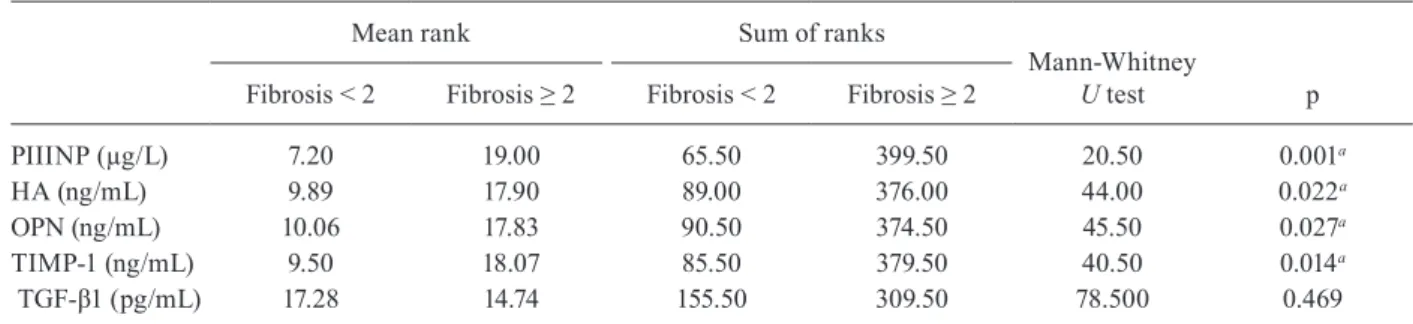
When comparing the stages of liver fibrosis among HCV patients, the levels of the direct biomarkers PII-INP, HA, OPN and TIMP-1 were significantly higher in
patients with a fibrosis stage ≥ 2 than in children with a
fibrosis stage < 2 (p < 0.05). No significant difference in
TGF-β1 was detected between the two groups (p > 0.05)
(Table III).
Table IV shows the diagnostic accuracy of the direct serum marker measurements in HCV patients with
non-significant (F < 2) or non-significant (F ≥ 2) fibrosis. The
cut-off value of PIIINP was 11.21 µg/L, with 48.55% Se, 100% Sp and a PPV of 100%. At the cut-off value of HA (24.69 ng/mL), 42% Se, 93.88% Sp and 92.54% PPV were recorded. The cut-off value of OPN was 101.56 ng/ mL, with a Se, Sp and PPV of 97.67%, 100% and 100%, respectively (Table IV). The cut-off value of TIMP-1 was 102.47 ng/mL, with a Se, Sp and PPV of 90.33%, 45.70%
and 75.76%, respectively. The cut-off value of TGF-β1
(6.53 pg/mL) corresponded with 85.70% Se, 42.70% Sp and 71% PPV.
The diagnostic accuracy of the indirect serum
mark-ers in HCV patients (2 > F ≥ 2) is shown in Table V. The
Se and Sp of AST were 96.90% and 45.12%, respectively, and ALT yielded values of 84.24% and 23.23%, respec-tively. The cut-off values of AST and ALT were 18 IU/L
and 20 IU/L, respectively, and their PPVs were 99.30% and 86.43%, respectively. Therefore, AST may be used as an indicator of liver fibrosis rather than ALT. The Se and Sp of albumin were 95% and 67.40%, respectively, and total bilirubin yielded values of 85.20% and 68.10%, respectively. The PPVs of albumin and total bilirubin were 70.10% and 67.40%, respectively. Therefore, both albumin and bilirubin appeared to be insufficient indica-tors of liver fibrosis.
DISCUSSION
Liver biopsy is regarded as the gold standard for the assessment of patients with liver fibrosis (Bianchi 2001). However, liver biopsy is an invasive procedure with po-tential complications and sampling error can result in substantial misdiagnosis and staging inaccuracies.
Recently, biochemical parameters were found to be good indicators of liver fibrosis, especially in children (Valva et al. 2011). Serum markers of liver fibrosis are divided into two categories: direct markers, which re-flect ECM turnover, and indirect markers, which rere-flect alterations in hepatic function, but not ECM metabolism
(Grigorescu 2006).
One of these direct markers is PIIINP, which can
be used as a measure of matrix deposition (Grigore -scu 2006). Walsh et al. (1999a) found a significant cor-relation between serum PIIINP levels and histological changes such as fibrosis, periportal necrosis and an
al-tered histological activity index. In addition, Fabris et al.
(1997) showed a correlation between PIIINP levels and the stages of hepatic fibrosis in alcoholic liver disease, viral hepatitis and primary biliary cirrhosis. In parallel with these observations, our results showed a significant increase in PIIINP levels (by 26.28%) in paediatric HCV patients compared with healthy subjects. PIIINP also showed a statistical association with more severe (high-stage) fibrosis. The cut-off value of PIIINP was 11.21 µg/L, with 48.55% Se and 100% Sp. The PPV was 100%, which emphasises the utility of PIIINP as an indicator of liver fibrosis and reduces the need for biopsy for fibrosis detection in HCV-infected children.
TABLE I
Serum levels of TGF-β1, TIMP-1, HA, PIIINP and OPN in HCV and healthy subjects
Parameters Healthy HCV p <
PIIINP (µg/L) 13.66 ± 2.11 17.25a ± 3.67 0.01
HA (ng/mL) 8.22 ± 0.98 12.87a ± 4.45 0.04
OPN (ng/mL) 18.23 ± 5.45 150.11a ± 11.28 0.0001
TIMP-1 (ng/mL) 82.75 ± 5.22 150.72a ± 9.78 0.0001
TGF-β1 (pg/mL) 3.11 ± 0.76 14.22a ± 3.27 0.0001
a: statistical analysis were done by Student’s t test where the level of significance between hepatitis C virus (HCV) and healthy group is at p < 0.05. Values are mean ± standard devia-tion of 30 subjects in each group. HA: hyaluronic acid; OPN: osteopontin; PIIINP: type III procollagen amino-terminal peptide; TGF-β1: transforming growth factor-β1; TIMP-1: tis -sue inhibitor of matrix metalloproteinase.
TABLE II
Serum levels of AST, ALT,
albumin and total bilirubin in HCV and healthy subjects
Parameters Healthy HCV p
AST (IU/L) 28.34 ± 4.45 142.15a± 13.47 0.0001
ALT (IU/L) 22.19 ± 5.22 80.72a± 12.44 0.0001
Albumin (g/dL) 3.22 ± 0.77 2.87a± 0.45 0.031
Total bilirubin (mg/dL) 0.81 ± 0.15 5.66a± 2.13 0.0001
The second matrix deposition marker is HA. HA is a glycosaminoglycan and a component of the ECM
synthe-sised by hepatic stellate cells (HSCs) (Grigorescu 2006).
Under normal circumstances, the endothelial cells of the liver sinusoid are the sites of HA uptake and degradation (Eriksson et al. 1983). Increased levels of HA are due to a decrease in hepatic removal, increased production or
both (Grigorescu 2006). Guéchot et al. (1995) postulated
high levels of serum HA in patients with liver diseases of different aetiologies and particularly in patients with cir-rhosis. Serum levels of HA have been shown to be related
not only to the stage of fibrosis (Guéchot et al. 1995), but
also to the degree of necroinflammation (Murawaki et al. 1995). Korner et al. (1996) added that the HA concen-tration has a good prognostic value for complications of liver cirrhosis: hepatic encephalopathy stages III and IV, refractory ascites and portal vein thrombosis. In agree-ment with these observations, the present study showed a significant increase in HA levels in paediatric HCV patients (56.56% increase) compared with healthy sub-jects. Additionally, HA was associated with more severe (high-stage) fibrosis. At a cut-off value of 24.69 ng/mL, HA yielded a Se of 42% and a Sp of 93.88%. The PPV reached 92.54%, which confirms the utility of HA for the identification of fibrosis, thus reducing the need for biopsy in these patients.
OPN is an ECM protein that is considered as the third marker for matrix deposition (Pan et al. 2003). Abu El Makarem et al. (2011) found a significant elevation in OPN levels in patients with HCV-related HCC. The same authors found that OPN levels are a potential di-agnostic marker for HCC, especially among high-risk groups of patients. Shang et al. (2012) monitored serum OPN levels for one year before the diagnosis of HCV-related HCC patients. Libra et al. (2005) postulated that OPN might play a role in lymphomagenesis, particularly in the context of HCV infection. In agreement with these results, the present study revealed a significant elevation in OPN levels (723.42% increase) that was significantly associated with the developmental stage of fibrosis. The results revealed that OPN is a good indicator of liver fi-brosis associated with HCV.
Regarding the markers associated with matrix deg-radation (metalloproteinases, MMPs and their inhibi-tors), MMPs show substrate Sp for interstitial collagen, whereas their tissue inhibitors (TIMPs) can irreversibly
bind to and inactivate MMPs (Friedman 1999). Although
several investigators have discussed the roles of MMPs in liver disease, most focused on their participation in cancer development and metastasis, whereas no study has assessed the role of MMPs in carriers of hepatitis vi-ruses (Youness et al. 2013). Excess production of TIMPs relative to MMPs may be an important factor in the pro-gression of liver fibrosis (Iredal et al. 1992). TIMPs are also observed in late-stage fibrosis, but not in the mild
TABLE III
Comparison of direct biomarkers between two stages of liver fibrosis
Mean rank Sum of ranks
Mann-Whitney
U test p
Fibrosis < 2 Fibrosis ≥ 2 Fibrosis < 2 Fibrosis ≥ 2
PIIINP (µg/L) 7.20 19.00 65.50 399.50 20.50 0.001a
HA (ng/mL) 9.89 17.90 89.00 376.00 44.00 0.022a
OPN (ng/mL) 10.06 17.83 90.50 374.50 45.50 0.027a
TIMP-1 (ng/mL) 9.50 18.07 85.50 379.50 40.50 0.014a
TGF-β1 (pg/mL) 17.28 14.74 155.50 309.50 78.500 0.469
a: significant level is at p < 0.05. Statistical analysis were done by Mann-Whitney U test. HA: hyaluronic acid; OPN: osteopontin; PIIINP: type III procollagen amino-terminal peptide; TGF-β1: transforming growth factor-β1; TIMP-1: tissue inhibitor of matrix metalloproteinase.
TABLE IV
Diagnostic accuracy of direct serum markers measurement in HCV patients
Parameters
HCV patients
Sea
(%) Spb
(%)
PPVc
(%)
NPVd
(%)
Cut-offe
PIIINP (µg/L) 48.55 100 100 48.55 11.21 HA (ng/mL) 42 93.88 92.54 45.77 24.69 OPN (ng/mL) 97.67 100 100 97.60 101.56 TIMP-1 (ng/mL) 80.33 45.70 75.76 61.43 102.47
TGF-β1 (pg/mL) 85.70 42.70 71 64.57 6. 53
stage (Walsh et al. 1999b). Additionally, Boeker et al. (2002) found a strong correlation between TIMP-1 lev-els and histological inflammatory scores and the serum level of TIMP-1 is associated with active inflammatory activity (Ueno et al. 1996). In line with the above ob-servations, our results revealed a significant increase in TIMP-1 levels (82.23% increase) compared with levels healthy children. In addition, TIMP-1 was significantly associated with high-stage fibrosis. The cut-off value of TIMP-1 was 102.47 ng/mL, with Se and Sp reaching 90.33% and 45.70%, respectively. The PPV of TIMP-1 was 75.76%, which indicated that this biomarker is a valuable tool for the detection of liver fibrosis.
In the present study, TGF-β1 levels were significantly
increased in paediatric HCV patients (357.23% increase)
compared with the control group. TGF-β1 is a cytokine
associated with hepatic fibrosis and is a homodimeric polypeptide that is secreted in an inactive form that
re-quires activation. In hepatic pathology, TGF-β1 is the
most important stimulus for the production of ECM by HSCs (Sasaki et al. 1992) and is also an inhibitor of hepa-tocyte growth and proliferation (Nakamura et al. 1985). In liver biopsies from patients with chronic liver disease,
TGF-β1 levels are correlated with type I collagen mRNA (Breitkopf et al. 2001). The value of serum TGF-β1 lev -els has certain limitations, including the potential
con-tamination of samples by TGF-β1 from platelets, interfer -ence by plasmin activity in the plasma that increases the
amount of TGF-β1, the binding of TGF-β1 to the ECM
and vascular endothelium at sites of injury, sequestra-tion by soluble proteins and the complicated clearance
of TGF-β1. These factors explain why plasma levels of TGF-β1 are unlikely to be of diagnostic value (Breitkopf
et al. 2001). In agreement with these observations and in
parallel with our study, serum TGF-β1 levels showed no
statistically significant differences among fibrosis stages
in paediatric patients. The cut-off value of TGF-β1 was
6.53 pg/mL, with a Se of 85.70%. Due to the low Sp (42.70%), the PPV of 71% and the possibility of
contami-nation of serum TGF-β1 with other TGF-β1 forms, we
cannot consider this protein as a marker for the diagnosis of liver fibrosis associated with HCV. Contradictory to our observation, certain studies showed a good
correla-tion between the serum levels of TGF-β1 and the rate of
fibrosis progression (Kanzler et al. 2001).
Although serum ALT levels generally reflect liver
injury (Grigorescu 2006), the correlation between ALT
levels, necroinflammation and the fibrosis score is poor,
especially in chronic HCV infection (Grigorescu 2006).
AST levels have a stronger correlation with hepatic
fibrosis than ALT levels (Gordon et al. 2000). The in -crease in ALT levels is related to mitochondrial dysfunc-tion and reduced clearance of AST by hepatic sinusoidal
cells (Grigorescu 2006). Kruger et al. (2011) confirmed
that ALT could not differentiate between the stages of disease or the grades of fibrosis. In agreement with the present study, AST and ALT levels were significantly increased in the HCV group (401.58% and 263.76% in-creases, respectively). In addition, AST and ALT values showed no statistical correlation with fibrosis stages. The Se and Sp of AST were 96.90% and 45.12%, respec-tively, and ALT yielded values of 84.24% and 23.23%, respectively. The cut-off values of AST and ALT were 18 IU/L and 20 IU/L, respectively, and their PPVs were 99.30% and 86.43%, respectively. Therefore, AST may be used as an indicator of liver fibrosis rather than ALT. A significant increase in total bilirubin levels in HCV-infected children (598.76% increase) vs. healthy individuals was recorded. In contrast, albumin levels showed a significant (10.86%) decrease. Albumin and to-tal bilirubin showed no statistical correlation with fibro-sis stage. The Se, Sp and PPVs of both albumin and total bilirubin appeared to be insufficient for detecting liver fibrosis. These observations were in accordance with the findings of Hongbo et al. (2007), who stated that routine serum markers, including AST, albumin and total biliru-bin, were independent predictors of significant fibrosis.
In conclusion, PIIINP, HA, OPN and TIMP-1 served as clinically useful biomarkers for predicting liver fibro-sis in HCV-infected children, whereas AST, ALT, albu-min and bilirubin appeared to be insufficient markers for liver damage detection. Due to the relatively limited number of cases in this study, further studies are needed to accurately determine whether to use these biomark-ers instead of biopsies, as the small sample size makes it difficult to evaluate the validity of the serum markers under investigation.
REFERENCES
Abu El Makarem MA, Abdel-Aleem A, Ahmed Ali A, Saber R, Shatat M, Abdel Rahem D, Sayed D 2011. Diagnostic significance of plasma osteopontin in hepatitis C virus-related hepatocellular carcinoma. Ann Hepatol10: 296-305.
Bianchi L 2001. Liver biopsy in elevated liver functions tests? An old question revisited. J Hepatol35: 290-294.
TABLE V
Diagnostic accuracy of indirect serum markers measurement in HCV patients
Parameters
HCV patients
Sea
(%) Spb
(%) PPVc
(%) NPVd
(%) Cut-offe
AST (IU/L) 96.9 45.12 99.30 41.24 18 ALT (IU/L) 84.24 23.23 86.43 42.50 20
Albumin (g/dL) 35 67.4 70.1 95 1.45
Total bilirubin (mg/dL) 85.2 68.1 67.4 85.7 0.42
Boeker RH, Haberkorn CI, Michels D 2002. Diagnostic potential of circullating TIMP-1 and MMP-2 as markers of liver fibrosis in patients with chronic hepatitis C. Clin Chim Acta316: 71-81.
Bravo A, Sheth S, Chopra S 2001. Liver biopsy. N Engl J Med344: 495-500.
Breitkopf K, Lahme B, Tag CG, Gressner AM 2001. Expression and
matrix deposition of latent transforming growth factor beta bing-ing proteins in normal and fibrotic rat liver and transdifferentiat-ing hepatic stellate cells in culture. Hepatology33: 387-396.
Dienstag J 2002. The role of liver biopsy in chronic hepatitis. Hepa-tology36 (Suppl.): S152-S160.
Doumas BT, Perry BW, Sasse EA, Straumfjord JV 1973. Standard-ization in bilirubin assays: evaluation of selected methods and stability of bilirubin solutions. Clin Chem19: 984-993.
El-Raziky MS, El-Hawary M, El-Koofy N 2004. Hepatitis C virus infection in Egyptian children: single centre experience. J Viral Hepatol 11: 471-576.
Eriksson S, Fraser JRE, Laurent TC 1983. Endothelial cells are a site
of uptake and degradation of hyaluronic acid in the liver. Exp Cell
Res144: 223-228.
Fabris C, Falleti E, Federico E 1997. A comparison of four markers of
fibrosis in diagnosis of cirrhosis. Ann Clin Biochem 34: 151-155.
Friedman SL 1999. Cytokines and fibrogenesis. Sem Liver Dis19: 129-140.
Gella FJ, Olivella T, Cruz PM, Arenas J, Moreno R, Durban R, Gomez
JA 1985. A simple procedure for routine determination of aspar-tate aminotransferase and alanine aminotransferase with pyri-doxal phosphate. Clin Chem Acta153: 241-247.
Gordon SC, Fang J W, Silverman AL 2000. The significance of base -line serum alanine aminotransferase on pretreatment disease characteristics and response to therapy in chronic hepatitis C.
Hepatology32: 400-404.
Grigorescu M 2006. Noninvasive biochemical markers of liver fibro -sis. J Gastrointestin Liver Dis5: 149-159.
Guéchot J, Poupon RE, Poupon R 1995. Serum hyaluronan as a mark -er of liv-er fibrosis. J Hepatol 22: 103-106.
Hongbo L, Xiaohui L, Hong K, Wei W, Yong Z 2007. Assessing rou-tine and serum markers of liver fibrosis in CHB patients using parallel and serial interpretation. Clin Biochem40: 562-566.
Iredal JP, Murhy G, Hembry RM 1992. Human hepatic lipocytes syn -thesize tissue inhibitor of metalloproteinase-1. Implications for regulation of matrix degradation in liver. J Clin Invest90: 282-287.
Jara P, Resti M, Hierro L, Giacchino R, Barbera C 2003. Chronic
hepatitis C virus infection in childhood: clinical patterns and evolution in 224 white children. Clin Infect Dis36: 275-280.
Kanzler S, Beumann M, Schirmacher P 2001. Prediction of progres-sive liver fibrosis in hepatitis C infection by serum and tissue
lev-els of transforming growth factor β. J Viral Hepatol8: 430-438.
Korner T, Kropf J, Gressner AM 1996. Serum laminin and hyaluro -nan in liver cirrhosis: markers of progression with high prognos-tic value. J Hepatol25: 684-688.
Kruger FC, Daniels CR, Kidd M, Swart G, Brundyn K, van Rensburg
C, Kotze M 2011. APRI: a simple bedside marker for advanced
fi-brosis that can avoid liver biopsy in patients with NAFLD/NASH.
S Afr Med J 101: 477-480.
Libra M, Indelicato M, De Re V, Zignego AL, Chiocchetti A,
Malapon-te G, Dianzani U, Nicoletti F, Stivala F, McCubrey JA, Mazzarino
MC 2005. Elevated serum levels of osteopontin in HCV-associated lymphoproliferative disorders. Cancer Biol Ther4: 1192-1194.
Manning D, Afdhal N 2008. Diagnosis and quantitation of fibrosis.
Gastroenterology134: 1670-1681.
Martínez SM, Crespo G, Navasa M, Forns X 2011. Noninvasive as -sessment of liver fibrosis. Hepatology53: 325-335.
Murawaki Y, Ikuta Y, Nishimura Y 1995. Serum markers for con-nective tissue turnover in patients with chronic hepatitis B and chronic hepatitis C: a comparative analysis. J Hepatol23: 145-152.
Murray K, Finn L, Taylor S, Seidel K, Larson A 2005. Liver histology
and alanine aminotransferase levels in children and adults with chronic hepatitis C infection. J Pediatr Gastroenterol Nutr41: 634-638.
Nakamura T, Tomita Y, Hirai R 1985. Inhibitory effect of
transform-ing growth factor-β on DNA synthesis of adult rat hepatocytes in
primary culture. Biochem Biophys Res Commun133: 1042-1050.
Pan HW, Ou YH, Peng SY, Liu SH, Lai PL, Lee PH, Sheu JC 2003. Overexpression of OPN is related with intrahepatic metastasis, early recurrence and poorer prognosis of surgically resected he-patocellular carcinoma. Cancer98: 119-127.
Parkes J, Guha IN, Roderick P, Harris S, Cross R 2011. Enhanced liver fibrosis (ELF) test accurately identifies liver fibrosis in patients
with chronic hepatitis C. J Viral Hepatol18: 23-31.
Pinzani M, Rombouts K, Colagrande S 2005. Fibrosis in chronic
liver diseases: diagnosis and management. J Hepatol45 (Sup-pl.): S22-S36.
Prati D 2006. Transmission of hepatitis C virus by blood transfu-sions and other medical procedures: a global review. J Hepatol 45: 607-616.
Saitou Y, Shiraki K, Yamanaka Y, Yamaguchi Y, Kawakita T 2005. Noninvasive estimation of liver fibrosis and response to interferon therapy by a serum fibrogenesis marker, YKL-40, in patients with HCV-associated liver disease. World J Gastroenterol28: 476-481.
Sanai F, Benmousa A, Al-Hussaini H, Ashraf S, Alhafi O 2008. Is se -rum alanine transaminase level a reliable marker of histological disease in chronic hepatitis C infection? Liver Int28: 1011-1014.
Sanvisens A, Serra I, Tural C, Tor J, Ojanguren I 2009. Hyaluronic acid, transforming growth factor-beta1 and hepatic fibrosis in patients with chronic hepatitis C virus and human immunodefi-ciency virus co-infection. J Viral Hepatol16: 513-518.
Sasaki H, Pollard RB, Schmitt D, Suzuki F 1992. Transforming growth factor-β in the regulation of immune response. Clin Im
-munol Immunopathol 65: 1-9.
Sebastiani G, Vario A, Guido M, Alberti A 2008. Performance of
noninvasive markers for liver fibrosis is reduced in chronic hepa-titis C with normal transaminases. J Viral Hepatol 15: 212-218.
Shang S, Plymoth A, Ge S, Feng Z, Rosen HR, Sangrajrang S, Hain -aut P, Marrero JA, Beretta L 2012. Identification of osteopontin as a novel marker for early hepatocellular carcinoma. Hepatology 55: 483-490.
Theise N, Bordenheimer H, Ferrel L 2007. Acute and chronic viral hepatitis. In AD Burt, BC Portmann, LD Ferrel (eds.), Mac
-Sween’s pathology of the liver, Vol. 8, 5th ed.,
Churchill-Living-stone, London, p. 418-419.
Ueno T, Tamaki S, Sugawara H 1996. Significance of serum tissue inhibitor of metalloproteinase-1 in various liver disease. J Hepa-tol24: 177-184.
Valva P, Casciato P, Carrasco JMD, Adrian Gadano A, Galdame O, Galoppo MC, Mullen E, De Matteo E, María Victoria Preciado
MV 2011. The role of serum biomarkers in predicting fibrosis progression in pediatric and adult hepatitis C virus chronic infec-tion. PLoS ONE6: e23218.
Walsh KM, Fletcher A, MacSween RN, Marris AJ 1999a. Compari -son of assays for N-terminal propeptide of type III procollagen
in chronic hepatitis C by using receiver operating characteristic analysis. Eur J Gastroenterol Hepatol 11: 827-831.
Walsh KM, Timms P, Campbell S 1999b. Plasma levels of matrix met-alloproteinase-2 (MMP-2) and tissue inhibitors of metalloprotei-nase-1 and-2 (TIMP-1 and TIMP) as noninvasive markers of liver disease in chronic hepatitis C. Comparison using ROC analysis.
Dig Dis Sci44: 624-630.
Youness ER, El Nemr M, El Azizy HM, Aly HF 2013. Significance of
apoptotic markers Bcl-2, CD4+ T cells, hepatocyte growth factor
and metalloproteinase-9 in infected patients with HCV. J Appl