J of Evidence Based Med & Hlthcare, pISSN- 2349-2562, eISSN- 2349-2570/ Vol. 2/Issue 44/Nov. 02, 2015 Page 8026
STUDY OF AGE, SEX AND ETIOLOGIC SPECTRUM OF PERICARDIAL
EFFUSION IN TERTIARY CARE HOSPITAL
P. Ravikaladhar Reddy1, Kekathi Vidya Sagar2, S. Chandrasekhar3, P. Nagaraja Ramesh4
HOW TO CITE THIS ARTICLE:
P. Ravikaladhar Reddy, Kekathi Vidya Sagar, S. Chandrasekhar, P. Nagaraja Ramesh. “Study of Age, Sex and Etiologic Spectrum of Pericardial Effusion in Tertiary Care Hospital”. Journal of Evidence based Medicine and Healthcare; Volume 2, Issue 44, November 02, 2015; Page: 8026-8034,
DOI: 10.18410/jebmh/2015/1078
ABSTRACT: Pericardial effusion is perhaps one of the most commonly overlooked clinical conditions and definite establishment of etiological agent is not always easy, successful or satisfactory. In this study, 50 cases of pericardial effusion admitted in Medical wards were analysed with emphasis on pattern of age and gender distribution, clinical presentation and etiology. The incidence of pericardial effusion common in age group between 21-40 years. The incidence of pericardial effusion is more in males. In the present study, the youngest patient is 15 year old and the oldest is 62 year old. Breathlessness being commonest symptom and raised JVP Is commonest sign. 60% of cases are of tuberculosis etiology, 15% are due to uremia and malignancy each, and 5% due to collagen vascular disease.
KEYWORDS: Pericardial Effusion, Tuberculosis, Uremia, Cardiac Tamponade.
INTRODUCTION: The pericardium has interested outstanding physicians from the biblical past to the current era. Knowledge of pericardial disease dates back to the time of GALEN [A.D 131-201], who gave the pericardium its name.
It is often involved by processes that affect the heart, but it may also be involved by diseases of adjacent tissues and may itself be a primary site of disease.1,2 Etiologic studies of
pericardial effusion have been done in the west and a viral etiology has been commonly reported but in tropics, etiologic spectrum could be much different because of different social, economic, nutritional and immunologic factors.3,4,5
AIMS AND OBJECTIVES:
1. To study the pattern of age, sex distribution, clinical presentation and etiology of moderate to large sized pericardial effusion in medical wards.
2. To study the prevalence of tubercular pericardial effusion 3. To study the incidence of cardiac tamponade.
MATERIALS AND METHODS: The patients admitted in the medical wards of Govt. General Hospital with the suspicion of pericardial effusion over period of 18 months were taken for study.
J of Evidence Based Med & Hlthcare, pISSN- 2349-2562, eISSN- 2349-2570/ Vol. 2/Issue 44/Nov. 02, 2015 Page 8027
In all cases:
1. Urine examination. 2. Hemogram.
3. ESR.
4. Blood sugar. 5. Blood urea. 6. Serum creatinine. 7. Serum electrolytes. 8. Chest x- ray.
9. Electrocardiography. 10.Echocardiography.
Wherever Necessary: 1. ASO titers.
2. RA factor.
3. Antinuclear antibodies, Anti ds DNA antibodies. 4. Serum cholesterol.
5. Thyroid profile. 6. Cardiac enzymes. 7. Mantoux test.
8. Pericardiocentesis and fluid analysis. 9. CT scan chest.
BASIS OF DIAGNOSIS: The diagnosis of the cause of pericardial effusion is not an easy one and requires a battery of investigations and sometimes may not be revealed even then.
In this study, routine urine and blood investigations, and specific investigations based on diagnostic suspicion were taken up. In all cases, electrocardiography and chest radiography were done, in those where the findings suggested a diagnosis of pericardial effusion, its presence was confirmed by echocardiography.
To find the etiology, pericardial fluid analysis was done including volume, colour, specific gravity, proteins, cell count, ADA levels, Grams and AFB staining and culture for pyogenic organisms and mycobacteria. The diagnosis of tuberculous effusion was based on increased erythrocyte sedimentation rate, lymphocytosis in blood, positive Mantoux test, exudative effusion on pericardial fluid analysis, increase in pericardial fluid ADA levels, with or without a positive AFB stain and mycobacterial culture. Other causes of effusion were simultaneously ruled out.
In one case of tuberculous effusion there was associated cervical lymphadenitis confirmed as tuberculosis on biopsy.
J of Evidence Based Med & Hlthcare, pISSN- 2349-2562, eISSN- 2349-2570/ Vol. 2/Issue 44/Nov. 02, 2015 Page 8028
thickening of pericardium on CECT chest. In another case patient had bronchogenic carcinoma with mediastinal lymphadenopathy.
The diagnosis of pericardial effusion in SLE was based on criteria for clinical diagnosis of SLE as per American rheumatology association, demonstration of antinuclear antibodies and anti- double standard DNA antibodies. In one case where pericardial effusion was associated with Ebsteins anomaly, the routine urine and blood investigations were normal, pericardial fluid was transudate, ANA, anti-ds DNA anti bodies were negative, no other systemic disease was made out, hence the effusion was categorized as Ebsteins anomaly associated with idiopathic effusion.
Pericardiocentesis was avoided in those cases where there was a contraindication in the form of prolonged bleeding time, as in uremic effusions and in those cases where the diagnosis was obvious on other investigations.
OBSERVATION AND DISCUSSION: Pericardial effusion is perhaps one of the most commonly overlooked clinical conditions and definite establishment of etiological agent is not always easy, successful or satisfactory. In this study, 50 cases of pericardial effusion were analysed with emphasis on age and gender distribution, clinical presentation and etiology.
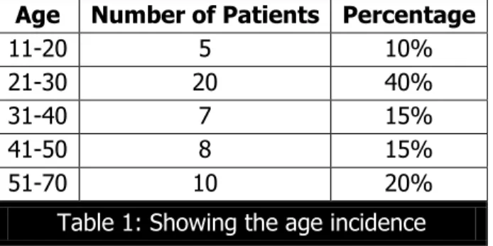
A. AGE AND SEX INCIDENCE IN PERICARDIAL EFFUSION: As shown in table number 1, the highest age incidence of pericardial effusion between 21-40 years. The youngest patient is 15 year old and the oldest is year old.
J. C. Banerjee et al6 in their study, observed that the peak age incidence was between
21-30 years. In the study of Pillay,7 the greatest incidence was between 20-40 years.
Age Number of Patients Percentage
11-20 5 10%
21-30 20 40%
31-40 7 15%
41-50 8 15%
51-70 10 20%
Table 1: Showing the age incidence
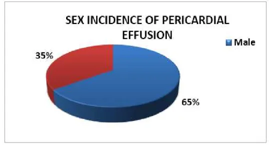
The incidence of pericardial effusion is more in males, that is 65%. Banerjee6 et al
reported 60% incidence in males in their study. Pillay7 reported 80% incidence in males in their
study of 40 cases.
Sex Number of Patients Percentage
Male 32 65%
Female 18 35%
J of Evidence Based Med & Hlthcare, pISSN- 2349-2562, eISSN- 2349-2570/ Vol. 2/Issue 44/Nov. 02, 2015 Page 8029
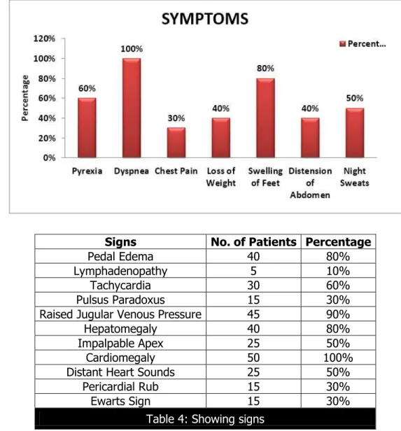
B. SYMPTOMATOLOGY: Breathlessness on exertion or rest is one of the earliest and most disabling symptom. In the present study, dyspnea is present in 100% of cases. The next common symptom is fever which is present in 60% of cases. John Hageman et al reported dyspnea as predominant symptom in their study.
Berry and Banerjee reported breathlessness in 65% of cases in their study. Banerjee reported fever in 60% of cases in his study. Hageman8 et al reported fever as a predominant
symptom in 73% of cases. Banerjee JC9 observed fever in 80% of their cases. Chest pain is
observed in 30% of cases in this study. Hageman reported chest pain in 39% cases. In the present study, swelling of feet is present in 80% of cases. In the study of Berry and Banerjee, swelling of feet was observed in 60% cases. Hageman et al reported swelling of feet in 55% of cases. Banerjee reported swelling of feet in 50% of cases.
Symptoms No. of Patients Percentage
Pyrexia 30 60%
Dyspnea 50 100%
Chest Pain 15 30%
Loss of Weight 20 40%
Swelling of Feet 40 80%
Distension of Abdomen 20 40%
Night Sweats 25 50%
J of Evidence Based Med & Hlthcare, pISSN- 2349-2562, eISSN- 2349-2570/ Vol. 2/Issue 44/Nov. 02, 2015 Page 8030
Signs No. of Patients Percentage
Pedal Edema 40 80%
Lymphadenopathy 5 10%
Tachycardia 30 60%
Pulsus Paradoxus 15 30%
Raised Jugular Venous Pressure 45 90%
Hepatomegaly 40 80%
Impalpable Apex 25 50%
Cardiomegaly 50 100%
Distant Heart Sounds 25 50%
Pericardial Rub 15 30%
Ewarts Sign 15 30%
Table 4: Showing signs
The commonest sign noted in the present study, is raised jugular venous pressure, seen in 100% of cases. Berry and Banerjee6 reported raised JVP in 55% cases. Banerjee reported
raised JVP in 56% of cases. Hageman8 reported raised JVP in 59% cases. In the present study,
hepatomegaly is observed in 80% of cases. Banerjee et al reported hepatomegaly in 60% cases. In the present study, pulsus paradoxus is noted in 30% cases, which coincides with the study of Banerjee.9,5
Hageman et al reported pulsus paradoxus in 45% cases in their series. In the present study, Ewarts sign is observed in 30% of cases. Banerjee6 observed Ewarts sign in 16% cases.
Hageman et al reported Ewarts sign in 20% cases. In the present study, pericardial rub is observed in 30% cases. Banerjee6 reported rub in 25% cases. Berry and Banerjee6,9 reported rub
J of Evidence Based Med & Hlthcare, pISSN- 2349-2562, eISSN- 2349-2570/ Vol. 2/Issue 44/Nov. 02, 2015 Page 8031
C. ETIOLOGY OF PERICARDIAL EFFUSION: The diagnosis of pericardial effusion is by demonstration of tubercle bacilli in the smear of pericardial effusion or by the growth of tubercle bacilli in culture.10 The diagnosis of tuberculous effusion is difficult because of rarity
of direct bacteriological proof. However the diagnosis may be suggested by Insidious onset, low grade fever, associated pleural effusion, tuberculous lung lesion, matted lymph nodes, sterile pericardial fluid, and response to anti tuberculous treatment.
In the present study, 60% of cases are of tuberculous etiology, 15% are due to uremia and malignancy each, and 5% due to collagen vascular disease.
In the series of Pillay,7 52.5% cases were of tuberculous etiology. In the series of
Banerjee, 62.5% cases were of tuberculous etiology. J.N Berry and Banerjee6 reported 60%cases
of tuberculous etiology in their series. Their incidence of malignant etiology was 5.56% and incidence of pyogenic etiology 2.78%.
In the present study of 50 cases of pericardial effusions of different etiology, the following signs and symptoms were noted.
Etiological Types No. of Cases Percentage
Tuberculosis 30 60%
Uremia 8 16%
Malignancy 7 14%
Collagen Vascular Disease 2 4%
Others 3 6%
J of Evidence Based Med & Hlthcare, pISSN- 2349-2562, eISSN- 2349-2570/ Vol. 2/Issue 44/Nov. 02, 2015 Page 8032
D. ELECTROCARDIOGRAPHIC FINDINGS: In 50% cases low voltage is observed. ST, T changes are seen in 40% cases. Normal electrocardiography is recorded in 16% cases.
E. PROPORTION OF LARGE AND MODERATE EFFUSIONS: An effusion is considered small, when the amount of fluid is less than 100 ml. When the amount of fluid is between 100-500 ml, it is considered a moderate effusion.11 If the pericardial fluid is more than 500 ml, it is
considered a large effusion. In this study, large effusions are 55% and small effusions 45%.
In majority of effusions, pericardial fluid is noted anteriorly, apically and posteriorly. Large effusions show greater accumulation of fluid anteriorly and apically. In the present study swinging motion of heart is seen in one patient who had a large effusion, but was not in cardiac tamponade.11 Swinging motion of heart is associated with striking undulant motion of anterior
J of Evidence Based Med & Hlthcare, pISSN- 2349-2562, eISSN- 2349-2570/ Vol. 2/Issue 44/Nov. 02, 2015 Page 8033
SUMMARY AND CONCLUSIONS:
1. It is common in the age group of 21-40 years in present study. Banerjee et al in their study observed that the peak age incidence was between 21-30 years. In the study of Pillay the greatest incidence was between 20-40 years.
2. Pericardial effusion is more common in males 65%.in present study. Banerjee et al reported 60% incidence in males in their study. Pillay reported 80% incidence in males in their study of 40 cases.
3. Breathlessness is the commonest symptom observed in pericardial effusion in 100% of cases and fever is present in 60% of cases. Hageman et al reported breathlessness as predominant symptom in their study. Banerjee reported breathlessness in 65% of cases in their study and fever in 60% of cases.
4. Raised jugular venous pressure is the commonest sign in 90%of cases. Banerjee reported raised JVP in 55% of cases. Hageman reported raised JVP in 59% cases.
5. Tuberculosis is the commonest etiological factor for pericardial effusion in India. In the present study 60% of cases are of tuberculous etiology. In the series of Pillay 52.5% cases were of tuberculous etiology. In the series of Banerjee 62.5% cases were of tuberculous etiology.
6. Uremia and malignancy are next to tuberculosis in causing pericardial effusion 15% each. In the series of J.N Berry and Banerjee incidence of malignant etiology was 5.56%.
BIBLIOGRAPHY:
1. Chaurasia BD. Pericardium and heart. Human anatomy regional and applied. New Delhi: CBS; 1999: 213-215.
2. Shabetai R, Mangiardi L, Bhargava V et al. The pericardium and cardiac function. Prog Cardiovasc Dis. 1979; 22: 107
3. Lewinter N Martin, Kabbani Samer. Pericardial diseases. Braunwalds heart disease, a textbook of cardiovascular medicine. Elsivier; 2006. 1759-1778.
4. Spodick DN. Pericardial diseases. In Braunwald E, Zipes D, Libby P(eds): Heart disease. Philadelphia, WB Saunders; 2001; 1823-1876.
5.
MEGREGORM. Pulsus paradoxus. N Engl J Med.1979; 301: 480
6. Berry and Banerjee. Diagnosis of pericarditis. J Assoc Physicians India. 1955; 1: 30.
7.
Pillay CRR. Etiology of pericarditis. Indian Heart J. 1955; 7: 65-70.
8. Hageman JH, d'Esopo ND, Glenn WWL. Tuberculosis of the pericardium: a long-term analysis of forty-four proved cases. N Engl J Med. 1964; 270: 327–332.
9. BANERJEE JC. Clinical aspects of pericarditis. Indian Heart J .1955; 7: 71-76.
10.Spodick DH. Pericardial rub. Prospective, multiple observer investigation of pericardial friction in 100 patients. Am. J Cardiol. 1975; 25: 357-362.
J of Evidence Based Med & Hlthcare, pISSN- 2349-2562, eISSN- 2349-2570/ Vol. 2/Issue 44/Nov. 02, 2015 Page 8034 4. Resident, Department of Medicine,
Kurnool Medical College, Government General Hospital.
NAME ADDRESS EMAIL ID OF THE CORRESPONDING AUTHOR:
Dr. Kekathi Vidya Sagar, Associate Professor, Department of Medicine, Kunrool Medical College, Government General Hospital, Kurnool, Andhra Pradesh.
E-mail: [email protected]
Date of Submission: 09/10/2015. Date of Peer Review: 10/10/2015. Date of Acceptance: 20/10/2015. Date of Publishing: 29/10/2015.
AUTHORS:
1. P. Ravikaladhar Reddy 2. Kekathi Vidya Sagar 3. S. Chandrasekhar 4. P. Nagaraja Ramesh
PARTICULARS OF CONTRIBUTORS:
1. Assistant Professor, Department of Medicine, Kurnool Medical College, Government General Hospital. 2. Associate Professor, Department of
Medicine, Kurnool Medical College, Government General Hospital. 3. Professor, Department of Medicine,