Use of focus assessed transthoracic echocardiography
(FATE) in the veterinary emergency room
Aplicabilidade da avaliação ecocardiográfica transtorácica focada (FATE) na sala de emergência veterinária Stephany Buba Lucina1 Ana Paula Sarraff-Lopes2 Marlos Gonçalves Sousa3*
ISSNe 1678-4596
INTRODUCTION
With recent advances in ultrasonography techniques the utility of echocardiography at the bedside in critically ill patients in the emergency room has been increased. Focus assessed transthoracic echocardiography (FATE), in these conditions, can be used to provide earlier and more accurate diagnosis, as well as to direct therapeutic choices by the medical staff (MOORE & COPEL, 2011). From this context, the American Society of Echocardiography (ASE) in conjunction with the American College of Emergency Physicians (ACEP) has developed a consensus for the use of echocardiography in the emergency rooms in man. The consensus was named as echocardiographic examination performed at
bedside - focused echocardiography (LABOVITZ et al., 2010). In veterinary medicine there is no standardization of FATE application in critically ill patients in the emergency room.
Cageside echocardiographic evaluation gives the veterinarian detailed information about the disease and direct diagnose and treatment of the emergency patient (TSE et al., 2013). FATE can be considered as an extension of the physical examination, and the term “visual stethoscope” has been coined. However, the obtained information should be interpreted in the context of each patient (MOORE & COPEL, 2011). According to the ASE, in well-trained hands, focused echocardiography can be regarded as an accurate
method, with reliable findings in the evaluation of
cardiac structure and function (SPENCER et al., 2013).
1Departamento de Medicina Veterinária, Universidade Federal do Paraná (UFPR), Curitiba, PR, Brasil. 2Escola de Ciências da Vida, Pontifícia Universidade Católica do Paraná (PUCPR), Curitiba, PR, Brasil.
3Departamento de Medicina Veterinária, Universidade Federal do Paraná (UFPR), 80050-540, Curitiba, PR, Brasil. E-mail: marlos98@ufpr.br. *Corresponding author.
ABSTRACT: The use of bedside focus assessed transthoracic echocardiography (FATE) is widespread in human medicine, and the potential
for veterinary medicine has only recently been investigated. Focused echocardiographic examination can be beneficial in critically ill patients compared to other bedside diagnostic methods, as well as facilitating rapid therapeutic approaches in the emergency room.The aim of this review is to discuss FATE and identify its main applications in veterinary medicine. In this context, FATE has proved to be benefical even when
carried out by a non-cardiologist physician or veterinarian. However, a few references on this subject exist in the veterinary literature and there is still a need for standardization of this technique for use in animals.
Key words: bedside, cardiology, cats, critical care, dogs.
RESUMO: A avaliação ecocardiográfica transtorácica focada (FATE) utilizada em pacientes à beira leito já é bastante difundida na medi
-cina, e apenas, recentemente tem se discutido a respeito deste assunto na medicina veterinária. Este exame pode trazer diversos benefícios aos pacientes críticos comparado aos outros métodos diagnósticos, além de facilitar e agilizar condutas terapêuticas na sala de emergência. O objetivo desta revisão é abordar a respeito desta modalidade de exame e identificar quais são suas principais aplicabilidades na medicina
veterinária. Dentro deste contexto, a FATE tem se mostrado benéfica inclusive quando realizada por médicos ou veterinários não cardiolo
-gistas. No entanto, poucas são as referêcias veteriárias a respeito deste assunto e ainda há necessidade de uma padronização desta técnica para uso em animais.
Palavras-chave:beira leito, cão, cardiologia, gato, intensivismo.
suspicion of heart failure, since physical examination
and thoracic radiography may not provide a definitive
diagnosis in these cases. This technique allows
the identification of left atrial enlargement, pleural
and pericardial effusions, left ventricular systolic dysfunction and morphological changes of the mitral valve (DEFRANCESCO, 2013).
The aim of this review was to address the differences between comprehensive echocardiography (CE) and FATE, describing how to perform the focused technique, exploring the possible
findings of these examinations, and thus, identifying
its main applications in veterinary medicine.
Comprehensive echocardiography versus FATE
For approximately 30 years transthoracic echocardiography has been considered the primary method for diagnosis and management of dogs and cats with cardiovascular disease (CHETBOUL, 2010). It is a non-invasive examination used to evaluate the anatomy and function of the heart and adjacent structures. Three echocardiographic modalities are used routinely in veterinary cardiology, M mode, two-dimensional echocardiography and Doppler, and each of these methods has its own application (WARE, 2011).
Despite the central role of transthoracic echocardiography in veterinary cardiology the echocardiographer must be aware of its limitations. Patient preparation (clipping and application of gel), a suitable table, as well as correct positioning of the animal, are important factors in producing the high quality images necessary for a reliable examination. Examination quality is also dependent on the correct positioning of the probe on the patient’s chest and machine set-up. Ultrasonographic examination is highly dependent on the acquisition and interpretation
of images by the echocardiographer, and findings
should always be correlated with the history and physical examination of the patient in order to obtain
a definitive diagnosis (OYAMA, 2004; CÔTÉ, 2005).
Echocardiographers require prolonged training to produce reliable echocardiographic examinations but are rarely part of the staff in an emergency setting. In addition, the intensive care environment is not condusive to CE. Environmental factors, such as inappropriate lighting, and the
fragile nature of the patients; i.e, respiratory distress,
hemodynamic instability, make examination challenging, and patients can be compromised by being
echocardiography in human medicine (LABOVITZ et
al., 2010; MOK, 2016). In the emergency room setting
echocardiography is not a comprehensive examination, rather it only includes two-dimensional and M mode echocardiography. Examination is abbreviated to get rapid information at the bedside in order to guide therapy and assist in patient monitoring (TSE et al., 2013).
FATE includes assessment of the relative size of the heart chambers, global cardiac function, volume status, and examination for the presence of pericardial effusion. Also, it can be used as a guide to invasive emergency room procedures such as pericardiocentesis. Diseases, such as heart tumors, thrombus, valvular dysfunction, abnormalities of heart wall motion, endocarditis and aortic dissection, can be diagnosed during FATE. However, it cannot replace CE as some parameters can only be examined during a complete echocardiography examination. In addition, whenever there is disagreement between the patient’s clinical signs and the information obtained by FATE, a CE should be performed (LABOVITZ et al., 2010).
FATE has some advantages over other techniques for cardiovascular evaluation, it is safe, noninvasive, accessible, does not expose the patient and the staff to ionizing radiation and does not require administration of contrast. It is usually performed with portable equipment, does not require patient displacement and the result are available immediately, which allows serial assessments (MEYER et al.,
2008; SPENCER et al., 2013; CASAROTO et al.,
2015). FATE can be performed in sternal recumbency, standing or lateral decubitus position while the patient receives oxygen and medications (Figure 1), providing
great benefits to veterinary patients (DEFRANCESCO,
2013). The time taken to complete such technique is
around six minutes, which is significantly faster than CE (BEAULIEU, 2007; CASAROTO et al., 2015).
The main error in interpretation of FATE is the failure to document left ventricular dysfunction. In general, emergency physicians tend to overestimate left ventricular function. In addition, non-cardiologists are unlikely to recognize other causes of hemodynamic compromise, such as cor pulmonale or valvular abnormalities (MELAMED et
al., 2009; CASAROTO et al., 2015).
FATE findings and execution
In medicine, FATE requires conventional
chambers images, with the visualization of the atriums and ventricles, interatrial and interventricular septum,
aorta and left ventricle outflow tract are performed.
Additionally, images in the parasternal long and short
axis are acquired (CHACKO & BRAR, 2014). In
veterinary medicine a single study was performed to verify whether training of veterinary emergency physicians would increase the accuracy of FATE in the interpretation of cardiovascular and
non-cardiovascular conditions in dogs in the emergency room. In this study veterinarians were trained to acquire images in the right parasternal short axis to visualize the left atrium and ventricle and the aorta,
and also the apical four and five chambers images.
After training, 90% of the participants were able to identify pleural effusion, 95% pericardial effusion and 86% left atrium enlargement. However, this training was not able to prepare veterinary physicians
the relative size of the heart chambers (LABOVITZ
et al., 2010), particularly the identification of left atrial
enlargement (DEFRANCESCO, 2013). In animals, the left atrium is usually compared to the aorta in the right parasternal short axis image at the heart base. In normal patients the left atrium: aorta ratio should be less than 1.3 in cats and 1.5 in dogs. Values greater than 2.0 are highly suggestive of volume overload and congestion, indicating the presence of congestive heart failure
(RISHNIW & ERB, 2000; ABBOTT & MACLEAN, 2006; BOON, 2011). In one study echocardiographic
measurement of left atrium diameter (>16.5mm) was shown to help differentiate cases of dyspnea due to congestive heart failure from respiratory disease
(SMITH & DUKES-MCEWAN, 2012).
Systolic function assessment is fundamental in hemodynamically unstable patients because an adequade amount of blood must be pumped from the heart to perfuse the peripheral tissues and meet the metabolic needs of the body (BOON, 2011). Moreover, the presence of hypotension, combined with a decrease
in ventricular function, requires optimization of fluid therapy or inotropic assistance (CHACKO & BRAR,
2014). The purpose of this examination is to facilitate
and expedite therapeutic choices; however, FATE
will differentiate normal or minimally compromised
patients from abnormal or significantly compromised
ones (LABOVITZ et al., 2010), since the systolic function is dependent on factors such as preload, afterload, contractility, distensibility, coordinated contraction and heart rate (BOON, 2011).
Systolic dysfunction is characterized by a decreased ability of the heart to pump blood and a consequent decrease in ejection fraction. Left ventricular systolic function can be visually evaluated (subjective evaluation) or by estimating the ejection fraction, which is the percentage of diastolic volume ejected during systole.
Quantification of the ejection fraction can be performed
in the same way in both people and animals, through the parasternal short axis image, from which the left ventricle internal diameter during systole and diastole can
be measured using M mode (BOON, 2011; CHACKO
& BRAR, 2014). However, serial echocardiographic studies are necessary to assess the response of patients to therapeutic interventions, since M mode estimates may
be affected by factors influencing preload and afterload.
Furthermore, ejection fraction may be over-estimated in patients with vasodilatation due to low systemic vascular
resistence (CHACKO & BRAR, 2014).
ventricular function can be visually assessed in man and animals (BOON, 2011). Furthermore, in people the ratio between the area of the right and left ventricle is used to estimate dilatation of the right ventricle, these measures can be obtained from the apical four chambers image. The interventricular septum should also be evaluated, in cases of overload volume in the right side of the heart the septum is deviated to the left. Under these conditions, the left ventricle loses its circular shape and assumes a “D” form that can be viewed in the parasternal
short axis image (CHACKO & BRAR, 2014). In
veterinary medicine right ventricle function is most commonly visually assessed (subjective evaluation). The movement of the tricuspid annulus is assessed by M mode, and pulmonary artery pressure estimation is by measurement of the velocity of tricuspid valve regurgitation. It is not possible to accurately estimate right ventricle ejection (BOON, 2011).
Blood volume and response to fluid
therapy must be monitored precisely in patients in the emergency room. Left ventricle obliteration during systole, visualized in the parasternal short axis view at the papillary muscle level, suggests hypovolemia
(CHACKO & BRAR, 2014). Blood volume can also
be estimated in people by measurement of the variation in diameter of the inferior vena cava (IVC) just below the diaphragm during respiratory movements. During inspiration the negative intrathoracic pressure causes a negative intraluminal pressure which reduces the diameter of the IVC and increases venous return to the heart. In patients with hypovolemia the IVC diameters vary more with inspiration and expiration compared to normovolemic patients (LABOVITZ et al., 2010). This parameter is called the IVC collapsibility index and it is calculated from the difference between maximum and minimum IVC diameter, divided by the maximum value with the M mode (VERMEIREN et al., 2015). Futhermore, in human patients an IVC diameter greater than 2cm in patients with spontaneous breathing indicates that the pressure on the right side of the heart
is increased (CHACKO & BRAR, 2014). There are
some limitations of this index, for example, in patients being mechanically ventilated, the IVC remains dilated and may not collapse (VERMEIREN et al., 2015). This technique is yet to be described in small animals.
Echocardiography is the most sensitive and
specific non-invasive method to diagnosis pericardial
animals. Very small quantities of fluid (from 15ml)
can be detected (BERG & WINGFIELD, 1984). Ultrasonographic examination of PE is characterized by the presence of an anechoic space between the
epicardium and the pericardium and, when the fluid
volume is great, the heart may seem to be “dancing” from one side to another within the pericardial space.
Pericardial effusion is easily identified by conventional transthoracic echocardiography (CHACKO & BRAR,
2014). However, it cannot be detected behind the right and left atrium, because in this region the parietal and visceral pleura are bonded more tightly to the heart base (BOON, 2011). Studies have also shown high sensitivity
and specificity in the detection of pericardial effusion by
focused echocardiography in traumatized patients in
the emergency room in people (MAYRON et al., 1988; MANDAVIA et al., 2001; JONES et al., 2003).
Cardiac tamponade is an emergency condition and a possible consequence of pericardial
effusion that can be identified by echocardiography by
the presence of diastolic collapse of the right atrium and
ventricle (CHACKO & BRAR, 2014). Additionally,
the lumen of the left ventricle may be reduced and its walls may appear thickened during diastole as a result of decreased venous return to the left cardiac chambers (“pseudohypertrophy”) (BOON, 2011). It is worth emphasizing that the diagnosis of cardiac tamponade is based on clinical signs, such as hypotension, tachycardia, paradoxical pulse and jugular distension. When emergency pericardiocentesis is indicated safety may be improved by performing it under echocardiographic guidance to show the best site for thoracic puncture
(LABOVITZ et al., 2010; BOON, 2011).
Pleural effusions can be easily identified
during FATE based on the presence of hypoechoic areas between the parietal and visceral pleura, although this is not the focus of this assessment. Thoracic ultrasound is more useful than radiography in cases of pleural
effusion because the fluid is an excellent medium for sound transmission. When there is fluid in the pleural
space the edges of the heart and the lungs are better
visualized; therefore, there is a better acoustic window
which allows a better evaluation of cardiac chambers, especially the left atrium. On thoracic radiographs the presence of pleural effusion obscures the cardiac
silhouette, pulmonary fields and vasculature.
Conversely, ultrasound helps to diagnose, quantify,
characterize the fluid and guide thoracocentesis (TIDWELL, 1992; DEFRANCESCO, 2013).
Clinical applications of FATE
There are several clinical conditions described in the medical literature in which FATE
has the potential to alter the therapeutic management of patients (LABOVITZ et al., 2010). In veterinary medicine this type of evaluation is still being developed as a preliminary diagnosis method and initial guide for the management of patients in the emergency room (TSE et al., 2013). Patient side ultrasonography
has been described in animals for identification of free fluid in the abdominal cavity (BOYSEN et al., 2004)
and the presence of pneumothorax, pleural effusion and pulmonary edema (LISCIANDRO et al., 2008).
Shock/hypotension
Shock is a clinical syndrome in which there is an inadequate oxygenation of tissues and cells
due to circulatory failure (VINCENT & BACKER, 2013). There are different classifications of shock
that vary according to the underlying disease, such as hypovolemic, cardiogenic, obstructive, distributive
and shock related to poisoning (JONES & KLINE,
2014). However, a fundamental characteristic of any type of shock is the presence of hypotension (SINGER et al., 2016). For patients with unkown cause of hypotension, the greatest advantage of FATE is the possibility to differentiate the types of shock. It is extremely important to differentiate cardiogenic shock from other types of shock, so that the appropriate therapy can be implemented as soon as possible (LABOVITZ et al., 2010).
Given the importance of proper and rapid
evaluation of patients in shock MOK (2016) has developed a simplified approach of FATE that is specifically directed to the management of different
types of shock. The M mode echocardiographic image of a dog with distributive shock caused by pancreatitis showing the impairment in cardiac
contractility can be seen in figure 2A.
Cardiac arrest
Since the focused echocardiography is a faster examination compared to CE it has been extensively used in acute conditions, in the management of critically ill patients and during resuscitation (OZEN et al., 2016). Patients with cardiac arrest require immediate diagnosis and prompt treatment, additionally these patients should be quickly evaluated to identify the cause of the cardiac arrest and whether it can be reversed. The purpose of FATE in patients with cardiac arrest is to improve cardiopulmonary resuscitation by the differentiation of asystole and electrical activity that does not generate pulse, determining the cause of cardiac arrest and to assist the
A number of factors influence success
of resuscitation, such as cardiac rhythm, access to
defibrillation, rapid chest compression, patient age,
presence of comorbidities and cardiopulmonary
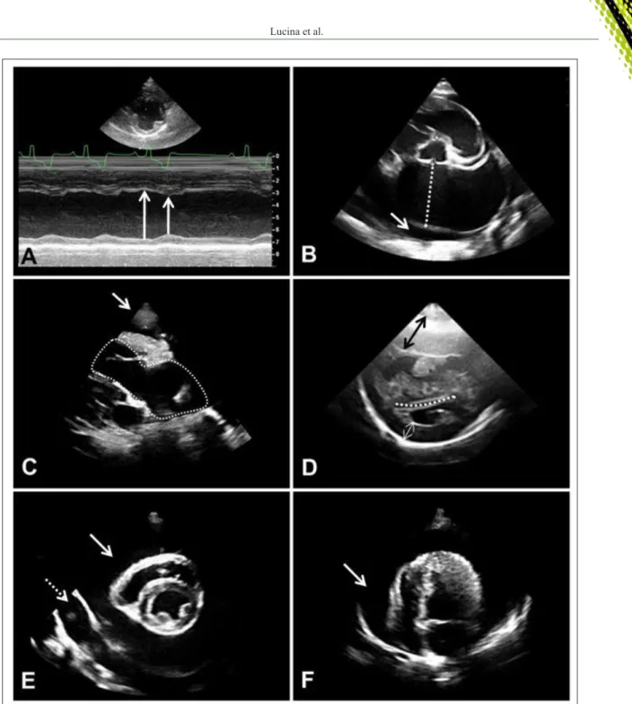
Figure 2 - Echocardiographic images showing (A) the impairment in cardiac contractility evidenced by the arrows, in which the left ventricle cavity does not suffer much change in its diameter comparing systole (short arrow) and diastole (long arrow),
in a dog with distributive shock (right parasternal short axis image at the level of chordae tendinea); (B) an enlarged
left atrium (dotted line) (left atrium-to-aorta ratio >2.0) and pericardial effusion (arrow) in a cat with hypertrophic
cardiomyopathy (right parasternal short axis image at the heart base); (C) pleural effusion (arrow) and a dilated right
atrium and ventricle (dotted line) in a dog with dyspnea and pulmonary hypertension (four chambers right paraesternal
long axis image); (D) hypertrophy of the right ventricular wall (black arrow), flattening of the interventricular septum
(dotted line) and pseudohypertrophy of the left ventricle (white arrow) in a dog with pulmonary hypertension (right
parasternal short axis image at the level of chordae tendinea); (E) pleural (dotted arrow) and pericardial (arrow) effusion in a dog admitted for dyspnea (right parasternal short axis image at the level of chordae tendinea); (F) pericardial
resuscitation time (SASSON et al., 2010; REA et
al., 2010). Several medical studies have discussed the application of FATE to decide when to cease cardiopulmonary resuscitation. Research has shown that the absence of movement of the ventricular wall during resuscitation implies a lower survival rate. This information can be used to decide when to end the resuscitation procedure (AICHINGER et al., 2012).
Respiratory distress
Evaluation of the size of the left atrium in animals with dyspnea (Figure 2B) can be useful, especially in cats, when physical examination and
thoracic radiography fail to provide a definitive diagnosis. Cats with heart failure have nonspecific
changes on auscultation, and the heart rate may be normal, increased or decreased (GOUTAL et al., 2010). Moreover, the prevalence of heart murmur in cats with cardiomyopathy (CM) is 55.7% (FERASIN et al., 2003), indicating that many affected cats do
not have an audible murmur. SMITH &
DUKES-MCEWAN (2012) reported that the absence of heart murmur was common in cats with heart failure, while arrhythmias and gallop rhythms were prevalent. These
findings differ from what is observed in dogs, where
there is an excellent correlation between the presence of the heart murmur and chronic degenerative valvular disease, and also the intensity of the murmur to the severity of the disease (HAGGSTROM et al., 1995).
Another peculiarity of cats is that cardiogenic pulmonary edema can manifest itself in a variety of ways and at different locations on the thoracic radiograph (RUSH et al., 2002). Radiographic
findings of cardiogenic pulmonary edema in cats are
more variable than in dogs because it has different
distribution in the lung field (LAMB, 2007). In a
study evaluating the radiographic pattern in cats with cardiogenic pulmonary edema, 17% had diffuse and uniform distribution, 61% had diffuse and non-uniform distribution, in 17% it was multifocal and in 4% focal (BENIGNI et al., 2009). Detection of pericardial effusion in cats with cardiomegaly and dyspnea is highly suggestive of heart failure (HALL et al., 2007).
Patients presenting with dyspnea, cyanosis and syncope without obvious changes in cardiac auscultation should also be investigated for pulmonary hypertension (PH). Primary etiologies of PH include left congestive heart failure, (post-capillary PH), thromboembolism, chronic respiratory disease, heartworm or right to left shunting
(pre-capillary PH) (JOHNSON, 2016).
Acute pulmonary embolism (PE) can lead
to a dramatic clinical deterioration and it is difficult to
diagnose because the clinical signs are nonspecific. The
gold standard for the diagnosis of PE is contrast computed tomography but in severely impaired patients CT may be inappropriate. Echocardiographic examination has been regarded as an excellent bedside diagnostic method in people with PE (GOLDHABER, 2002).
In patients with PH the echocardiographic examination may show dilatation of the pulmonary artery, hypertrophy and enlargement of the right
ventricle, rectified interventricular septum with
paradoxical movement, systolic dysfunction of the right ventricle, right atrium dilatation and decreased right ventricle lumen (Figure 2C e 2D).
All these changes can be seen by FATE; however,
the Doppler modality is the most used to identify PH. Using Doppler it is possible to estimate the pulmonary artery pressure gradient by measuring the velocity of tricuspid valve regurgitation, thereby PH can be graded as mild, moderate or severe. PH is diagnosed once the maximum velocity of tricuspid valve regurgitation is greater than 2.8m/s-1 (BOON, 2011).
Pleural effusion is another potential
diagnosis in patients with dyspnea and muffled heart
and lung sounds on cardiopulmonary auscultation and
can also be easily identified during FE (Figure 2E)
(DEFRANCESCO, 2013).
Thoracic trauma
DEFRANCESCO (2013) stated that all dogs and cats that have suffered thoracic trauma should be evaluated by ultrasound before radiographic examination. Patient side thoracic ultrasound is
indicated to identify fluid around the heart or in the
thoracic cavity which may indicate post traumatic bleeding. FATE aims to evaluate the presence of pericardial effusion and potential myocardium injury that require immediate surgical intervention (LABOVITZ et al., 2010).
In human patients with blunt and penetrating thoracic traumas focused echocardiography has been used for over 20 years (LABOVITZ et al., 2010). The use of FATE has reduced the time to diagnosis, thereby allowing rapid institution of emergency thoracotomy and laparotomy when required, thus decreasing morbity and mortality of thoracic trauma
(SYMBAS et al., 1999; ROZYCKI et al., 1999).
of critically ill patients is already widespread in the intensive care units of human hospitals. However, there are few references on this subject in the intensive care of small animals. In this context FATE, when used at the patient side, has many advantages compared to other diagnostic modalities - even when carried out by a non-cardiologist doctor or veterinarian. Despite the fact that this method seems to be promising, more studies are need
to define the necessary adaptations and standardizations
of FATE for use in animals in the emergency room.
ACKNOWLEDGEMENTS
The authors acknowledge the financial support of
Coordenação de Aperfeiçoamento de Pessoal de Nível Superior (CAPES).
REFERENCES
ABBOTT, J.A.; MACLEAN, H.N. Two-dimensional echocardiographic
assessment of the feline left atrium. Journal of Veterinay Internal
Medicine, v.20, p.111-119, 2006. Available from: <http://onlinelibrary.
wiley.com/doi/10.1111/j.1939-1676.2006.tb02830.x/epdf>. Accessed: Sept. 10, 2016. doi: 10.1111/j.1939-1676.2006.tb02830.x.
AICHINGER, G. et al. Cardiac movement identified on prehospital
echocardiography predicts outcome in cardiac arrest patients. Prehospital Emergency Care, v.16, p.251-255, 2012. Available from: <http://dx.doi.org/10.3109/10903127.2011.640414>. Accessed: Sept. 10, 2016. doi: 10.3109/10903127.2011.640414.
BEAULIEU, Y. Bedside echocardiography in the assessment of the critically ill. Critical Care Medicine, v.35, supl.5, p.235-249, 2007. Available from: <http://www.scielo.br/scielo.php?script=sci_artte xt&pid=S1679-45082015000400644>. Accessed: Sept. 10, 2016. doi: 10.1590/S1679-45082015MD3271.
BENIGNI, L. et al. Radiographic appearance of cardiogenic pulmonary oedema in 23 cats. Journal of Small Animal Practice, v.50, p.9-14, 2009. Available from: <http://onlinelibrary.wiley. com/doi/10.1111/j.1748-5827.2008.00655.x/pdf>. Accessed: Sept. 14, 2016. doi: 10.1111/j.1748-5827.2008.00655.x.
BERG, R. J.; WINGFIELD, W. Pericardial Effusion in the Dog:
A review of 42 cases. Jounal of the American Animal Hospital
Association, v.20, n.5, p.721-730, 1984.
BOON, J.A. Veterinary echocardiography. 2ed. Ames: Wiley-Blackwell, 2011. 767p.
BOYSEN, S.R. et al. Evaluation of a focused assessment with
sonography for trauma protocol to detect free abdominal fluid in
dogs involved in motor vehicle accidents. Journal of American
Veterinary Medical Association, v.225, n.8, p.1198-1204, 2004.
Available from: <https://www.researchgate.net/publication/8199944_ Evaluation_of_a_Focused_Assessment_With_Sonography_for_ Trauma_Protocol_to_Detect_Free_Abdominal_Fluid_in_Dogs_
conformed algorithm. Critical Care Medicine, v.15, supl.5, p.150-161, 2007. Available from: <www.emergencyultrasoundteaching. com/assets/.../Code_2007_Breitkreutz_CCM.pdf>. Accessed: Sept. 10, 2016. doi: 10.1097/01.CCM.0000260626.23848.FC.
CASAROTO, E. et al. Bedside echocardiography in critically ill patients. Medical Developments, v.13, n.4, p.644-646, 2015. Available from: <http://www.scielo.br/scielo.php?script=sci_artte xt&pid=S1679-45082015000400644>. Accessed: Sept. 14, 2016. doi: 10.1590S1679-45082015MD3271.
CHACKO, J.; BRAR, G. Bedside ultrasonography: applications
in critical care: part I. Indian Journal of Critical Care Medicine, v.18, n.5, p.301-309, 2014. Available from: <http://www.ijccm.org/
article.asp?issn=0972-5229;year=2014;volume=18;issue=5;spage =301;epage=309;aulast=Chacko>. Accessed: Sept. 14, 2016. doi:
10.4103/0972-5229.132492.
CHETBOUL, V. Advanced techniques in echocardiography in small animals. Veterinary Clinics of North America: Small Animal
Practice, v.40, p.529-543, 2010. Available from: <http://dx.doi.
org/10.1016/j.cvsm.2010.03.007>. Accessed: Sept. 10, 2016. doi: 10.1016/j.cvsm.2010.03.007.
CÔTÉ, E. Echocardiography: common pitfalls and practical
solutions. Clinical Techniques in Small Animal Practice, v.20, p.156-163, 2005. Available from: <http://www.sciencedirect.com/ science/article/pii/S1096286705000344>. Accessed: Sept. 10, 2016. doi: 10.1053/j.ctsap.2005.05.003.
DEFRANCESCO, T.C. Management of Cardiac Emergencies in Small Animals. Veterinary Clinics of North America: Small
Animal Practice. v.43, p.817-842, 2013. Available from: <http://
dx.doi.org/10.1016/j.cvsm.2013.03.012>. Accessed: Sept. 10, 2016. doi: 10.1016/j.cvsm.2013.03.012.
FEIGENBAUM, H. Echocardiography. 5ed. Philadelphia: Lea & Febinger, 1994. 659p.
FERASIN, L. et al. Feline idiopathic cardiomyopathy: a retrospective study of 106 cats (1994-2001). Journal of Feline
Medicine and Surgery. v.5, n.3, p.151-159, 2003. Available from:
<http://dx.doi.org/10.1016/S1098-612X(02)00133-X>. Accessed: Sept. 14, 2016. doi: 10.1016/S1098-612X(02)00133-X.
GOLDHABER, S.Z. Echocardiography in the management of pulmonary embolism. Annals of Internal Medicine, v.136, n.9, p.691– 700, 2002. Available from: <http://annals.org/aim/article/715263/ echocardiography-management-pulmonary-embolism>. Accessed: Sept. 10, 2016. doi: 10.7326/0003-4819-136-9-200205070-00012.
GOUTAL, C.M. et al. Evaluation of acute congestive heart failure in dogs and cats: 145 cases (2007-2008). Journal of Veterinary
Emergency and Critical,v.20, n.3, p.330–337, 2010. Available from:
<http://onlinelibrary.wiley.com/doi/10.1111/j.1476-4431.2010.00524.x/ full>. Accessed: Sept. 10, 2016. doi: 10.1111/j.1476-4431.2010.00524.x.
HAGGSTROM, J. et al. Heart sounds and murmurs: changes related to severity of chronic valvular disease in the Cavalier King
doi/10.1111/j.1939-1676.1995.tb03276.x/pdf>. Accessed: Sept. 10, 2016. doi: 10.1111/j.1939-1676.1995.tb03276.x.
HALL, D.J. et al. Pericardial effusion in cats: a retrospective study of clinical findings and outcome in 146 cats. Journal of Veterinary
Internal Medicine, v.21, p.1002–1007, 2007. Available from:
<http://onlinelibrary.wiley.com/doi/10.1111/j.1939-1676.2007. tb03056.x/epdf>. Accessed: Sept. 10, 2016. doi: 10.1111/j.1939-1676.2007.tb03056.x.
JOHNSON, L.R. Cor pulmonale and pulmonary thromboembolysm. In:SMITH JR. et al. Manual of canine and feline cardiology. 5 ed. St. Louis: Elsevier, 2016. Cap.10, p.181-197.
JONES A.E. et al. Focused training of e mergency medicine
residents in goal-directed echocardiography: a prospective study.
Academic Emergency Medicine, v.10, p.1054-1058, 2003.
Available from: <http://onlinelibrary.wiley.com/doi/10.1197/S1069-6563(03)00346-4/pdf>. Accessed: Sept. 10, 2016. doi: 10.1197/ S1069-6563(03)00346-4/pdf.
JONES, A.E.; KLINE, J.E. Shock. In: MARX, J. A. et al. Rosen’s
emergency medicine: concepts and clinical practice. 8ed.
Philadelphia: Saunders, 2014. Cap. 6.
LABOVITZ, A.J. et al. Focused Cardiac Ultrasound in the Emergent
Setting: A Consensus Statement of the American Society of Echocardiography and American College of Emergency Physicians.
Journal of the American Society of Echocardiography, v.23,
p.1225-1230, 2010. Available from: <http://dx.doi.org/10.1016/j. echo.2010.10.005>. Accessed: Sept. 10, 2016. doi: 10.1016/j. echo.2010.10.005.
LAMB, C.R. The canine and feline lung. In: THRALL, D.E.
Textbook of Veterinary Diagnostic Radiology. 5ed. Philadelphia:
Saunders, 2007. Cap.34, p.591-608.
LISCIANDRO, G.R. et al. Evaluation of a thoracic focused assessment with sonography for trauma (TFAST) protocol to detect pneumothorax and concurrent thoracic injury in 145 traumatized dogs. Journal of Veterinary Emergency and Critical Care, v.18, p.258-269, 2008. Available from: <http://onlinelibrary.wiley.com/ doi/10.1111/j.1476-4431.2008.00312.x/epdf>. Accessed: Sept. 14, 2016. doi: 10.1111/j.1476-4431.2008.00312.x.
MANDAVIA, D.P. et al. Bedside echocardiography by emergency physicians. Annals of Emergency Medicine, v.38, p.377-382, 2001. Available from: <http://www.annemergmed.com/article/ S0196-0644(01)17352-X/fulltext>. Accessed: Sept. 10, 2016. doi: 10.1067/mem.2001.118224.
MAYRON R. et al. Echocardiography performed by emergency physicians: impact on diagnosis and therapy. Annals of Emergency
Medicine, v.17, p.150-154, 1988. Available from: <http://
www.annemergmed.com/article/S0196-0644(88)80301-9/pdf>. Accessed: Sept. 10, 2016. doi: 10.1016/S0196-0644(88)80301-9.
MELAMED, R. et al. Assessment of the left ventricular function by intensivists using hand-held echocardiography. Chest, v.135, n.6, p.1416-1420, 2009. Available from: <http://www.sciencedirect. com/science/article/pii/S001236920960341X>. Accessed: Sept. 10, 2016. doi: 10.1378/chest.08-2440.
MEYER, S. et al. Review article: non-invasive assessment of cardiac output with portable continuous-wave doppler ultrasound. Emergency
Medicine Australasia, v.20, n.3, p.201-208, 2008. Available from:
<http://dx.doi.org/10.1053/j.jvca.2012.03.022>. Accessed: Sept. 10, 2016. doi: 10.1053/j.jvca.2012.03.022.
MOK, K. L. Make it SIMPLE: enhanced shock management
by focused cardiac ultrasound. Journal of Intensive Care, v.4, n.51, p.1-17, 2016. Available from: <https://jintensivecare. biomedcentral.com/articles/10.1186/s40560-016-0176-x>. Accessed: Sept. 14, 2016. doi: 10.1186/s40560-016-0176-x.
MOORE C.L.; COPEL J.A. Point of care ultrasonography.
New England Journal of Medicine, v.364, n.8, p.749–757,
2011. Available from: <http://www.nejm.org/doi/full/10.1056/
NEJMra0909487>. Accessed: Sept. 10, 2016. doi: 10.1056/ NEJMra0909487.
OYAMA, M.A. Advances in echocardiography. Veterinary
Clinics: Small Animal Practice, v.34, n.5, p.1083-1104, 2004.
Available from: <http://dx.doi.org/10.1016/j.cvsm.2004.05.004>. Accessed: Sept. 10, 2016. doi: 10.1016/j.cvsm.2004.05.004.
OZEN, C. et al. Assessment of ventricular wall motion with echocardiography during cardiac arrest to predict survival. Turkish
Journal of Emergency Medicine, v.16, n.1, p.12-16, 2016. Available
from: <https://www.ncbi.nlm.nih.gov/pmc/articles/PMC4882209/>. Accessed: Sept. 10, 2016. doi: 10.1016/j.tjem.2015.08.001.
REA, T.D. et al. Predicting survival after out-of-hospital cardiac arrest: role of the Utstein data elements. Annals of Emergency
Medicine, v.55, p.249-257, 2010. Available from: <http://www.
annemergmed.com/article/S0196-0644(09)01556-X/fulltext>. Accessed: Sept. 10, 2016. doi: 10.1016/j.annemergmed.2009.09.018.
RISHNIW, M.; ERB, H.N. Evaluation of four 2-dimensional
echocardiographic methods of assessing left atrial size in dogs.
Journal of Veterinary Internal Medicine, v.14, n.4,
p.429-435, 2000. Available from: <http://onlinelibrary.wiley.com/ doi/10.1111/j.1939-1676.2000.tb02252.x/pdf>. Accessed: Sept. 10, 2016. doi: 10.1111/j.1939-1676.2000.tb02252.x.
ROZYCKI, G.S. et al. The role of ultrasound in patients with possible
penetrating cardiac wounds: a prospective multi-center study. Journal
of Trauma and Acute Care Surgery, v.46, p.543-551, 1999.
RUSH, J.E. et al. Population and survival characteristics of cats with
hypertrophic cardiomyopathy: 260 cases (1990-1999). Journal of the
American Veterinary Medical Association, v.220, p.202-207, 2002.
Available from: <http://dx.doi.org/10.2460/javma.2002.220.202>. Accessed: Sept. 14, 2016. doi: 10.2460/javma.2002.220.202.
SASSON, C. et al. Predictors of survival from out-of-hospital cardiac arrest: a systematic review and meta-analysis. Circulation: Cardiovascular Quality and Outcomes, v.3, p.63-81, 2010. Available from: <http://dx.doi. org/10.1161/CIRCOUTCOMES.109.889576>. Accessed: Sept. 10, 2016. doi: 10.1161/CIRCOUTCOMES.109.889576.
SINGER, M. et al. The third international consensus definition
for sepsis and septic shock (sepsis-3). Journal of The American
Medical Association, v.315, n.8, p.801-810, 2016. Available from:
<https://www.ncbi.nlm.nih.gov/pmc/articles/PMC4968574/>. Accessed: Sept. 10, 2016. doi: 10.1001/jama.2016.0287.
SMITH, S.; DUKES-MCEWAN, J. Clinical signs and left atrial size in
cats with cardiovascular disease in general practice. Journal of Small
Animal Practice, v.53, n.1, p.27-33, 2012. Available from: <http://
SYMBAS, NP. et al. Blunt cardiac rupture: the utility of emergency department ultrasound. Annals of Thoracic Surgery, v.67, n.5, p.1274-1276, 1999. Available from: <http://dx.doi.org/10.1016/S0003-4975(99)00169-1>. Accessed: Sept. 10, 2016. doi: 10.1016/S0003-4975(99)00169-1.
TIDWELL, A.S. Diagnostic Pulmonary Imaging. Problems in
Veterinary Medicine, v. 4, n.2, p.239-264, 1992.
TSE, Y.C. et al. Evaluation of a training course in focused
echocardiography for noncardiology house officers. Journal of
Veterinary Emergency and Critical Care, v.23, n.3, p.268-273,
preload for the “non-cardiologist”. Anaesthesiology Intensive
Therapy, v.47, p.89-104, 2015. Available from: <https://com-anest.
sites.medinfo.ufl.edu/files/2015/06/2016-Cardiac-Ultrasound.pdf.>.
Accessed: Sept. 10, 2016. doi: 10.5603/AIT.a2015.0074.
VINCENT, J.L.; BACKER, B. Circulatory shock. New England
Journal of Medicine, v.369, p.1726-1734, 2013. Available from:
<http://www.nejm.org/doi/full/10.1056/nejmra1208943#t=article>. Accessed: Sept. 10, 2016. doi: 10.1056/nejmra1208943.
WARE, W.A. Cardiovascular Disease in Small Animal