17
p-ISSN:2231-6140,eA Study of Clinical Spectru
Dr. Gargi Pathak1, Dr. Anuya Ch
1
Professor and Head of Unit, 2 Assis Medical College, Ahmedabad.
Abstract:
Introduction: Dengue v cause of arboviral, disease in th dengue fever from paediatric d designed to document the prese infection in children. Methodolo october 2015 and total of 126 pa years. A detailed history, carefu done in all the patients. Results
marker than thrombocytopenia an not found in the previousstudies Majority of patients had platelet dengue IgM and 44.4% had test like hepatitis (20.6%), myocard Dengue haemorrhagic fever (4.7% in serotype and epidemiology of frequency subsequent to myocard increased cases of coinfections li of 126 patients 6 patients expired Keyword: Bradycardia, Dengue f
Introduction:
Dengue is an acute vira fatal complications. The first cli epidemics occurred almost simu and North America in the 1780 report dates from 1789 of 1780 e by Benjamin Rush, who coined th because of the symptoms of myal first outbreak reported in Madras Dengue viruses (DV) bel and there are four serotypes of th 1, DV-2, DV-3 and DV-4. DV
*
Corresponding Author:
Dr. Malvika Nagpure,
E-mail: [email protected]
e-ISSN:2395-7859 Origi
trum of Dengue Fever in A Tertiary Care
Chauhan2, Dr. Malvika Nagpure3*, Dr. Kinjal Kath sistant Professor, 3,4 Resident doctor, Department of Pa
viruses, of the family Flaviviridae, are the m the world. We report a clinico-epidemiological department of civil hospital Ahmedabad. Thi esenting features, laboratory results and outcom ology: A prospective study was carried from O patients were studied from age group between 1 eful clinical examination and laboratory investi
ts and Conclusions: We documented Leucopeni
and were seen in more numbers (47%) in our stud ies. Most common symptom was fever with body
let count between 50000-1 lakh. 55% had tested ested positive for dengue NS1. Wide variety of c arditis (14.2%), dengue shock (11.1%), encepha
.7%), ARDS (2.3%) were seen, which might indic of the Dengue. Interestingly Bradycardia was seen arditis with simultaneously raised CPK-MB levels s like malaria, enteric, hepatitis, UTI, not seen pre
ed.
e fever, Myocarditis.
iral infection with potential clinically recognized dengue ultaneously in Asia, Africa, 780s. The first clinical case 0 epidemic in Philadelphia is d the term “break bone fever” yalgia and arthralgia. In India ras in 1780.
belong to family Flaviviridae f the virus referred to as
DV-V is a positive-stranded encapsulated RNA v composed of three structural protein g encode the nucleocapsid or core (C membrane-associated (M) protein, an om
iginal Article
re Centre.
atheriya4 Paediatrics, B. J.
most common al study of the his study was ome of dengue October
1 month to 12 stigations were enia as an early tudy which was dy ache (73%). ted positive for complications phalitis (4.7%),
dicate a change een in increased els. There were previously .Out
18
p-ISSN:2231-6140,e-ISSN:2395-7859 Original Article (E) glycoprotein and seven non-structural (NS) proteins. It is transmitted mainly by Aedes aegypti mosquito and also by Aedes albopictus. All four serotypes can cause the full spectrum of disease from a subclinical infection to a mild self-limiting disease, the dengue fever (DF) and a severe disease that may be fatal, the dengue haemorrhagic fever/dengue shock syndrome (DHF/DSS)1.The WHO estimates 50 million dengue infections occur annually and almost half the world’s population lives in countries where Dengue infection is endemic (W.H.O., 2008). Over the last 10 -15 years Dengue Haemorrhagic Fever has become a leading cause of hospitalization and death among children in SEAR countries following Diarrheal and acute respiratory infections (W.H.O., 1999). Dengue Fever has been reported from India over a long time but DHF was first reported in 1963 from Calcutta city (Dengue report, 2007) 1,2,3. The exact clinical profile is important for patient management and thus crucial for saving lives. The present study is an attempt to describe the clinical as well as laboratory findings of serologically confirmed hospitalised cases of dengue fever during the study period.Materials and Methods:
This is a prospective observational study, carried out from October 2014 to October 2015 in paediatric department of civil hospital Ahmedabad. A written informed consent was obtained from the child’s parents. Total 126 patients were studied.
Inclusion criteria: All laboratory confirmed cases of dengue fever within age group of 1month to 12years were included (NS1/IgM positive)
Exclusion criteria: Patients <1 month & > 12 years were excluded.
A detailed history was taken and a careful clinical examination was performed. The laboratory investigations like haemoglobin (Hb), the total count and the differential leucocyte counts (TLC and DLC), platelet count, haematocrit (Hct), liver function tests (LFT), urea, creatinine, chest X-ray, Dengue IgM NS1 and ultrasonography of abdomen were done in all the patients . Other relevant investigations were performed according to the clinical condition of the patient. All patients were treated according to guidelines by National Vector Borne Control Programme.
Results:
19
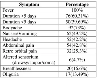
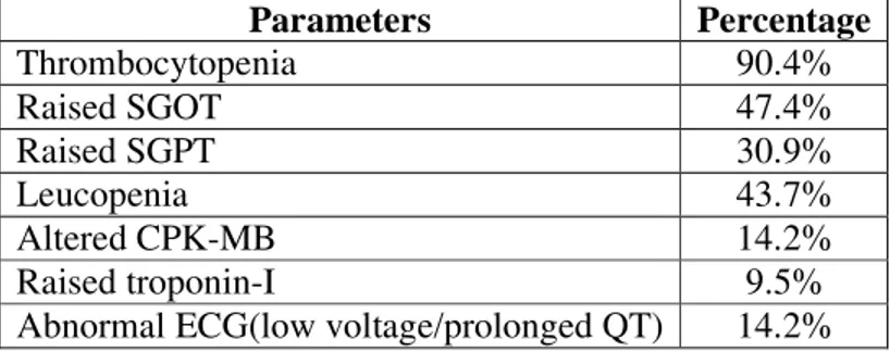
p-ISSN:2231-6140,e-ISSN:2395-7859 Original Article of myocarditis in most patients having the same which was not seen in the previous studies4,5,6. On investigation, leucopenia was seen in 43.5% of patients and thrombocytopenia were seen in 90.4% of patients, however leucopenia was seen earlier than thrombocytopenia and in more number of patients in the disease course. Among the liver enzymes, SGOT was elevated in a larger proportion (47.42%) of patients when compared to alanine aminotransferase (SGPT) (30.92%) and was more specific marker for dengue. Maximum patients had platelet in the range of 50,000-100000 (33.2%), followed by platelet count of 20,000-50,000 (30.3%). Parameters like prothrombin time (PT) and activated partial thromboplastin time (aPTT) were abnormal in 19 (15%) patients and was carried out only in those who had signs of bleeding (22.1%)) .One of the important findings of dengue was raised haematocrit which was seen in 34.02% of the cases. 55.5% patients tested positive for dengue IgM and 44.4% of them for NS1Ag. Tourniquet test was found to be negative in the majority of patients (Only 11.9% were positive). 14.2% of patients had elevated CPK MB and (9.5%) of them had elevated troponin-I levels. ECG changes in form of either prolonged QT interval or low voltage waves were seen in all patients with myocarditis (14.2%).Most common USG finding seen in the study was pleural effusion in 26.4% cases. Other findings were hepatomegaly (21.9%), gall bladder edema (15.3%). Most common complication seen in the study was hepatitis (20.6%) followed by myocarditis (14.2%), dengue shock(11.1%) encephalitis (4.7%), dengue haemorrhagic fever (4.7%) and ARDS(2.3%).Fluid accumulation in the form of pleural effusion (26.9%) ascites (15.8%), oedema(11.6%) were seen. Right sided effusion (20.6%) was most commonly seen followed by bilateral effusion (6.18%). During this study period coinfections were more seen in form of malaria (9.12%), enteric fever (5.39%), hepatitis (2.48%), UTI (3.73%). Among 126 patients, 119(94.4%) of the cases needed intravenous fluids. Dopamine was required in 11 (8.7%). Platelet concentrate was required in 13 patients in severe dengue cases; FFP was given to 19(15%) patients, PCV to 11patients. In our study 120 patients recovered and 6 patients (4.7%) expired. The causes of mortality were complications in form of ARDS, Encephalitis, dengue shock syndrome and Dengue haemorrhagic fever.Table 1: Symptoms at the time of presentation
Symptom Percentage
Fever 100%
Duration >5 days 76(60.31%) Duration <5 days 50(39.69%)
Bodyache 92(73%)
Nausea/Vomiting 62(49.2%)
Headache 52(42.2%)
Abdominal pain 54(42.8%) Retro orbital pain 32(25.3%) Altered sensorium
(drowsy/stupor/coma) 6(4.7%) Breathlessness 20(16.6%)
20
p-ISSN:2231-6140,e-ISSN:2395-7859 Original Article Almost all patients presented with generalised symptoms such as fever, nausea, vomiting and body ache, very few presented with retro-orbital pain, altered sensorium or oliguria.Table 2: Clinical signs
Sign Percentage Tachycardia 46(36.5%) Hepatomegaly 27(21.5%) Pleural effusion 26(21%) Skin rash/Petechiae 23(18.2%) Hypotension 21(17%) Ascites 20(15.8%) Bradycardia 18(14.2%)
Oedema 15(11.6%)
Jaundice 8(6.6%)
Tachycardia and hepatomegaly were among the most common signs but relatively higher percentage of patients had bradycardia not reported previously4,5,6.
Table 3: Complications
Complications Percentage
Hepatitis 26(20.6%)
Myocarditis 18(14.2%)
Dengue shock 14(11.1%)
Encephalitis 6(4.7%)
Dengue Haemorrhagic fever 6(4.7%)
ARDS 3(2.3%)
Hepatitis was amongst the most common complication.
Table 4:Laboratory parameters
Parameters Percentage
Thrombocytopenia 90.4%
Raised SGOT 47.4%
Raised SGPT 30.9%
Leucopenia 43.7%
Altered CPK-MB 14.2%
Raised troponin-I 9.5%
Abnormal ECG(low voltage/prolonged QT) 14.2%
Discussion:
21
p-ISSN:2231-6140,e-ISSN:2395-7859 Original Article revealed that fever was the most common presenting symptom seen in all patients (100%). Body ache was seen in (73%) of the patients followed by vomiting (49.2%), abdominal pain (42.8%), headache (41.2%) and retro orbital pain (25.3%) Breathlessness (16.6%), oliguria(13.49%). This goes with previous study of Hema et al4 and Tiple et al6 study. Most common sign was found to be tachycardia(36.5%) followed by, hepatomegaly(21.9%),pleural effusion(21%),skin rash(18.2%) and hypotension(17%) ascites (15.8%).Positive tourniquet test were seen in only 11.9&% as opposed to Hema et al4 study having 33.3% positive cases .There were increased incidences of fluid accumulation in form of pleural effusion(26.9%),ascites(15.8%),oedema(11.6%) which was not seen previously4,5,6,. Bradycardia was seen as the early sign of myocarditis which was not seen in the previous studies4,5,6 along with simultaneously raised CPK-MB levels and ECG changes. SGOT was also elevated in all patients having myocarditis. The most common bleeding manifestations in both severe and non severe dengue were petechiae, purpura, and ecchymosis that goes with previous studies4,5,6. Gastrointestinal bleeding was significantly seen in severe dengue cases. Two patients in the severe dengue group had convulsion due to encephalitis. In our study thrombocytopenia was seen in 90% patients and Leucopenia in 43%. Arul Kumar et al5 showed thrombocytopenia (96%) and Leucopenia (38%) i.e. more percentage of Leucopenia was seen as. Elevation of SGOT was significantly more compared to SGPT in the present study as compared to Amrita et. al8 that showed almost equal percentage of rising SGPT and SGOT. R kumar et al9 had similar findings and it was found in our study that raised levels were associated more with severity of infection and could be used as a significant pointer of the same . Very high levels of SGOT and SGPT indicate severity of the disease along with morbidity and mortality 9,10 . Rise in PT/aPTT also depicts severity of disease and was seen in 15% of the patients. 14.2% of patients had elevated CPK MB and 9.5% of them had elevated troponin-I. Associated coinfections in form of malaria (9.12%), enteric fever (5.39%) hepatitis (2.48%), UTI(3.73%) were seen similar to previous study4.Complications were less compared to Tiple et al6 and more compared to Hema et al4 Selvan et al10.In our study 120 dengue cases recovered There was less mortality in the present study group, may be to due to increasing awareness and early seeking of medical attention and improved health services.Conclusion:
22
p-ISSN:2231-6140,e-ISSN:2395-7859 Original Article References:1. WHO. Geneva: World Health Organization; 2009. Dengue: guidelines for diagnosis, treatment, prevention and control – New Ed. Available from
www.who.int/tdr/publications/documents/dengue-diagnosis.pdf.
2. W.H.O., 2008. Dengue and Dengue Haemorrhagic Fever. Fact sheet No 117, revised May 2008, Geneva, WHO, (http://www.who.int/media centre/fact sheets/fs 117/en1) WHO. 2009. Prevention and control of Dengue and Dengue Haemorrhagic Fever. Comprehensive guidelines.
3. W.H.O. SEARO, New Delhi; 1999. Guidelines for treatment of dengue fever/dengue haemorrhagic fever in small hospitals.
(who.int/iris/bitstream/10665/69230/1/WHO_FCH_CAH_05.13_eng.pdf)
4. Mittal H, Faridi M.M, Arora SK, Patil R: clinico-haematological profile and platelet trends in children with Dengue during 2010 epidemic in North India, the Indian journal of paediatrics, April 2012.79: 467-471.
5. Arunagirinathan AK, Thirunavukarasu BK, Narayanaswamy DK, Raghavan A, Raghavendhran VD: clinical profile and outcome of Dengue fever cases in children by adopting revised WHO guidelines: A hospital based study. International Journal of Scientific Study, May 2015.3(2)181-185.
6. Nishikant T, Kisan P, More S S. Dengue fever in a tertiary care hospital. International Journal of recent trends September 2015; 16(2): 358-362.
7. S. Ahmed, F. Arif, Y. Yahya. “Dengue fever outbreak in Karachi 2006-a study of profile and outcome of children under 15 years of age,” Journal of the Pakistan Medical Association, 2008: 58:4–8.
8. Roy A, Sarkar D, Chakraborty S, Chaudhuri J, Ghosh P. Profile of Hepatic Involvement by Dengue Virus in Dengue Infected Children N Am J Med Sci. 2013 August; 5(8): 480–484.
9. Kumar R, Tripathi P, Tripathi S, Kanodia A, Pant S, Venkatesh V. Prevalence and Clinical Differentiation of Dengue Fever in Children in Northern India,`Infection, jan 2008. 36:444-449.