Lactamase-Producing
Escherichia coli
: Association with
Epidemiological and Clinical Features
Jesu´s Rodrı´guez-Ban˜o1,2*, Jesu´s Mingorance3, Natalia Ferna´ndez-Romero3, Lara Serrano1, Lorena Lo´pez-Cerero1, Alvaro Pascual1,4, the ESBL-REIPI group"
1Unidad Clı´nica de Enfermedades Infecciosas y Microbiologı´a, Hospital Universitario Virgen Macarena, Sevilla, Spain,2Servicio de Microbiologı´a, Hospital Universitario La Paz - IdiPAZ, Madrid, Spain,3Departamento de Medicina, Universidad de Sevilla, Sevilla, Spain,4Departamento de Microbiologı´a, Universidad de Sevilla, Sevilla, Spain
Abstract
There is scarce data about the importance of phylogroups and virulence factors (VF) in bloodstream infections (BSI) caused by extended-spectrumb-lactamase-producingEscherichia coli(ESBLEC). A prospective multicenter Spanish cohort including 191 cases of BSI due to ESBLEC was studied. Phylogroups and 25 VF genes were investigated by PCR. ESBLEC were classified into clusters according to their virulence profiles. The association of phylogropus, VF, and clusters with epidemiological features were studied using multivariate analysis. Overall, 57.6%, 26.7%, and 15.7% of isolates belonged to A/B1, D and B2 phylogroups, respectively. By multivariate analysis (adjusted OR [95% CI]), virulence cluster C2 was independently associated with urinary tract source (5.05 [0.96–25.48]); cluster C4 with sources other than urinary of biliary tract (2.89 [1.05– 7.93]), and cluster C5 with BSI in non-predisposed patients (2.80 [0.99–7.93]). Isolates producing CTX-M-9 group ESBLs and from phylogroup D predominated among cluster C2 and C5, while CTX-M-1 group of ESBL and phylogroup B2 predominantes among C4 isolates. These results suggest that host factors and previous antimicrobial use were more important than phylogroup or specific VF in the occurrence of BSI due to ESBLEC. However, some associations between virulence clusters and some specific epidemiological features were found.
Citation:Rodrı´guez-Ban˜o J, Mingorance J, Ferna´ndez-Romero N, Serrano L, Lo´pez-Cerero L, et al. (2012) Virulence Profiles of Bacteremic Extended-Spectrumb -Lactamase-ProducingEscherichia coli: Association with Epidemiological and Clinical Features. PLoS ONE 7(9): e44238. doi:10.1371/journal.pone.0044238
Editor:Axel Cloeckaert, Institut National de la Recherche Agronomique, France
ReceivedJune 11, 2012;AcceptedJuly 30, 2012;PublishedSeptember 7, 2012
Copyright:ß2012 Rodrı´guez-Ban˜o et al. This is an open-access article distributed under the terms of the Creative Commons Attribution License, which permits unrestricted use, distribution, and reproduction in any medium, provided the original author and source are credited.
Funding:This study was funded by the Ministerio de Ciencia e Innovacio´n, Instituto de Salud Carlos III - co-financed by European Development Regional Fund ‘‘A way to achieve Europe’’ ERDF, Spanish Network for Research in Infectious Diseases (REIPI RD06/0008), Fondo de Investigacio´n Sanitaria (grants 070190, 10/02021, 10/01955, and 10/00795), and Junta de Andalucı´a (grants 0048/2008, and CTS-5259). The funders had no role in the study design, data collection and analysis, decision to publish, or preparation of the manuscript.
Competing Interests:The authors have read the journal’s policy and have the following conflicts: J. Rodrı´guez-Ban˜o has been a consultant for Wyeth, Merck, and Pfizer, has served as speaker for Wyeth, Merck, Pfizer, Astra-Zeneca and GlaxoSmithKline, and has received research support from Merck and Wyeth. J. Mingorance has received research support from Roche and Pfizer. A. Pascual has been a consultant for Merck and Pfizer, has served as speaker for Wyeth, Astra-Zeneca, Merck, and Pfizer and has received research support from Merck and Pfizer and Wyeth. All other authors had no conflict of interest. This does not alter the authors’ adherence to all the PLoS ONE policies on sharing data and materials.
* E-mail: [email protected]
"Membership of the ESBL-REIPI group is provided in the Acknowledgments.
Introduction
Most extraintestinal infections due toEscherichia coliare caused by isolates derived from the so-called virulent phylogenetic groups (PG) B2 and D, which exhibit more virulence factors (VF) than other PGs such as A and B1, hence considered ‘‘low virulence’’ or ‘‘commensal’’ PGs [1,2]. Specifically, in studies on bloodstream infections (BSI) caused byE. coli,.70% of the isolates belonged to PGs B2 (which was predominant) and D [3–8]. Isolates from PG B2 have been associated with BSI with a urinary tract source [4,5,7] and in non-predisposed patients [7,9], while PG A has been found with increased frequency in nosocomial BSI, compromised hosts [4,6,10] and in BSI caused by antibiotic-resistant isolates [5,6,8].
Some VFs have been assigned a pathogenic role in extraintes-tinal infections based on comparisons with rectal isolates, association with infections in non-predisposed patients, and animal models [11]. In studies dealing with BSI, several VF has been
found to be associated with specific epidemiologic features, but it is papGII that has been more consistently associated with urinary tract sepsis as opposed to other sources, and in patients without predisposing factors [4,9,12].
Extended-spectrum beta-lactamase-producing Escherichia coli (ESBLEC) are increasing worldwide as a cause of community and nosocomial BSI, frequently affecting patients with predispos-ing conditions [12,13]. There are scarce data about the distribution of PGs and VF in BSI due to ESBLEC [14] and their association with antimicrobial resistance. Also, to the best of our knowledge, the impact of specific VF in the epidemiology of BSI due to ESBLEC has not been studied. Finally, there is some controversy about the real virulence of ESBLEC, including isolates producing CTX-M-15 belonging to the worldwide spread clone ST131 [16–18].
(individ-ually or in clusters) were associated with the epidemiology, patients’ features and source of BSI.
Methods
Study Design and Patients
Data and isolates from a prospective cohort including 191 cases of BSI due to ESBLEC from 13 Spanish hospitals were used for this analysis. The epidemiology, clinical features, outcomes, types of ESBL and susceptibility data of this cohort were previously reported [13,14]. Briefly, all monomicrobial BSI in patients with sign or symptoms of systemic infection caused by ESBLEC diagnosed in the participating hospitals between October 2004 and January 2006 were included. The cases were detected by daily review of microbiological results of blood cultures at each center. Data collected included demographics, acquisition classified as community, healthcare-associated or nosocomial [13], chronic underlying diseases, severity of underlying condition according to Charlson index [19], invasive procedures, exposure to antibiotics in the preceding 2 months, and source of BSI according to clinical and microbiological criteria.
For this analysis, patients with any of the following were considered to have systemic predisposing features for BSI: diabetes mellitus, liver cirrhosis, chronic renal insufficiency, inmunosup-presive therapy, and neutropenia. Patients with a procedure-associated BSI (including vascular catheter, urinary catheter, endoscopic procedures and surgery), or any urinary or biliary tract BSI in the presence of obstructive diseases of these tracts were considered to have local predisposing factors for BSI. The study was approved by the Ethics Committee of Hospital Universitario Virgen Macarena which waived the need to obtained consent because all data were analysed anonymously and the observational nature of the study.
Microbiological Studies
Methods for bacterial identification, susceptibility studies and ESBL confirmation and characterization were previously reported [13,14]. Briefly, ESBL production and susceptibility by micro-dilution to cefuroxime, cefotaxime, ceftazidime, cefepime, amox-icillin-clavulanic acid, piperacillin-tazobactam, ciprofloxacin, gen-tamycin, tobramycin, amikacin, ertapenem, imipenem, merope-nem, trimethoprim-sulfamethoxazol, fosfomycin, and tigecycline were studied according to CLSI recommendations [20]; a resistance score (number of antimicrobials to which the isolate was resistant) was calculated for each isolate. b-lactamase characterization was carried out by isoelectric focusing, PCR of theblagenes, and sequencing. ST131 clone was studied by O25b typing [17] and analysis for allele 3 ofpabB[21]; the phylogenetic group was determined by multiplex PCR [22].
Twenty-five genes codifying for putative VF were studied, including adhesins (papC, papGI, papGII, papGIII, fimH, sfaD/E, afaB/C, iha); toxins (cnf1, cdtB, sat, hlyA); related to iron acquisition (iucD, iroN, iutA, ireA,andfyuA); protectins (kps MTII, traT, cvaC,and ompT); and miscellaneous (ibeA, maIX, svg,andusp). The presence/ absence of VF genes was studied by PCR using previously described primers [2,23–31]. Total DNA was purified from each strain with the UltraClean Microbial DNA purification kit (MO BIO Laboratories Inc., Carlsbad, CA). DNAs were distributed in 96-well master plates and PCRs were done in 50mL mixtures
containing 5ml (20 ng) template DNA, 0.2mM of each primer,
0.2mM mix dNTPs and 1U DNA polymerase (Biotools S. L., Spain) in 1X buffer with MgCl2. PCR conditions were as follows:
5 min at 95uC, followed by 30 cycles of 30 s at 95uC, 30 s at annealing temperature of each primer pair, 1 min at 72uC, and a
final 5 min incubation at 72uC. The PCR products were analyzed by electrophoresis in 96-well agarose gels (VG-FAST, Fisher Bioblock Scientific) stained with GelRedTM (Biotium Inc.). A virulence score (number of VF genes) was calculated for each isolate. The similarity of the isolates according to their VF genotypes was studied by constructing a dendogram using the binary patters (0, 1) of VF for each isolate; clusters of isolates were identified using the Dice similarity coefficient. After reviewing the data obtained, a 70% similarity threshold was used after reviewing the data obtained (a 60% threshold was not discriminative enough, since only one cluster included 72.2% isolates; and a 80% threshold found only 4 clusters with.5 cases including 32.4% of the isolates).
Statistical Analysis
Percentages were compared using the chi squared test or the Fisher exact test, as appropriate, and continuous variables using the Mann-Whitney U test. Multivariate analysis were performed by logistic regression; variables with a univariate p value ,0.1 were introduced in the models, and selected using a stepwise backward process; 0.1 was set as the limit for removal of terms. All tests were performed using SPSS 18.0.
Results
Among the 191 ESBLEC bacteremic isolates, 55 (28.8%) belong to PG A, 55 (28.8%) to PG B1, 51 (26.7%) belong to PG D, and 30 (15.7%) to B2. The median (IQR) virulence score were 10 (9–12) for B2, 8 (7–9) for D, 5 (4–6) for B1, and 4 (2–6) for A. Among the B2 isolates, 21 (70% of B2, 10.9% of the whole series) were O25b andpabB3 positive and were considered as belonging to ST131. For easier understanding, PGs A and B1 were analyzed together because both showed similar virulence scores, frequency of VF (onlyfimH,iucD, andiroNwere significantly more frequent among B1 than among A), ESBLs (predominance of CTX-M-14), and resistance patterns.
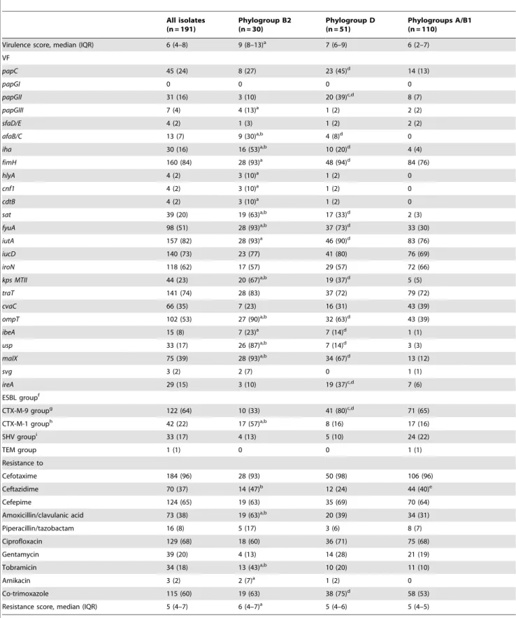
The frequency of VF genes according to PG is shown in table 1. In summary, papC, afaB/C, iha, fimH, sat, fyuA, iutA, kps MTII, ompT, ibeA, usp, and maIXwere more frequent among in B2 and D than in A/B1; additionally,papGIII, hlyA, cnf1, and cdtB,although infrequent, were more prevalent in B2 than in A/B1; andpapGII andireAwere more frequent in D than in A/B1. Finally,sfaD/E, afaB/C, sat, fyuA, kps MTII, ompT, usp, and maIX were more frequent in B2 than in D, while onlypapGIIwas more frequent in D than in B2.
Table 1.Virulence factor genes, ESBL groups and antimicrobial resistance of 191 ESBL-producingE. coliisolates causing BSI according to phylogroups.
All isolates (n = 191)
Phylogroup B2 (n = 30)
Phylogroup D (n = 51)
Phylogroups A/B1 (n = 110)
Virulence score, median (IQR) 6 (4–8) 9 (8–13)a 7 (6–9) 6 (2–7)
VF
papC 45 (24) 8 (27) 23 (45)d 14 (13)
papGI 0 0 0 0
papGII 31 (16) 3 (10) 20 (39)c,d 8 (7)
papGIII 7 (4) 4 (13)a 1 (2) 2 (2)
sfaD/E 4 (2) 1 (3) 1 (2) 2 (2)
afaB/C 13 (7) 9 (30)a,b 4 (8)d 0
iha 30 (16) 16 (53)a,b 10 (20)d 4 (4)
fimH 160 (84) 28 (93)a 48 (94)d 84 (76)
hlyA 4 (2) 3 (10)a 1 (2) 0
cnf1 4 (2) 3 (10)a 1 (2) 0
cdtB 4 (2) 3 (10)a 1 (2) 0
sat 39 (20) 19 (63)a,b 17 (33)d 2 (3)
fyuA 98 (51) 28 (93)a,b 37 (73)d 33 (30)
iutA 157 (82) 28 (93)a 46 (90)d 83 (76)
iucD 140 (73) 23 (77) 41 (80) 76 (69)
iroN 118 (62) 17 (57) 29 (57) 72 (66)
kps MTII 44 (23) 20 (67)a,b 19 (37)d 5 (5)
traT 141 (74) 28 (83) 37 (72) 79 (72)
cvaC 66 (35) 7 (23) 16 (31) 43 (39)
ompT 102 (53) 27 (90)a,b 32 (63)d 43 (39)
ibeA 15 (8) 7 (23)a 7 (14)d 1 (1)
usp 33 (17) 26 (87)a,b 7 (14)d 3 (3)
maIX 75 (39) 28 (93)a,b 34 (67)d 13 (12)
svg 3 (2) 2 (7) 0 1 (1)
ireA 29 (15) 3 (10) 19 (37)c,d 7 (6)
ESBL groupf
CTX-M-9 groupg 122 (64) 10 (33) 41 (80)c,d 71 (65)
CTX-M-1 grouph 42 (22) 17 (57)a,b 8 (16) 17 (16)
SHV groupi 33 (17) 4 (13) 5 (10) 24 (22)
TEM group 1 (1) 0 0 1 (1)
Resistance to
Cefotaxime 184 (96) 28 (93) 50 (98) 106 (96)
Ceftazidime 70 (37) 14 (47)b 12 (24) 44 (40)e
Cefepime 124 (65) 19 (63) 35 (69) 70 (64)
Amoxicillin/clavulanic acid 73 (38) 19 (63)a,b 20 (39) 34 (31)
Piperacillin/tazobactam 16 (8) 5 (17) 3 (6) 8 (7)
Ciprofloxacin 129 (68) 18 (60) 36 (71) 75 (68)
Gentamycin 39 (20) 4 (13) 14 (28) 21 (19)
Tobramicin 34 (18) 13 (43)a,b 10 (20) 11 (10)
Amikacin 3 (2) 2 (7)a 1 (2) 0
Co-trimoxazole 115 (60) 19 (63) 38 (75)d 58 (53)
Resistance score, median (IQR) 5 (4–7) 6 (4–7)a 5 (4–6) 5 (4–5)
aHigher in B2 vs A/B1 (p
,0.05).
bHigher in B2 vs D (p
,0.05).
cHigher in D vs B2 (p
,0.05).
dHigher in D vs A/B1 (p
,0.05).
eHigher in A/B1 vs D (p
(81% vs. 22.2%, p = 0.004), and less frequentlypapGII(0 vs. 33.3, p = 0.02) andireA(0 vs. 33.3%, p = 0.02).
The features of the patients according to PG are shown in table 2. Isolates from PG B2 and D did not seem to be related to lower frequency of predisposing features for invasive infections than isolates from PG A/B1. The only significant difference was cancer, which was less frequent among patients with B2 isolates than among those with A/B1. Also, there were not significant differences in the epidemiological features or sources of BSI. Even when B2 and D isolates were grouped, the only significant difference with A/B1 isolates was that the former more frequently occurred in nursing home residents (9/81 [11.1%] vs 3/110 [2.7%], p = 0.03).
The association of all 25 specific VF genes studied with predisposing factors for BSI, type of acquisition, previous antibiotic use, or source of BSI was studied. Overall, no association was found (data not shown) with 2 exceptions: papGIIwas more frequent in patients without any predisposing factor (local or systemic) than in patients with them (25% vs 12%, p = 0.02), while the opposite occurred withsat (25% vs 40%, p = 0.03). We also performed stratified analysis according to source. Among patients with a urinary tract source of BSI, those without any local or systemic predisposing feature had isolates with a higher prevalence ofpapCandpapGIIthan those with any predisposing factor (46% vs 10%; p = 0.01, and 36% vs 7%; p = 0.001, respectively). No significant associations were found between VF and other sources of BSI.
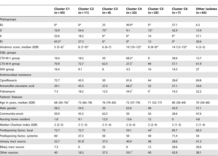
The profiles of VF genes were extremely diverse: the 191 strains showed 159 different profiles, of these 134 were unique, 21 profiles appeared twice, 2 appeared three times, one profile was repeated four times and another one appeared five times. Such diversity prompted us to classify them in clusters; 29 clusters were found using a 70% similarity threshold; 6 clusters arbitrarily named C1– C6 grouped 128 isolates (67%). PGs, ESBLs, antimicrobial resistance, and epidemiological data according to cluster are shown in Table 3; distribution of VF among the clusters are shown in Figure 1. In summary, isolates from C1 caused infections in younger patients; those in C2 were associated with higher frequency of urinary tract source; C4 isolates showed higher frequency of CTX-M-1 group of ESBLs, resistance to amoxicillin/ clavulanic acid and tobramycin, and bacteremia from sources other than urinary or biliary tracts; those in C5 had the lower frequency of local predisposing factors; and C6 isolates showed less frequent resistance for ciprofloxacin. Sixteen of the 22 isolates from C4 (73%) belonged to ST131; also, 76% isolates from ST131 belonged to C4.
To further investigate the association of C2 with urinary tract source, multivariate analysis were performed. We introduced the following variables: age, gender, acquisition, local predisposing factor, systemic predisposing factor, PG, cluster,papGII, VF score, ESBLs, and antimicrobial resistance score. C2 was independently associated with urinary tract source after controlling for age, local and systemic predisposing factors, while PGs,papGII, VF score or antimicrobial resistance were not (table 4). We did the same to
investigate the association of C4 with sources other than urinary or biliary tracts. C4 was independently associated, while again PGs, specific VF, VF score, and antimicrobial resistance score were not (table 4).
Finally, we analyzed the association between different microbi-ological features and absence of predisposing systemic and local features for BSI. In the univariate analysis, cluster 5,papC, papGII, sat,female gender, lower age, community, source, and no receipt of previous antimicrobial use showed a p value ,0.1 and were introduced in the multivariate analysis. The variables selected as independent predictors of BSI in non-predisposed patients were lower age, community-acquired BSI, no receipt of previous antimicrobials, and cluster 5 (table 5).
Discussion
Our study showed that the phylogenetic background or virulence profiles of ESBLEC causing BSI in Spain were different to what would be expected for bacteremicE. coli. The fact that isolates from the so-called ‘‘low virulent’’ PGs A and B1 predominated as caused of BSI is in contrast with previous studies including mainly non-ESBL-producing isolates, in which B2 ad D were predominant [3–8]. As a consequence, the prevalence of all VF studied was much lower among ESBLEC isolates than among previous collections of blood isolates ofE. coliexcept foriutA, iroN, traT, and cvaC[2–4,6,7,12]. There are, to our knowledge, scarce previous data on collections of blood ESBLEC isolates. In a study from The Netherlands including 41 ESBLEC blood isolates, only 22% of isolates belong to A or B1 PGs [15]. Similar to our results, PG A was predominant in the subgroup of ESBL-producers from a French study; however, only 19 ESBLEC were included [8].
Three facts may explain our results. First, most cases occurred in patients with local or systemic predisposing factors for BSI; hence, less virulence factors would be required to cause invasive infection in such patients. Second, previous antibiotic treatment was common, which would have selected for ESBLEC because of their multidrug-resistant nature regardless their virulence profile. Although antimicrobial resistance has been frequently shown to be more frequent among isolates from the A and B1 PGs than among B2 isolates [11], B2 isolates were more frequently resistant to several antimicrobials (particularly amoxicillin-clavulanic acid and tobramycin) than isolates from other PGs. This reflects the resistance profile of isolates of ST131 producing CTX-M-15 [32,33], which comprised most B2 isolates in our series. And third, in a recent study performed in France, non-ST131 B2 E. coli isolates were found to rarely produce CTX-M enzymes [34]; this, together with the fact that ST131 was not predominant in our series, would provide an additional explanation for the low rate of B2 isolates.
Even though ESBLEC from the B2 and D PGs showed, as expected, a much higher content in VF, we did not find B2 and D isolates to have caused infections in clearly less predisposed patients than A/B1 isolates, with the exception of cancer (less frequent among B2). A recent study on non-ESBL-producingE. f7 isolates produced
.1 ESBL.
gMainy CTX-M-14. hMainly CTX-M-15. iMainly SHV-12.
Data are presented as number of isolates (percentage) except where specified. doi:10.1371/journal.pone.0044238.t001
coli found that B2 isolates were predominant as cause of bacteremia and spontaneous peritonitis in patients with liver cirrhosis [35]; of note, liver cirrhosis was more frequent among patient with B2 isolates than those with D or A/B1 isolates in our series, but the differences did not reach statistical significance. Also, we found that PGs or specific VFs were not independently associated to source of BSI. In previous studies of E. coli bacteremic isolates, those from PG B2 had been associated with urinary tract source of BSI [4,5,7]. As regards specific VF, several studies have investigated their association with BSI sources; the studies were different in populations, definitions, and VF studied, making it difficult to draw clear conclusions [4,9,12]. However,
papGIIhas been more consistently associated with urinary tract source in these studies. We did not find such association, although papGII was more frequent in crude analysis among non-predisposed patients with urinary tract BSI.
Overall, these results suggest that host factors and previous antimicrobial use were more important than phylogroup back-ground, virulence score or specific VF in the occurrence of BSI due to ESBLEC. However, by investigating the existence of clusters of isolates according to their VF content, we found some associations between virulence background and some specific epidemiological features. Thus, cluster C2 (mainly PG D, CTX-M-14 producers) was independently associated with urinary tract Table 2.Comparison of predisposing features according to phylogroup among 191 patients with bacteremia due to ESBL-producingE. coli.
All isolates (n = 191)
Phylogroup B2 (n = 30)
Phylgroup D (n = 51)
Phylogroups A/B1 (n = 110)
Age in years, median (IQR) 71 (55–78) 72 (58–82) 71 (58–78) 69 (54–77)
Male gender 107 (56) 20 (66.7) 28 (54.9) 59 (53.6)
Acquisition
Community 23 (12) 4 (13.3) 3 (5.9) 16 (14.5)
Healthcare-associated 72 (37.6) 12 (40) 24 (47.0) 36 (32.7)
Nosocomial 96 (50.2) 14 (46.7) 24 (47.9) 58 (52.7)
Nursing home resident 12 (6.2) 2 (6.7) 7 (13.7) 3 (2.7)
Charlson index, median (IQR) 2 (1–4) 2.5 (1–4) 2 (1–5) 2 (1–4)
Diabetes mellitus 52 (27.2) 9 (30) 12 (23.5) 31 (28.2)
Chronic pulmonary disease 34 (17.8) 4 (13.3) 9 (17.6) 21 (19.1)
Cancer 55 (28.7) 4 (13.3)a,b 15 (29.4) 36 (32.7)
Liver cirrhosis 18 (9.4) 5 (16.7) 4 (7.8) 9 (8.2)
Chronic renal insufficiency 28 (14.6) 3 (10) 5 (9.8) 20 (18.2)
Inmunosuppresive therapy 27 (14.1) 4 (13.3) 9 (17.6) 14 (12.7)
Obstructive urinary disease 43 (22.5) 7 (23.3) 7 (13.7)c 29 (26.4)
Biliary tract disease 18 (9.4) 2 (6.7) 3 (5.9) 13 (11.8)
Neutropenia 10 (5.2) 1 (3.3) 3 (5.9) 6 (5.5)
Urinary catheter 66 (34.5) 13 (43.3) 17 (33.3) 36 (32.7)
Central venous catheter 53 (27.7) 5 (16.7) 12 (23.5) 36 (32.7)
Mechanical ventilation 8 (4.1) 1 (3.3) 3 (5.9) 4 (3.6)
Previous surgery 44 (20.9) 6 (20) 14 (27.5) 24 (21.8)
Predisposing factor, local 122 (63.8) 19 (63.3) 30 (58.8) 73 (66.4)
Predisposing factor, systemic 100 (52.3) 16 (53.3) 24 (47.1) 60 (54.5)
Predisposing factors, systemic or local 158 (82.7) 24 (80) 41 (80.4) 93 (84.5)
Previous antibiotic use, any 107 (56) 18 (60) 28 (54.9) 61 (55.5)
Fluoroquinolones 49 (25.6) 7 (23.3) 12 (23.5) 30 (27.3)
Cephalospororins 53 (27.7) 9 (30) 15 (29.4) 29 (26.4)
Amoxicillin/clavulanic acid 20 (10.4) 3 (10) 3 (5.9) 14 (12.7)
Source
Urinary tract 90 (47.1) 12 (40) 25 (49) 53 (48.2)
Biliary tract 24 (12.5) 3 (10) 5 (9.8) 16 (14.5)
Othersd 77 (40.3) 15 (50) 21 (41.2) 41 (37.3)
aP value for B2 vs A/B1 = 0.03. bP value for B2 vs D = 0.09. cP value for D vs A/B1 = 0.07.
All other comparisons, P value$0.1.
dOther sources were: unkown, 25 patients; intraabdominal (non-biliar), 24; respiratory tract, 10; catheter-related, 9; miscellaneous, 8.
BSI; C4 (mostly B2 and ST131, CTX-M-15 producers) with non-urinary or biliary tract sources; and C5 (mostly D and CTX-M-14 producers) with BSI in non-predisposed patients. All these clusters had moderate to high virulence scores. The classification of isolates into clusters according to VF had been previously carried out by
Johnson et al. according to clonal groups [36]; however, we constructed the clusters by considering exclusively the VF content of the isolates and without taking into account neither the phylogroups nor any other clonal relationship among isolates because our aim was to specifically investigate the influence of FV
Figure 1. Distribution of virulence factors according to clusters.Percentage of isolates: white: 0–25%; pale grey, 26–50%; dark grey, 51–75%; black,.75%.
content by itself in the epidemiology of the infections. Hypothetical implications from our data are that vaccines developed against specific VFs might not be efficacious in avoiding invasive infections due to ESBLEC in predisposed patients, and that reducing the
antibiotic pressure in such patients might be a more important measure to try and reduce such infections in these patients.
Strengths of our study include its multicenter nature, clinical data are comprehensive and were prospectively collected, and isolates are well characterized. However, it has some limitations: we could not compare the ESBLEC profiles with a control group of non-ESBL producers and thus used collections from other studies as a reference; we studied the genes codifying for VF, but Table 3.Phylogroups, virulence score, ESBLs, selected antimicrobial resistance, and associated patients’ features of ESBL-producingE. coliisolates causing BSI according to virulence profile clusters.
Cluster C1 (n = 55)
Cluster C2 (n = 11)
Cluster C3 (n = 8)
Cluster C4 (n = 22)
Cluster C5 (n = 25)
Cluster C6 (n = 7)
Other isolates (n = 63)
Phylogroups
B2 0* 0* 25 90.9* 0* 57.1 6.3
D 10.9 54.4 75* 9.1 72* 42.9 15.9
A 23.6 18.2 0* 0* 16 0* 57.1
B1 65.5* 27.3 0* 0* 12 0* 20.6
Virulence score, median (IQR) 5 (5–6)* 8 (7–9)* 6 (6–7) 10 (10–12)* 8 (8–9)* 14 (12–15)* 4 (2–5)
ESBL groups
CTX-M-1 group 16.4 18.2 50 68.2* 8 28.6 12.7
CTX-M-9 group 70.9 72.7 62.5 27.3* 84 57.1 61.9
SHV group 16.4 9.1 0 4.5 16 14.3 27
Antimicrobial resistance
Ciprofloxacin 72.7 45.5 50 81.8 64 28.6* 69.8
Amoxicillin-clavulanic acid 29.1 45.5 37.5 68.2* 32 57.1 34.0
Tobramycin 7.3 18.2 12.5 54.5* 0* 14.3 22.2
Patients’ features
Age in years, median (IQR) 68 (50–76)* 73 (66–78) 76 (70–83) 72 (57–79) 71 (52–77) 80 (58–84) 70 (58–80)
Male gender 58.2 54.5 50 63.6 48 42.9 57.1
Community-onset 50.9 45.5 62.5 50 56 28.6 47.6
Nursing home resident 1.8 9.1 12.5 13.6 12 0 4.8
Median Charlson index (IQR) 2 (2–5) 2 (1–5) 2 (1–4) 2 (2–4) 3 (2–4) 3 (1–3) 2 (1–4)
Predisposing factor, local 72.7 72.7 75 59.1 44* 85.7 60.3
Predisposing factor, systemic 60 37.3 50 50 40 71.4 54
Urinary tract source 52.7 81.8* 37.5 40.9 48 28.6 41.3
Biliary tract source 7.3 0 25 0 12 28.6 20.6
Other sources 40 18.2 37.5 59.1* 40 42.9 38.1
*P values,0.05 in comparison with isolates not included in the cluster.
Data are presented as percentage of isolates in each cluster except where specified. doi:10.1371/journal.pone.0044238.t003
Table 4.Multivariate analysis of variables associated with specific sources of bloodstream infection.
OR (95% CI) P
Urinary tract source
Age (per year) 1.02 (1.00–1.04) 0.009
Local predisposing factor 2.10 (1.11–3.98) 0.002
Systemic predisposing factor 0.57 (0.31–1.04) 0.07
Cluster C2 5.05 (0.96–26.48) 0.05
Non urinary or biliary tract sources
Age (per year) 0.96 (0.94–0.98) 0.001
Local predisposing factor 0.27 (0.14–0.54) ,0.001
Systemic predisposing factor 2.75 (1.41–5.36) 0.003
Cluster C4 2.89 (1.05–7.93) 0.03
doi:10.1371/journal.pone.0044238.t004
Table 5.Multivariate analysis of variables associated with absence of local or systemic predisposing conditions.
OR (95% CI) P
Absence of systemic and local predisposing factors
Age (per year) 0.97 (0.94–1.00) 0.04
Community-onset BSI 2.62 (1.02–6.76) 0.04
No previous antibiotics 5.69 (2.24–14.45) ,0.001
Cluster 5 2.80 (0.99–7.93) 0.05
this does not necessarily reflect the expression of these VF during infection; and results might not be applicable to areas with a different epidemiology of ESBLEC.
In conclusion, bacteremic ESBLEC more frequently belonged to PGs A and B1 and thus had a lower virulent content than expected; neither PGs or specific VF were consistently associated with predisposing features or sources of BSI; and some clusters of isolates identified according to their virulence profile were identified and associated with specific source or acquisition of BSI in the absence of predisposing factors.
Acknowledgments
Other participants from the ESBL-REIPI/GEIH group are: Paloma Gijo´n (Hospital Universitario Gregorio Maran˜o´n, Madrid, Spain), Jose´ Ramo´n Herna´ndez (Hospital Universitario Virgen Macarena, Sevilla, Spain), Jose M. Cisneros (Hospital Universitario Virgen del Rocı´o, Sevilla, Spain), Carmen Pen˜a (Hospital Universitario de Bellvitge, Barcelona, Spain),
Manuel Almela (Hospital Clinic, Barcelona, Spain), Benito Almirante (Hospital Universitario Vall d’Hebro´n, Barcelona, Spain), Fabio Grill (Hospital Universitario Ramo´n y Cajal, Madrid; present address, Hospital Universitario La Paz, Madrid, Spain), Javier Colomina (Hospital de la Ribera, Alzira, Valencia, Spain), Monserrat Gime´nez (Hospital Germans Trias i Pujol, Badalona, Spain), Antonio Oliver (Hospital Son Espases, Palma de Mallorca, Spain), Juan Pablo Horcajada (Hospital Universitario Marque´s de Valdecilla, Santander; present address, Hospital del Mar, Barcelona, Spain), Gemma Navarro (Corporacio Sanitaria Parc Taulı´, Sabadell, Spain), Ana Coloma (Hospital Santa Creu i San Pau, Barcelona, Spain).
Author Contributions
Conceived and designed the experiments: JRB JM AP. Performed the experiments: NFR LS LLC. Analyzed the data: JRB JM LLC AP. Contributed reagents/materials/analysis tools: JRB NFR LS LLC. Wrote the paper: JRB JM AP.
References
1. Picard B, Sevali J, Gouriou S, Duriez P, Brahimi N, et al. (1999) The link between phylogeny and virulence inEscherichia coli extraintestinal infection. Infect Immun 67: 546–553.
2. Johnson JR, Stell AL (2000) Extended virulence genotypes of Escherichia coli
strains from patients with urosepsis in relation to phylogeny and host compromise. J Infect Dis 181: 261–272.
3. Sannes MR, Kuskowski MA, Owens K, Gajewski A, Johnson JR (2004) Virulence factor profiles and phylogenetic background ofEscherichia coliisolates from veterans with bacteremia and uninfected control subjects. J Infect Dis 190: 2121–2128.
4. Moreno E, Planells I, Prats G, Planes AM, Moreno G, Andreu A (2005) Comparative study ofEscherichia colivirulence determinants in strains causing bacteremia versus strains causing pyelonephritis and other sources of bacteremia. Diag Microbiol Infect Dis 53: 93–99.
5. Bukh AS, Schønheyder HC, Emmersen JM, Søgaard M, Bastholm S, et al. (2009)Escherichia coliphylogenetic groups are associated with site of infection and level of antibiotic resistance in community-acquired bacteraemia: a 10 year population-based study in Denmark. J Antimicrob Chemother 64: 163–168. 6. Cooke NM, Smith SG, Kelleher M, Rogers TR (2010) Major differences in
frequencies of virulence factors and multidrug resistance between community and nosocomialEscherichia colibloodstream isolates. J Clin Microbiol 48: 1099– 1104.
7. Lefort A, Panhard X, Clermont O, Woerther P-L, Branger C, et al. (2011) Host factors and portal of entry outweight bacterial determinants to predict the severity ofEscherichia colibacteremia. J Clin Microbiol 49: 777–783. 8. Courpon-Claudinon A, Lefort A, Pahnard X, Clermond O, Dornic Q, et al.
(2011) Bacteraemia caused by third-generation cephalosporin-resistant Esche-richia coliin France: prevalence molecular epidemiology, and clinical features. Clin Microbiol Infect 17: 557–565.
9. Johnson JR, Kuskowski MA, O’Bryan TO, Maslow JN (2002) Epidemiological correlates of virulence genotype and phylogenetic background amongEscherichia coliblood isolates from adults with diverse-source bacteremia. J Infect Dis 185: 1439–1447.
10. Moreno E, Prats G, Planells I, Planes AM, Pe´rez T, et al. (2006) Characterization ofEscherichia coliisolates derived from phylogenetic groups A and B1 causing extraintestinal infection. Enferm Infecc Microbiol Clin 24: 483– 489.
11. Johnson JR, Russo TA (2005) Molecular epidemiology of extraintestnal pathogenic (uropathogenic)Escherichia coli.Int J Med Microbiol 295: 383–404. 12. Wang MC, Tseng CC, Chen CY, Wu JJ, Huang JJ (2002) The role of bacterial
virulence and host factors in patients withEscherichia colibacteremia who have acute cholangitis or upper urinary tract infection. Clin Infect Dis 35: 1161–1166. 13. Rodrı´guez-Ban˜o J, Pico´n E, Gijo´n P, Herna´ndez JR, Ruı´z M, et al. (2010) Community-onset bacteremia due to extended-spectrum beta-lactamase-pro-ducingEscherichia coli: risk factors and prognosis. Clin Infect Dis 50: 40–48. 14. Rodrı´guez-Ban˜o J, Pico´n E, Gijo´n P, Herna´ndez JR, Cisneros JM, et al. (2010)
Risk factors and prognosis of nosocomial bloodstream infections caused by extended-spectrumb-lactamase-producingEscherichia coli. J Clin Microbiol 48: 1726–1731.
15. van der Bij AK, Peirano G, Goessens WH, van der Vorm ER, van Westreenen M, et al. (2011) Clinical and molecular characteristics of extended-spectrum-beta-lactamase-producingEscherichia colicausing bacteremia in the Rotterdam Area, Netherlands. Antimicrob Agents Chemother 55: 3576–3578.
16. Lavigne JP, Blanc-Potard AB, Bourg G, Moreau J, Chanal C, et al. (2006) Virulence genotype and nematode-killing properties of extra-intestinalEscherichia coliproducing CTX-M beta-lactamases. Clin Microbiol Infect 12: 1199–1206.
17. Clermont O, Lavollay M, Vimont S, Deschamps C, Forestier C, et al. (2008) The CTX-M-15-producingEscherichia colidiffusing clone belongs to a highly virulent B2 phylogenetic subgroup. J Antimicrob Chemother 61: 1024–1028. 18. Lavigne JP, Vergunst AC, Goret L, Sotto A, Combescure C, et al. (2012)
Virulence potential and genomic mapping of the worldwide cloneEscherichia coli
ST131. PLoS One 7: e34294.
19. Charlson ME, Pompei P, Ales KL, MacKenzie CR (1987) A new method of classifying prognostic co-morbidity in longitudinal studies: development and validation. J Chron Dis 40: 373–383.
20. Clinical and Laboratory Standard Institute (CLSI) (2009) Performance Standards for Antimicrobial Susceptibility Testing; 19th Informational Supple-ment. CLSI document M100-S19. CLSI, Wayne, PA, USA.
21. Clermont O, Dhanji H, Upton M, Gibreel T, Fox A, et al. (2009) Rapid detection of the O25b-ST131 clone ofEscherichia coliencompassing the CTX-M-15-producing strains. J Antimicrob Chemother 64: 274–277.
22. Clermont O, Bonacorsi S, Bingen E (2000) Rapid and simple determination of theEscherichia coliphylogenetic group. Appl Environ Microbiol 66: 4555–4558. 23. Le Bouguenec C, Archambaud M, Labigne A (1992) Rapid and specific detection of thepap, afa,andsfaadhesin-encoding operons in uropathogenic
Escherichia colistrains by polymerase chain reaction. J Clin Microbiol 30: 1189– 1193.
24. Karkkainen UM, Kauppinen J, Ikaheimo R, Katila ML, Siitonen A (1998) Rapid and specific detection of three different G adhesin classes of P-fimbriae in uropathogenicEscherichia coliby polymerase chain reaction. J Microbiol Methods 34: 23–29.
25. Blanco M, Blanco JE, Alonso MP, Mora A, Balsalobre C, et al. (1997) Detection ofpap, sfaandafaadhesin-encoding operons in uropathogenicEscherichia coli
strains: relationship with expression of adhesins and production of toxins. Res Microbiol 148: 745–755.
26. Yamamoto S, Teai A, Yuri K, Kurazono H, Takeda Y, et al. (1995) Detection of urovirulence factors inEscherichia coliby multiplex polymerase chain reaction. FEMS Immunol Med Microbiol 12: 85–90.
27. Ananias M, Yano T (2008) Serogroups and virulence genotypes ofEscherichia coli
isolated from patients with sepsis. Braz J Med Biol Res 41: 877–883. 28. Nakano M, Yamamoto S, Terai A, Ogawa O, Makino SI, et al. (2001)
Structural and sequence diversity of the pathogenicity island of uropathogenic
Escherichia coliwhich encodes the USP protein. FEMS Microbiol Lett 205: 71–76. 29. Russo TA, Carlino UB, Johnson JR (2001) Identification of a new iron-regulated virulence gene,ireA, in an extraintestinal pathogenic isolate ofEscherichia coli. Infect Immun 69: 6209–6216.
30. Girardeau JP, Lalioui L, Said AM, De Champs C, Le Bouqie´nec C (2003) Extended virulence genotype of pathogenicEscherichia coliiIsolates carrying the
afa-8operon: evidence of similarities between isolates from humans and animals with extraintestinal infections. J Clin Microbiol 41: 218–226.
31. Bidet P, Metais A, Mahjoub-Messai F, Durand L, Dehem M, et al. (2007) Detection and identification by PCR of a highly virulent phylogenetic subgroup among extraintestinal pathogenic Escherichia coli B2 strains. Appl Environ Microbiol 72: 2373–2377.
32. Nicolas-Chanoine MH, Blanco J, Leflon-Guibout V, Demarty R, Alonso MP, et al. (2008) Intercontinental emergence ofEscherichia coliclone O25:H4-ST131 producing CTX-M-15. J Antimicrob Chemother 61: 271–281.
33. Pitout JDD, Gregson DG, Campbell L, Laupland KB (2009) Molecular characteristics of extended-spectrum-b-lactamase-producing Escherichia coli
isolates causing bacteremia in the Calgary Health Region from 2000 to 2007: emergence of clone ST131 as a cause of community-acquired infections. Antimicrob Agents Chemother 53: 2846–2851.
B2 isolates, except clone ST131, rarely produce CTX-M enzymes. J Clin Microbiol (in press).
35. Bert F, Johnson JR, Ouattara B, Leflon-Guibout V, Johnston B, et al (2010) Genetic diversity and virulence profiles of Escherichia coli isolates causing spontaneous bacterial peritonitis and bacteremia in patients with cirrhosis. J Clin Microbiol 48: 2709–2714.
36. Johnson JR, O’Bryan TT, Kuskowski M, Maslow JN (2001) Ongoing horizontal and vertical transmission of virulence genes andpapAalleles amongEscherichia coli