Tiago André Fontoura de MELO(a)
Grasiela Sabrina Longhi GRÜNDLING(a)
Francisco MONTAGNER(b)
Alcione Luiz SCUR(c)
Liviu STEIER(d)
Roberta Kochenborger SCARPARO(a)
José Antônio Poli de FIGUEIREDO(a)
Fabiana Vieira VIER-PELISSER(a)
(a)Pontifícia Universidade Católica do Rio Grande do Sul – PUCRS, School of Dentistry, Clinical Department, Porto Alegre, RS, Brazil.
(b)Pontifícia Universidade Católica do Rio Grande do Sul – PUCRS, School of Dentistry, Endodontics Division, Porto Alegre, RS, Brazil.
(c)Private Practice, Gramado, RS, Brazil.
(d)University of Warwick, Warwick Medical School, Institute of Clinical Education, Coventry, United Kingdom.
LPS levels in root canals after the
use of ozone gas and high frequency
electrical pulses
Abstract: The present study aims to verify the effect of ozone gas (OZY®
System) and high frequency electric pulse (Endox® System) systems
on human root canals previously contaminated with Escherichia coli
lipopolysaccharide (LPS). Fifty single-rooted teeth had their dental crowns removed and root lengths standardized to 16 mm. The root
canals were prepared up to #60 hand K-iles and sterilized using
gamma radiation with cobalt 60. The specimens were divided into
the following ive groups (n = 10) based on the disinfection protocol
used: OZY® System, one 120-second-pulse (OZY 1p); OZY® System, four
24-second-pulses (OZY 4p); and Endox® System (ENDOX). Contaminated
and non-contaminated canals were exposed only to apyrogenic water and used as positive (C+) and negative (C-) controls, respectively. LPS (O55:B55) was administered in all root canals except those belonging to group C-. After performing disinfection, LPS samples were collected from the canals using apyrogenic paper tips. Limulus Amoebocyte Lysate (LAL) was used to quantify the LPS levels, and the data obtained was analyzed using one-way ANOVA. The disinfection protocols used
were unable to reduce the LPS levels signiicantly (p = 0.019). The use
of ozone gas and high frequency electric pulses was not effective in eliminating LPS from the root canals.
Keywords: Endodontics; Endotoxins; Microbiology; Root Canal Therapy.
Introduction
Microorganisms and their byproducts, such as bacterial LPS (endotoxin), play a key role in the development of apical periodontitis.1,2,3
LPS is released during multiplication or death of gram-negative bacteria, and is associated with many biological effects, such as release of
pro-inlammatory mediators4 and induction of periapical bone resorption.5
The endotoxins can adhere to mineralized tissues6 and disseminate
through the dentinal tubules,7 making root canal sterilization dificult
if only chemo-mechanical preparation is used.8 Although the effects of
different disinfection strategies9,10,11,12 on LPS have already been examined, none of them are fully effective as yet.
Electrofulguration and ozone gas equipment were tested for microorganisms, with satisfactory results.13,14,15,16 Electrofulguration
system such as Endox®, deliver electrical pulses into the root canal via a
stainless steel surgical needle that works as an active electrode,15 thereby
Declaration of Interests: The authors certify that they have no commercial or associative interest that represents a conflict of interest in connection with the manuscript.
Corresponding Author: Tiago André Fontoura de Melo E-mail: [email protected]
DOI: 10.1590/1807-3107BOR-2016.vol30.0019
Submitted: Jun 27, 2015
eliminating organic and inorganic content through steaming.17 Ozone is a very reactive gas that has
the ability to oxidize cell walls and the cytoplasmic membrane of microorganisms.14
Till date, there have been no studies investigating the effects of this equipment on LPS. Thus, the objective of the present study was to assess in vitro the effect of ozone gas and high frequency electrical pulses on LPS levels in infected root canals.
Methodology
The present study was approved by the Ethics and Research Committee of Pontifícia Universidade Católica do Rio Grande do Sul - PUCRS (Protocols
no. 5859 and 811.207).
All plates and materials used in this study were sterilized using cobalt 60 gamma radiation (20 kGy for 6 hours) (Empresa Brasileira de Radiações - EMBRARAD, Cotia, Brazil), as previously described.2
Sample selection and preparation
Fifty single-root premolars had their dental crowns sectioned in such a way that the root length was standardized to 16 mm. A 15 mm working length (WL) was established. The canals were manually prepared using the serial technique up to #60
hand K-iles (Dentsply/Maillefer Instruments S.A.,
Ballaigues, Switzerland), and irrigated using 2%
sodium hypochlorite (Iodontosul, Porto Alegre, Brazil).
The smear layer was removed usi ng 17%
trisodium EDTA (Iodontosul, Porto Alegre, Brazil)
for 5 minutes after agitation in the root canal with a
#60 hand K-ile for one minute. Final irrigation was
performed using 2 mL of 2% sodium hypochlorite, and the root canals were dried using sterilized
paper points (Dentsply/Maillefer Instruments S.A.,
Ballaigues, Switzerland).
The teeth were randomly fixed in 12-wells cult ure plates (Kasvi, Curitiba, Brazil) with Durepoxi® (Henkel, Düsseldorf, Germany). Each
plate contained two teeth from each of the five experimental groups (Table).
Specimen contamination
This protocol was performed according to Signoretti et al.18 The teeth were inoculated with
30 μL of a solution containing the endotoxin
Escherichia coli O55:B5 (Lonza, Walkersville, USA)
inside a laminar low chamber with the help of a
micro pipette.
A solution containing LPS (80 EU/mL), previously diluted in apyrogenic water (50.37 EU/mL), was
used to contaminate of all specimens except those belonging to group C- that were inoculated with
30 μL of apyrogenic water.
Apyrogenic cotton pellets were placed in the cervical portion of the canals in all samples. The plates containing the samples were sealed and incubated for 24 hours at 37°C temperature.
Desinfection Procedure
Prior to performing the clinical protocols, all root
canals were illed with 10 μL of apyrogenic water.
The disinfection protocols used in each experimental group were as follows:
a. Group C+: the LPS contaminated canals did not undergo any disinfection protocol.
b. Group C-: the canals had no previous LPS inoculation and did not undergo any disinfection protocol.
c. Group OZY 1p: the tip of the OZY® system was
introduced up to the working length of the root canal and one 120-second-long pulse was delivered, as described by Kustarsi et al.16
Table. Descriptive chart for the experimental groups and disinfection protocols used.
Experimental Group n Contamination/LPS Clínic Protocol
C+ 10 Yes Without treatment
C- 10 No Without treatment
OZY 1p 10 Yes OZY® System (1 pulse - 120 s)
OZY 4p 10 Yes OZY® System (4 pulses - 24 s each)
d. Group OZY 4p: the OZY® system was
introduced up to the working length of the root canals and four 24-second pulses were delivered, according to Case et al.14 There was a
5 second interval between pulses.
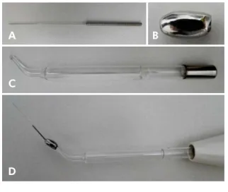
An adapter was developed for the already available “Oto” tip to compensate for the unavailability of a
speciic tip for use with the OZY® system (Endox SRL,
Italy). It was made of 420 surgical steel and had 0.20 mm
diameter at the end and 30 mm length (Figure 1). The OZY® system was operated at 5N intensity
in accordance with the manufacturer’s instructions. The only variation between the OZY 1p and OZY 4p groups was in the device activation time.
a. Group ENDOX: the protocol used for the Endox®
system (Lysis srl, Milan, Italy) was the same as that
described by Lendini et al.17 The black probe of the
device (measuring 30 mm in length and 0.20 mm in diameter) was introduced into the root canal. Two pulses were administered in the medium third of the root canal (5 mm short of WL), and two additional pulses in the apical third (in WL). Thus, four 600 kHz pulses were delivered, with standard
time for each application being 1/10 of a second.
Determination of LPS Levels
The canals content were collected by holding three apyrogenic #60 paper points (Tanari®, Manaus,
Brazil) in position for 10 seconds. They were then transferred into glass tubes, sealed, and stored at
-20°C until LPS levels quantiication was perfomed. Quantiication of an apyrogenic water sample
and the paper tips used was previously assessed in order to check the accuracy of LPS level estimation
The glass tubes containing the paper tips were
illed with 1 mL apyrogenic water, warmed at 37°C
for one hour, and finally centrifuged (Phoenix, Araraquara, Brazil) for 1 minute.
The chromogenic kinetic test of the turbidimetric LAL (Pyrogent 5000®, BioWhitaker, Cambrex Co.,
Walkersville, USA) was used to quantify the LPS levels in the root canals, as already described and applied in some studies.2,11
The samples collected from the canals were mixed with the LAL reagent and automatically monitored over time with the help of a photometer until turbidity developed. The increase in optical density was measured by the reaction time, which is inversely proportional to the amount of LPS present in the sample.
A curve was drawn using the endotoxins supplied by the kit, with known concentration as a parameter for estimating LPS levels present in the root canals All
the samples collected for analysis and quantiication
were diluted ten times.
For test validation, the assays were performed
twice in distinct wells in a 96-well microplate
(Corning Costar, Cambridge, USA). One hundred
μL of apyrogenic water was added to the negative control, 100 μL for standard endotoxin at different concentrations for the curve, and 100 μL from each sample for quantiication. LPS level was measured
according to the manufacturers’ instructions. The microplate was incubated in enzyme immunoassay reader (Ultramark, Bio-Rad Laboratories
Inc., Hercules, USA) for 10 minutes at 37 ± 1°C, and
the reader was coupled with a computer with Wink QCL software version 4 (BioWittaker, Cambrex Co. Walkersville, EUA).
After incubation, 100 μL of LAL chromogenic
kinetic reagent (Sigma Chemical Company St. Louis, USA) was added to each plate well to start
quantiication of LPS levels.
A B
C
D
Figure 1. Adapter at the “Oto” tip of the OZY® system: (A) “endodontic” tip, (B) adapter, (C) “Oto” tip and
Statistical analysis
EU/mL measures were logarithmically transformed
to reduce asymmetry and heteroscedasticity. The data were reported as geometric mean, maximum, and minimum values. The groups were compared using-way variance analysis (one-way ANOVA) on the logarithms,
and the signiicance level was set at 5% (p≤ 0.05).
Results
The standard curve for LAL assay validation
followed the linearity criteria (r = 1). The LAL assay
showed that LPS was present in 100% of the root canals initially contaminated.
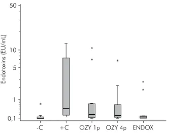
Results are shown in Figure 2. There were no
signiicant differences (p = 0.019) in the reduction
of endotoxin between the experimental groups undergoing disinfection and group C+. However, a significant difference was observed when the endotoxin concentration of the group C- specimens was compared with the other groups.
Discussion
Successful endodontic treatment of infected teeth involves elimination of microorganisms as well as inactivation of endotoxins and other toxic products.2
Thus, considering the results observed when using the ozone gas system14,19 and high frequency electrical pulses13,15 on microbial strains, analysis of these systems
on bacterial LPS was also thought to be pertinent.
The endotoxin used in the present study was obtained from Escherichia coli as it is the standard endotoxin employed in most research studies.12,20
LAL assay was used to quantify LPS levels as it is extremely sensitive to measurement.2,10
The turbidimetric LAL assay indicated that the endotoxins were present in all previously contaminated samples. The concentrations ranged from 0.1 to
12.8 EU/mL in the positive control group, and this
was in accordance with Jacinto et al.21 who reported
endotoxin concentrations ranging between 2.3 and
22.1 EU/mL. This range in values can be attributed to
the sensitivity of the test that allows detection of slight variations, and to the differences in the dental anatomy of species. However, the LPS concentration obtained
was inferior to the approximate value of 50.37 EU/mL injected into each root canal. This can be justiied by
the fact that the samples were collected from the main canal and not from the deeper dentinal tubular region. According to Horiba et al.,7 the endotoxin is capable of
penetrating into the dentinal tubules four times deeper
than that of the microorganisms, reaching 800 μm in
depth, mainly due to its low molecular weight. Thus, there is a need for auxiliary strategies that complement mechanical instrumentation to perform effective root canal disinfection.
None of the clinical protocols performed herein
promoted signiicant reduction of endotoxin levels
in relation to the positive control group. This can be
justiied by the fact that the principle anti-microbial
action14,15 of the two systems involves causing damage
to the structure of the microbial coating. However, LPS does not present a coating structure and it is mainly composed of specific polysaccharide O, a central nucleus, and a lipid component A,22 thereby yielding
these systems ineffective against it. This was conirmed
by Cardoso et al.23 who showed that ionized water was
unable to neutralize the root canal endotoxins. The results suggest presence of endotoxins in the ozone water group as well the saline solution control group. Another factor that may corroborate the results obtained in this study is the ephemeral ozone gas half-life.24 The
only techniques that have been tested so far and have showed a certain degree of effectiveness against LPS are (a) the use of calcium hydroxide as an intra-canal medication12,25,26 as it hydrolyses lipid A and changes
Endotoxins (EU/mL)
00
10
5
1
0,1
-C +C OZY 1p OZY 4p ENDOX *
* *
*
* *
Figure 2. Boxplot distribution of endotoxin concentration in EU/mL for each experimental group.
it into chains of fatty acid and nontoxic sugars,27 and
(b) the application of laser Nd:YAG.9
The results obtained in this study reinforce the importance of instrumentation in addition to any disinfection protocol,11,28,29 especially with regard to bacterial LPS that strongly adheres to the dentinal wall6 and makes it necessary to use endodontic
instruments for its removal. Some studies have reported endotoxin decrease up to 44.4%,30 59.99%,28
and 57.98%10 after chemo-mechanical preparation.
Martinho et al.11 noticed 98.06% decrease in the
endotoxin after performing canal preparation with NiTi (Mtwo®) rotatory tools under irrigation with 2.5%
sodium hypochlorite solution, and 96.27% decrease when
the rotatory preparation was associated with irrigation using apyrogenic saline solution. According to the authors, a basic factor that must be taken into account when performing chemo-mechanical preparation to decrease bacterial LPS is the enlargement the apical third. Enlargements over size 30 instruments are recommended for removal of endotoxin as well as
infected dentin. Furthermore, this approach allows for deeper irrigation and increases the probability of opening secondary canals and apical deltas. However, this was contradicted by Martinho et al.29 who tested different systems with different designs, conicities, and tapers such as WaveOne®, Reciproc®, Protaper®, and Mtwo® and
did not observe any statistically signiicant differences
in the decrease of bacterial LPS. The percentage of LPS
reduction observed with each system was 95.15%, 96.21%, 97.98%, and 96.34%, respectively.
Conclusion
Based on the results of this study, the clinical protocols used for disinfection were unable to reduce LPS levels. The use of ozone gas and high frequency electric pulses was not effective in the elimination of LPS in root canals.
Acknowledgments
This study was supported by grants from Fundaçãode Amparo à Pesquisa do Estado do Rio Grande do Sul - FAPERGS
(process no. 12/0439-0 / edital no. 13/2011).
1. Kakehashi S, Stanley HR, Fitzgerald RJ. The effects of surgical exposures of dental pulps in germ-free and conventional
laboratory rats. Oral Surg Oral Med Oral Pathol. 1965;20:340-9. doi:10.1016/0030-4220(65)90166-0
2. Marinho ACS, Martinho FC, Zaia AA, Ferraz CCR, Gomes BPFA. Monitoring the effectiveness of root canal procedures on endotoxin levels found in teeth with chronic
apical periodontitis. J Appl Oral Sci. 2014;22(6):490-5. doi:10.1590/1678-775720130664
3. Khabbaz MG, Anastasiadis PL, Sykaras SN. Determination of endotoxins in the vital pulp of human carious teeth: association with pulpal pain. Oral Surg Oral Med Oral Pathol Oral Radiol Endod. 2001;91(5):587-93.
doi:10.1067/moe.2001.113831
4. Agarwal S, Piesco NP, Johns LP, Riccelli AE. Differential
expression of IL-1 beta, TNF-alpha, IL-6, and IL-8 in
human monocytes in response to lipopolysaccharides from different microbes. J Dent Res. 1995;74(4):1057-65.
doi:10.1177/00220345950740040501
5. Hong CY, Lin SK, Kok SH, Cheng SJ, Lee MS, Wang TM, et al. The role of lipopolysaccharide in infectious bone resorption
of periapical lesion. J Oral Pathol Med. 2004;33(3):162-9. doi:10.1111/j.0904-2512.2004.00045.x
6. Barthel CR, Levin LG, Reisner HM, Trope M. TNF-alpha release in monocytes after exposure to calcium hydroxide
treated Escherichia coli LPS. Int Endod J. 1997;30(3):155-9. doi:10.1046/j.1365-2591.1997.00066.x
7. Horiba N, Maekawa Y, Matsumoto T, Nakamura H. A study of the distribution of endotoxin in the dentinal
wall of infected root canals. J Endod. 1990;16(7):331-4. doi:10.1016/S0099-2399(06)81944-8
8. Byström A, Sundqvist G. Bacteriologic evaluation of the efficacy of mechanical root canal instrumentation in
endodontic therapy. Scand J Dent Res. 1981;89(4):321-8. doi:10.1111/j.1600-0722.1981.tb01689.x
9. Archilla JR, Moreira MS, Miyagi SP, Bombana AC, Gutknecht N, Marques MM. Single session of Nd:YAG laser intracanal irradiation neutralizes endotoxin in dental root dentin. J Biomed
Opt. 2012;17(11):118002. doi:10.1117/1.JBO.17.11.118002
10. Gomes BPFA, Martinho FC, Vianna ME. Comparison of 2.5% sodium hypochlorite and 2% chlorhexidine gel on oral bacterial lipopolysaccharide reduction from
primarily infected root canals. J Endod. 2009;35(10):1350-3. doi:10.1016/j.joen.2009.06.011
11. Martinho FC, Chiesa WMM, Marinho ACS, Zaia AA, Ferraz CCR, Almeida JFA, et al. Clinical investigation of the efficacy of chemomechanical preparation with rotary nickel-titanium files for removal of endotoxin from
primarily infected root canals. J Endod. 2010;36(11):1766-9. doi:10.1016/j.joen.2010.08.019
12. Si lva L, Nel s on-Fi l ho P, L e on a rdo M R, Ro s si MA, Pansani CA. Effect of calcium hydroxide on
bacterial endotoxin in vivo. J Endod. 2002;28(2):94-8. doi:10.1097/00004770-200202000-00011
13. Aranda-Garcia AR, Guerreiro-Tanomaru JM, Faria-Júnior NB, Chavez-Andrade GM, Leonardo RT, Tanomaru-Filho M, et al. Antibacterial effectiveness of several irrigating solutions
and the Endox Plus system - an ex vivo study. Int Endod J. 2012;45(12):1091-6. doi:10.1111/j.1365-2591.2012.02069.x
14. Case PD, Bird PS, Kahler WA, George R, Walsh LJ. Treatment of root canal biofilms of Enterococcus faecalis with ozone gas and passive ultrasound activation. J Endod. 2012;38(4):523-6.
doi:10.1016/j.joen.2011.12.020
15. Cassanelli C, Marchese A, Cagnacci S, Debbia EA. Alteration of membrane permeability of bacteria and yeast by high frequency alternating current (HFAC). Open Microbiol J. 2008;2:32-7.
doi:10.2174/1874285800802010032
16. Kuştarci A, Sümer Z, Altunbaş D, Koşum S. Bactericidal effect of KTP laser irradiation against Enterococcus faecalis compared with gaseous ozone: an ex vivo study. Oral Surg
Oral Med Oral Pathol Oral Radiol Endod. 2009;107(5):e73-9. doi:10.1016/j.tripleo.2009.01.048
17. Lendini M, Alemanno E, Migliaretti G, Berutti E. The effect of high-frequency electrical pulses on organic
tissue in root canals. Int Endod J. 2005;38(8):531-8. doi:10.1111/j.1365-2591.2005.00983.x
18. Signoretti FGC, Gomes BPFA, Montagner F, Barrichello Tosello
F, Jacinto RC. Influence of 2% chlorhexidine gel on calcium
hydroxide ionic dissociation and its ability of reducing endotoxin. Oral Surg Oral Med Oral Pathol Oral Radiol
Endod. 2011;111(5):653-8. doi:10.1016/j.tripleo.2010.11.016 19. Halbauer K, Prskalo K, Jankovic B, Tarle Z, Panduric V,
Kalenic S. Efficacy of ozone on microorganisms in the tooth
root canal. Coll Antropol. 2013;37(1):101-7. PMID: 23697257.
20. Oliveira LD, Carvalho CAT, Valera MC, Koga-Ito CY, Jorge AOC. Diffusion ability of endotoxin through
dentinal tubules. Braz Oral Res. 2005;19(1):5-10. doi:10.1590/S1806-83242005000100002
21. Jacinto RC, Gomes BPFA, Shah HN, Ferraz CC, Zaia AA, Souza-Filho FJ. Quantification of endotoxins in necrotic root
canals from symptomatic and asymptomatic teeth. J Med Microbiol. 2005;54:777-83. doi:10.1099/jmm.0.45976-0 22. Mohammadi Z. Endotoxin in endodontic infections: a review.
J Calif Dent Assoc. 2011;39(3):152-5. PMID: 21563594.
23. Cardoso MG, Oliveira LD, Koga-Ito CY, Jorge AO. Effectiveness of ozonated water on Candida albicans, Enterococcus faecalis, and endotoxins in root canals. Oral Surg Oral Med Oral Pathol Oral Radiol Endod.
2008;105(2):e85-91. doi:10.1016/j.tripleo.2007.10.006
24. Farac RV, Pizzolitto AC, Tanomaru JM, Morgental RD, Lima
RK, Bonetti-Filho I. Ex-vivo effect of intracanal medications
based on ozone and calcium hydroxide in root canals contaminated with Enterococcus faecalis. Braz Dent J.
2013;24(2):103-6.doi:10.1590/0103-6440201301992
25. Tanomaru JM, Leonardo MR, Tanomaru Filho M, Bonetti
Filho I, Silva LA. Effect of different irrigation solutions and calcium hydroxide on bacterial LPS. Int Endod J. 2003;36(11):733-9. doi:10.1046/j.1365-2591.2003.00717.x 26. Adl A, Motamedifar M, Shams MS, Mirzaie A. Clinical
investigation of the effect of calcium hydroxide intracanal dressing on bacterial lipopolysaccharide reduction from infected root canals. Aust Endod J. 2015;41(1):12-6.
doi:10.1111/aej.12054
27. Safavi KE, Nichols FC. Effect of calcium hydroxide on
bacterial lipopolysaccharide. J Endod. 1993;19(2):76-8. doi:10.1016/S0099-2399(06)81199-4
28. Martinho FC, Gomes BPFA. Quantification of endotoxins and cultivable bacteria in root canal infection before and after chemomechanical preparation with 2.5% sodium hypoch lorite. J Endod. 2008;34(3):268-72.
doi:10.1016/j.joen.2007.11.015
29. Martinho FC, Gomes BP, Fernandes AM, Ferreira NS, Endo
MS, Freitas LF, Camões IC. Clinical comparison of the
effectiveness of single-file reciprocating systems and rotary systems for removal of endotoxins and cultivable bacteria
from primarily infected root canals. J Endod. 2014;40(5):625-9. doi:10.1016/j.joen.2013.12.006
30. Vianna ME, Horz HP, Conrads G, Zaia AA, Souza-Filho FJ, Gomes BP. Effect of root canal procedures on endotoxins