Activation of Intestinal Epithelial Stat3
Orchestrates Tissue Defense during
Gastrointestinal Infection
Nadine Wittkopf1, Geethanjali Pickert2, Ulrike Billmeier1, Mousumi Mahapatro1, Stefan Wirtz1, Eva Martini1, Moritz Leppkes1, Markus Friedrich Neurath1, Christoph Becker1*
1Department of Medicine 1, Friedrich-Alexander-University, 91052 Erlangen, Germany,2Institute of Translational Immunology, Johannes Gutenberg-University, 55131 Mainz, Germany
*christoph.becker@uk-erlangen.de
Abstract
Gastrointestinal infections with EHEC and EPEC are responsible for outbreaks of diarrheal diseases and represent a global health problem. Innate first-line-defense mechanisms such as production of mucus and antimicrobial peptides by intestinal epithelial cells are of utmost importance for host control of gastrointestinal infections. For the first time, we directly dem-onstrate a critical role for Stat3 activation in intestinal epithelial cells upon infection of mice withCitrobacter rodentium–a murine pathogen that mimics human infections with
attach-ing and effacattach-ingEscherichia coli.C.rodentiuminduced transcription of IL-6 and IL-22 in gut samples of mice and was associated with activation of the transcription factor Stat3 in intes-tinal epithelial cells.C.rodentiuminfection induced expression of several antimicrobial pep-tides such as RegIIIγand Pla2g2a in the intestine which was critically dependent on Stat3 activation. Consequently, mice with specific deletion of Stat3 in intestinal epithelial cells showed increased susceptibility toC.rodentiuminfection as indicated by high bacterial load, severe gut inflammation, pronounced intestinal epithelial cell death and dissemination of bacteria to distant organs. Together, our data implicate an essential role for Stat3 activa-tion in intestinal epithelial cells duringC.rodentiuminfection. Stat3 concerts the host re-sponse to bacterial infection by controlling bacterial growth and suppression of apoptosis to maintain intestinal epithelial barrier function.
Introduction
Bacterial infections of the gastrointestinal tract are a frequent cause of diarrhea followed by de-hydration and are estimated to cause 2.5 million deaths per year, especially in underdeveloped countries [1]. Among the diarrhea-inducing bacteria, pathogenicEscherichia colisuch as en-teropathogenicE.coli(EPEC) and enterohaemorrhagicE.coli(EHEC) frequently cause out-breaks of diarrheal diseases [2,3]. The largest outbreak of EHEC infection ever seen in Europe occurred in Germany in spring 2011: more than 2,900 cases of acute gastroenteritis, more than
a11111
OPEN ACCESS
Citation:Wittkopf N, Pickert G, Billmeier U, Mahapatro M, Wirtz S, Martini E, et al. (2015) Activation of Intestinal Epithelial Stat3 Orchestrates Tissue Defense during Gastrointestinal Infection. PLoS ONE 10(3): e0118401. doi:10.1371/journal. pone.0118401
Academic Editor:Bernhard Ryffel, French National Centre for Scientific Research, FRANCE
Received:October 31, 2014
Accepted:December 14, 2014
Published:March 23, 2015
Copyright:© 2015 Wittkopf et al. This is an open access article distributed under the terms of the Creative Commons Attribution License, which permits unrestricted use, distribution, and reproduction in any medium, provided the original author and source are credited.
Data Availability Statement:All relevant data are within the paper.
850 cases of haemolytic uraemic syndrome (HUS) and at least 50 related deaths in three months were listed [4].
EHEC and EPEC infections cause non-specific gastroenteritis and the benefit of antibiotic agents for treatment of EHEC and EPEC infections is a long-standing matter of debate [5]. Not only might current antibiotics shape the natural course of the disease, but affect the pathogen as well as the beneficial commensal gut flora. Accordingly, there is an urgent need for new ther-apeutic approaches to combat pathogens and prevent common side-effects such as antibiotic-associated diarrhea. Interestingly, the gut itself also possesses antibacterial defense mechanisms such as antimicrobial peptides (AMPs) produced by specialized intestinal epithelial cells, desig-nated Paneth cells [6,7]. Therefore, studies make efforts to investigate the role of AMPs during infections with intestinal pathogens. For example, mice overexpressing the antimicrobial pep-tide CRAMP (cathelicidin-related antimicrobial peppep-tide) were demonstrated to be protected from oral infection withCitrobacter rodentium(C.rodentium) [8], a naturally occurring mouse pathogen that is commonly used to mimic human infections with EHEC and EPEC.
The body’s own antimicrobial defense mechanisms in the gut are of utmost importance for maintenance of a healthy intestine. Using a mouse model of inflammatory bowel disease, we recently suggested a role of Stat3 (signal transducer and activator of transcription 3) activation in intestinal epithelial cells (IECs) for tissue homeostasis. Interestingly, among the genes which were differentially regulated in Stat3 deficient IECs, several were previously reported to show antimicrobial functions. Consequently, Gene Ontology analysis indicated that Stat3 might be involved in antimicrobial defense [9]. Stat3 is a transcription factor whose increased activation in colonic epithelial cells has been observed in some patients with active inflammatory bowel disease (IBD) [10,11]. Moreover, genome-wide association studies have linked polymorphisms in the allele encoding Stat3 with an increased risk for the development of IBD [12,13]. Stat3 is activated by binding of certain ligands such as IL-6, IL-10 and IL-11 to their specific receptors; activated Stat3 then transfers to the nucleus and induces the transcription of target genes includingBCL2,MYCandBIRC5. It has recently been demonstrated that IL-22 induces activation of Stat3 and that IL-22 is crucial for production of AMPs in the intestine [14]. Ac-cordingly, IL-22 is important to combat most intestinal infections. While IL-22 production is increased after infection withListeria monocytogenes, it is not required for clearance of the in-fection [15]. Yet, IL-22 deficient mice develop severe colitis after infection withC.rodentium
and show high mortality [14]. Therefore, although IL-22 expression is increased during colitis, IL-22 is hypothesized to exert protective functions via activation of Stat3 in the intestinal epi-thelium. Although IL-22 has been demonstrated to protect the host from gastrointestinal infec-tions, the functional role of Stat3 in the epithelium has not been demonstrated. Using intestinal epithelial cell specific Stat3 deficient mice (Stat3ΔIEC), we demonstrate a critical role for Stat3 activation in IECs duringC.rodentiuminfection. Our data show that mice unable to activate Stat3 in IECs are severely impaired in their ability to defend againstC.rodentium. Mechanisti-cal data indicate that Stat3 orchestrates the intestinal epithelial response to bacterial infection by production of antimicrobial peptides and suppression of apoptosis.
Materials and Methods
Mice
We obtained C57BL/6 wildtype mice from the animal facility of the University of Mainz. Mice carrying both loxP-flanked Stat3 alleles (Stat3fl/fl) and the Cre-recombinase under control of the Villin-promoter were described before (Stat3ΔIEC) [9]. All mice were housed in individually ventilated cages. To exclude an effect of different mouse microbiota in separated cages, Stat3ΔIECmice and control (Stat3fl/fl) littermates were housed in mixed cages. The protocol was
carried out in strict accordance with the recommendations in the Guide for the Care and Use of Laboratory Animals of the National Institutes of Health. The protocol was approved by Landesuntersuchungsamt Koblenz (Permit Number: 2.3 177–07/G 07–1–006). Mouse endos-copy and IVIS analysis were performed using isoflurane anesthesia, and all efforts were made to minimize suffering.
Citrobacter rodentium
infection and endoscopy
For bacteria-induced colitis, mice were infected with an erythromycin resistant and lumines-cent strain ofCitrobacter rodentium(C.rodentiumstrain ICC169 was kindly provided by C. Riedel [29]). Bacteria were grown in sterile LB-Medium supplemented with 500μg / ml
erythromycin at 37°C and resuspended in sterile PBS to a final concentration of approximately 4 x 10⁹bacteria per 200μl. Mice were fastened for eight hours and 200μl of bacteria suspension
were given to each animal using a feeding needle. For quantification of the applied numbers ofC.rodentium, the bacterial suspension was plated on CASO-blood agar plates (Heipha Dr. Müller GmbH, Eppelheim, Germany). For monitoring of luminescentC.rodentiumgrowth and distribution in live mice, mice were anesthetized by gassing with isoflurane (1.5–2% mixed with air; Abbott).The abdomen of the mice was depilated and luminescence was measured using an IVIS Lumina II system (Caliper Life Science, Waltham, Massachusetts, USA). Quanti-fication of luminescence was performed using the IVIS-associated software“Living Image”
(Caliper Lifescience). For follow up development of colitis and endoscopic scoring, the Colo-view high resolution mouse endoscopic system (Storz, Tuttlingen, Germany) was used as previ-ously described [30]. Health of mice was monitored by weighing mice every other day. At the end of the experiments, mice were sacrificed by cervical dislocation. Tissue samples were col-lected and either instantly frozen in liquid nitrogen or fixed in Histofix (Roth, Karlsruhe, Germany).
Organoid culture
Small intestinal crypts were isolated and cultured as previously described by Sato et al [31]. After 6 days of organoid growth, organoids were treated with IL-22 (100 ng / ml; Peprotech, Rocky Hill, New Jersey, USA) for 24 hours or left untreated. Organoids were harvested, washed twice with PBS and embedded in Histogel (Thermo Scientific, Waltham, Massachusetts, USA) according to manufacturer’s recommendations. After solidifying, solid Histogel-organoid sam-ples were fixed in Histofix (Roth).
Expression analysis
mRNA was isolated using the NucleoSpin RNA II kit (Macherey-Nagel, Düren, Germany) and reverse transcribed into complementary DNA using the iScript cDNA Synthesis Kit (Bio-Rad, Hercules, California, USA) as recommended by the manufacture. Quantitative PCR was per-formed using cDNA-specific Quantitect Primer assays (Qiagen, Hilden, Germany) and SsoFast EvaGreen Supermix (Bio-Rad) according to the manufacturer’s recommendations. Samples were either normalized to the general housekeeping geneHprtor the intestinal epithelial housekeeping geneVillin.
Histological examination
slices were fixed on glass slides using 4% PFA and paraffin-embedded tissue sections were dewaxed and rehydrated. pStat3 (Cell Signaling, Danvers, Massachusetts, USA), anti-MPO (Abcam, Cambridge, United Kingdom), anti-CD4 (BD Pharmingen, San Jose, California, USA), anti-CD11c (BD Bioscience, San Jose, California, USA) and anti-RegIIIγ(antibodies on-line GmbH, Aachen, Germany) were used as primary antibodies. A biotinylated secondary anti-rabbit-antibody (Dianova, Hamburg, Germany) was used together with the TSA Cy3 sys-tem (Perkin Elmer, Waltham Massachusetts, USA) in accordance with the manufacturer’s protocol. Dying cells were detected using TUNEL assay (In situ Cell death Detection Kit Fluo-rescein, Roche, Basel, Switzerland) as recommended by the manufacturer. Bacteria present in tissues were detected by fluorescencein situhybridization (FISH) of bacterial RNA as previous-ly described [32]. Nuclei were counterstained with Hoechst (Invitrogen, Darmstadt, Germany). Images were obtained using the bright-field / fluorescence microscope Olympus IX70 (Olym-pus, Hamburg, Germany) or Leica DMI4000 B (Leica, Wetzlar, Germany).
Statistical analysis
Statistical analysis was performed using the two-tailed student’s t-test.
p0.05,
p0.01,
p0.001, n.s. = not significant. Percentages of cells in immunofluorescence pictures were
calculated using ImageJ (National Institute of Health) [33].
Results
To examine the role of Stat3 for antimicrobial defense mechanisms during gastrointestinal in-fectionsin vivo, we used theC.rodentiuminduced infectious colitis mouse model. After oral in-fection of C57BL/6J wildtype mice with 4 x 10⁹luminescentC.rodentium, the development of infection was monitored using anin vivoimaging system. In agreement with a transient infec-tion, we observed a continuous increase in luminescence in the gut over seven days and a de-crease to background levels afterwards (Fig. 1a). Endoscopic analysis of infected mice revealed signs of intestinal inflammation such as bowel-wall thickening, granularity of the mucosa, and fibrin formation (Fig. 1b). Upon screening for the expression of cytokines known to regulate intestinal defense mechanisms, IL-6 and IL-22 revealed strong induction after bacterial infec-tion (Fig. 1c). Our finding is in line with recent data published by Zheng et al. [14], showing that IL-22 was strongly increased afterC.rodentiuminfection. IL-22 has been reported to be produced by several cell types including dendritic cells [9], neutrophils [16], Th17 cells [17,18] and subsets of NK cells [19,20]. However, recent data suggest RORγt+innate lymphoid cells (ILCs) to be the main producers of IL-22 in the gut [21]. Both IL-6 and IL-22 can induce the Stat3 signaling pathway in epithelial cells upon binding to their receptors. To analyze Stat3 ac-tivation in the intestinal epithelium in response to bacterial infections, gut samples from wild-type mice were harvested at different time-points and stained for phosphorylated Stat3. While colonic epithelial cells of uninfected mice showed no phosphorylated Stat3, the transcription factor was activated after infection withC.rodentium(Fig. 1d). Notably, the number of IECs showing activation of Stat3 increased with progression of infection. In striking similarity to
C.rodentiumderived luminescence signals, the strongest phosphorylation of Stat3 was ob-served around day 6. These results suggest activation of Stat3 as a biological response of the in-testinal epithelium to bacterial infection.
Fig 1. Infection of wildtype mice withC.rodentiumactivates Stat3 in the intestinal epithelium.A) LuminescentC.rodentiumwas visualized and quantified in live wildtype mice at different time points after infection using IVIS. Exemplary pictures on day three (left) and day seven (right) are shown. Data show mean values + SEM (n = 4 mice). B) Representative colonoscopy pictures demonstrate absence of colitis in unchallenged mice (top) and inflammation in the colon ofC.rodentiuminfected mice (bottom, day six after infection). C) Transcription of Stat3-inducing cytokines in the colon ofC.rodentiuminfected wildtype mice (n = 4) and unchallenged wildtype mice (n = 5). Gene transcription is normalized toHprt. Data show mean values + SEM. D) Colon cross-sections of wildtype mice were stained for phosphorylated Stat3 (pStat3, red) at different time points ofC.rodentiuminfection. Nuclei are shown in blue. Scale bars: 100μm. The experiment was performed four times with similar results.
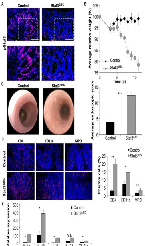
propria and colonic epithelial cells as demonstrated above, phosphorylated Stat3 was detectable in lamina propria cells but not IECs of infected Stat3ΔIECmice (Fig. 2a). IEC-specific Stat3 con-ditional knockout mice were highly susceptible toC.rodentiuminfection. Stat3ΔIECmice signif-icantly lost more body weight than controls and showed a general deterioration in health of these animals, as evidenced by a lack of activity and ruffled fur (Fig. 2b). While control mice de-veloped mild intestinal inflammation, infected Stat3ΔIECmice showed severe signs of colonic inflammation with high granularity, strong formation of fibrin and diarrhea (Fig. 2c). The dif-ference in extent of colitis was confirmed by statistical analysis of endoscopic colitis scores (Fig. 2c). In agreement with endoscopic analysis, immunofluorescence stainings of gut cross-sections revealed prominent infiltrations of CD4 positive and CD11c positive immune cells into the lamina propria of infected Stat3ΔIECmice (Fig. 2d). Moreover, Stat3ΔIECmice showed increased expression of inflammatory cytokines such as IL-1α, IL-1β, IL-6 and TNF-α
(Fig. 2e). Together, our results show that a defect in activating Stat3 in intestinal epithelial cells leads to the development of a strikingly more severe intestinal inflammation after infection withC.rodentiumas compared to wildtype mice.
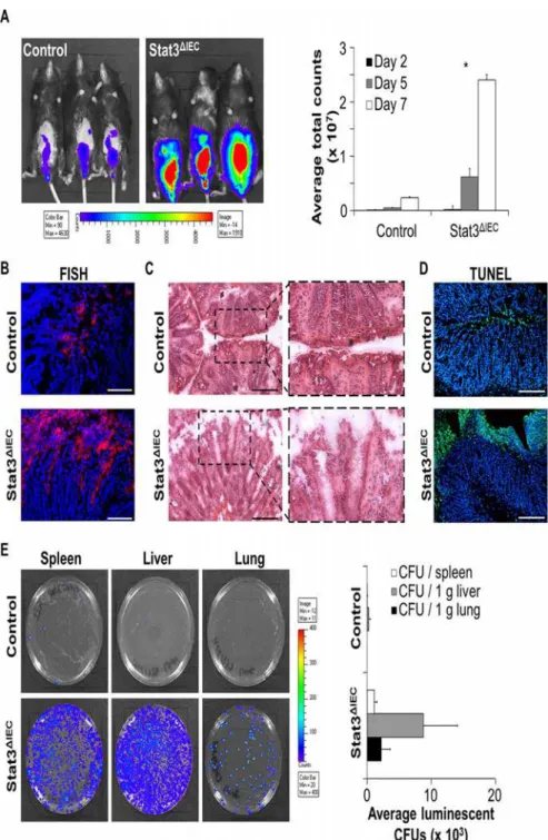
In line with increased intestinal inflammation in Stat3ΔIECmice, we detected enlarged infec-tion loads ofC.rodentiumbyin vivoimaging in the gut of these mice (Fig. 3a). To investigate bacterial colonization of the gut in a more detailed fashion, we performed FISH (fluorescence in situ hybridization) of colonic cross-sections and observed an increased amount of bacteria directly adherent to the intestinal epithelial surface (Fig. 3b). Remarkably, in contrast to control mice, where bacteria remained on the surface of the epithelial brush border, bacteria in infected Stat3ΔIECdeficient mice penetrated deeply into the crypts. These data suggest the inflammatory phenotype observed in infected Stat3ΔIECmice to be aggravated by the excessively increased amount of bacteria adherent to the bowel wall and indicate defects in intestinal epithelial barrier function.
In fact, we demonstrate a disrupted intestinal surface epithelium in the colon of infected Stat3ΔIECmice by H&E staining (Fig. 3c). SinceC.rodentiumcan induce apoptosis in intestinal epithelial cell lines [22], we investigated whetherC.rodentiuminfected Stat3ΔIECmice show in-creased epithelial cell death. TUNEL (TdT-mediated dUTP nick-end labeling) staining re-vealed excessive cell death of intestinal epithelial cells inC.rodentiuminfected Stat3ΔIECmice (Fig. 3d). Cell death predominantly occurred within the surface epithelium ofC.rodentium in-fected Stat3ΔIECmice directly exposed to the lumen, suggesting that Stat3 is required to control
C.rodentiuminduced epithelial cell death and to maintain barrier integrity.
A serious complication of human gastrointestinal infections is systemic spread. To investi-gate whether the proposed barrier defects lead to translocation of bacteria from the lumen into the bowel wall and to distant sites, we harvested spleen, liver, and lung tissue and plated ho-mogenized samples on LB-agar plates. AlthoughC.rodentiumhas been reported to be a non-invasive pathogen [23], invasion ofC.rodentiuminto all investigated organs of Stat3ΔIECmice was observed (Fig. 3e). In line with this observation, Stat3ΔIECmice not only showed severe in-testinal inflammation but signs of sepsis such as dramatic weight loss, ruffled fur and lack of physical activity.
Collectively, these data demonstrate the essential requirement of Stat3 activation in IECs for the control ofC.rodentiumgrowth and for maintaining intestinal epithelial barrier function to prevent systemic spreading of bacteria.
Fig 2. Stat3ΔIECmice are hypersusceptible toC. infection and develop severe colitis.A)
the gut and showed significantly decreased transcription of Pla2g5 compared to unchallenged control mice (Fig. 4a). Infection withC.rodentiumsignificantly induced the transcription of all the investigated antimicrobial peptide genes in the small intestine of control mice. However, induction of most AMPs was abrogated in infected Stat3ΔIECmice (Fig. 4a). Since RegIIIγwas recently described to promote host defense againstC.rodentium, we next performed immuno-histochemistry to investigate RegIIIγexpression in control and conditional Stat3 deficient mice. RegIIIγwas predominantly expressed at the crypt bottom of wildtype mice but was also detected in enterocytes and its expression strongly increased after infection withC.rodentium
(Fig. 4b). In contrast, almost no expression of RegIIIγin the gut of Stat3ΔIECmice was observed under unchallenged conditions and only small amounts were detected after infection (Fig. 4b). Thus, we hypothesize that Stat3 activation in IECs may drive the expression of AMPs and thereby controls growth of luminal bacteria.
We recently demonstrated Stat3 in intestinal epithelial cells to be activated via stimulation with IL-22 [9]. In agreement with the IL-22—pStat3—pathway driving RegIIIγexpression, phosphorylated Stat3 was detected in organoid epithelial cells simultaneously with high amounts of secreted RegIIIγafter IL-22 treatment (Fig. 4c). In fact, RegIIIγexpression in epi-thelial organoids was more than 300 fold increased after treatment with IL-22 (Fig. 4d). These data clearly show that IL-22 induces phosphorylation of Stat3 in intestinal epithelial cells fol-lowed by production of RegIIIγ.
The data obtained in the present study show that a defect in Stat3 activation leads to an im-paired transcription of antimicrobial peptides thereby facilitating overgrowth ofC.rodentium. The activation of Stat3 in the intestinal epithelium via IL-22 maintains the epithelial barrier function under stress conditions and prevents translocation ofC.rodentiumto systemic sites.
Discussion
Intestinal infections lead to production of various cytokines and chemokines by lamina propria immune cells. The secreted cytokines not only influence other immune cells but provide con-stant information about the gut health status to the epithelial barrier and induce adequate epi-thelial responses [24]. Maintenance of intact intestinal epithelial barrier function is known to be crucial for combating intestinal infections. Recently, IL-22 has been found to play a major role in infections with the EHEC/EPEC like pathogenC.rodentiumin mice [14]. In line with this report, we also demonstrated strong induction of IL-22 in the gut of mice infected with
C.rodentium. However, our data go beyond the role of IL-22 and for the first time directly demonstrate a functional role of Stat3 signaling in intestinal epithelial cells for control of gas-trointestinal infectionsin vivo. Our study collectively shows that epithelial Stat3 concerts the host response to bacterial infection by controlling bacterial growth and suppression of apopto-sis to sustain intestinal epithelial barrier function. Interestingly, while our data together with the study published by Zheng et al. demonstrate the IL-22—Stat3 axis to be important in relative to day 0. Data show mean values +/- SEM (n = 4 control mice, n = 6 Stat3ΔIEC
mice). C) Left: Endoscopic pictures ofC.rodentiuminfected control and Stat3ΔIECmice show severe signs of colonic inflammation in infected Stat3ΔIECmice. Right: Severity of colitis was quantified by endoscopic scoring. Data show mean values + SEM (n = 3 mice in each group). D) Left: Representative pictures of colonic cross-sections derived from infected control and Stat3ΔIEC
mice stained for CD4 (red), CD11c (red) and MPO (red). Nuclei were stained with Hoechst (blue). Scale bars: 200μm. Right: Software-based calculation of the proportion of CD4+cells, CD11c+cells, and MPO+cells in immunofluorescence pictures stained for the designated cells. Data show mean values + SEM. E) Transcription of cytokines in the colon ofC.rodentium infected control and Stat3ΔIEC
mice. Data were calculated relative to infected control and show mean values + SEM (n = 4 mice in each group).Hprtwas used as internal standard. Four independent experiments were performed with similar results on each occasion.
Fig 3. Breakdown of intestinal epithelial barrier in infected Stat3ΔIECmice facilitates systemic spread
ofC.rodentium.A) Left: Visualization of luminescentC.rodentiumin live mice by IVIS on day six after infection. Right: Quantification ofC.rodentiuminfestation by measuringC.rodentiumluminescence at different time points after infection of control and Stat3ΔIECmice. Data show mean values + SEM (n = 5 mice in each group). B) Fluorescencein situhybridization (FISH) of bacterial RNA (red) present in colonic cross-section. Nuclei are shown in blue. Scale bars: 50μm. C) H&E staining of colonic cross-sections from infected control and Stat3ΔIECmice. Scale bars: 200μm. D) Colon cross-sections derived fromC.rodentiuminfected control and Stat3ΔIEC
mice were stained for cell death by TUNEL (green). Nuclei were counterstained with Hoechst (blue). Scale bars: 200μm. E)C.rodentiuminfestation of different organs in infected control and Stat3ΔIECmice was assessed by plating organ lysates on agar plates (luminescent CFU indicateC.rodentiumcolonies) (left) and counting luminescent CFUs (right) the next day. Data show mean values + SEM (n = 5 control mice, n = 4 Stat3ΔIEC
mice). Representative pictures are shown.
Fig 4. Impaired expression of AMPs in Stat3ΔIECmice.A) Quantitative transcription analysis was
combatingC.rodentiuminfections, IL-22 was shown to be detrimental in case ofSalmonella enterica serovar Typhimuriuminfection [25]. It was concluded that IL-22 induced upon
S.Typhimuriuminfection can be exploited by this pathogen to suppress the commensal bacte-rial flora by induction of antimicrobial peptides responsible for metal ion starvation. A strong impact of antimicrobials induced by Stat3 activation on the gut flora is also supported by our study as unlike control mice,C.rodentiumsensitive Stat3ΔIECmice showed no induction of RegIIIγ, RegIIIβand Pla2g2a after infection. Remarkably, expression of RegIIIγand RegIIIβ
was completely diminished in Stat3ΔIECmice, indicating that Stat3 is a key regulator of these AMPs. Although these data might indicate impaired killing ofC.rodentiumin Stat3ΔIECmice, an alternative hypothesis is thatC.rodentiumin Stat3ΔIECmice can capture ecological niches that might have developed from altered AMP expression in Stat3 deficient mice, similarly to the study by Behnsen and colleagues [25]. In support of the hypothesis, Zheng and coworkers demonstrated that administration of exogenous mouse or human RegIIIγ, which acts on gram-positive bacteria [26], to IL-22 deficient mice infected with gram-negativeC.rodentium, partly protected these mice from gastroenteritis [14]. Interestingly, some patients with inflammatory bowel disease also show decreased expression of some AMPs [27]. However, it is still not clear, whether impaired AMP expression is cause or consequence of the disease. Thus, the present study suggests that at least for infections with gastrointestinal pathogens in mice, deficiency in the production of AMPs predispose to a severe course of the disease.
We hypothesize that the altered expression of antimicrobial peptides in the gut of Stat3ΔIEC mice favoredC.rodentiumovergrowth and further might facilitate deep penetration of bacteria into the crypt lumen. Indeed, Stat3ΔIECmice showed abundant bacterial invasion into the lower crypt compartment. Usually, the crypt lumen constitutes a rather bacterial free environment [28] whose colonization would cause significant dangers. Our data support this conclusion since spread ofC.rodentiumto distant organs followed by a septic phenotype was observed in Stat3ΔIECmice after infection. SinceC.rodentiumis described as a non-invasive pathogen [23], we suggested death of Stat3 deficient IECs to cause breakdown of the intestinal epithelial barrier integrity allowing invasion ofC.rodentium. However, since increased apoptosis was only de-tected in infected but not unchallenged Stat3ΔIECmice, increased death of Stat3 deficient intesti-nal epithelial cells might only occur under challenged conditions such asC.rodentiuminfection. In fact,C.rodentiumhas been described to induce cell death in intestinal epithelial cell lines [22].
In conclusion, we showed that activation of Stat3 in the intestinal epithelium is a key regula-tor during gastrointestinal infection. Stat3 activation ameliorates intestinal inflammation and prevents systemic spreading of bacteria by controlling growth of bacteria via regulation of AMP expression and by maintaining intestinal epithelial barrier function (Fig. 5). Thus, Stat3 orchestrates the epithelial response to gastrointestinal infections on multiple levels.
Acknowledgments
The authors thank Dr. Christian Riedel (University Ulm) for kindly providing luminescent
C.rodentiumstrain ICC169, Julia Schuster and Christina Lindner for excellent technical assistance. C.rodentiuminfected Stat3ΔIEC
mice). B) Representative pictures of small intestinal cross-sections derived from unchallenged and infected control and Stat3ΔIECmice were stained for RegIIIγ(red). Nuclei were stained with Hoechst (blue). Scale bars: 50μm. C) Representative pictures of wildtype organoids treated with or without IL-22 for 24 hours and stained for pStat3 (red) and RegIIIγ(green) using immunohistochemistry. Nuclei were stained with Hoechst (blue). Scale bars: 50μm. D) Transcription of RegIIIγin organoids treated with IL-22 for 24 hours or left untreated (n = 6 from 3 different experiments). Gene transcription is normalized toHprt. Data show mean values + SEM.
Author Contributions
Conceived and designed the experiments: NW CB. Performed the experiments: NW GP UB MM EM ML. Analyzed the data: NW MFN CB. Contributed reagents/materials/analysis tools: SW MM ML. Wrote the paper: NW CB.
References
1. Kosek M, Bern C, Guerrant RL. The global burden of diarrhoeal disease, as estimated from studies be-tween 1992 and 2000. Bull World Health Organ. 2003; 81: 197–204. PMID:12764516
2. Clarke SC, Haigh RD, Freestone PP, Williams PH. Virulence of enteropathogenic Escherichia coli, a global pathogen. Clin Microbiol Rev. 2003; 16: 365–378. PMID:12857773
3. Kaper JB, Nataro JP, Mobley HL. Pathogenic Escherichia coli. Nat Rev Microbiol. 2004; 2: 123–140.
PMID:15040260
4. Robert Koch-Institut. Abschließende Darstellung und Bewertung der epidemiologischen Erkenntnisse im EHEC O104:H4 Ausbruch, Deutschland 2011. Available:http://www.rki.de/DE/Content/InfAZ/E/ EHEC/EHEC_O104/EHEC-Abschlussbericht.html. Accessed 26 May 2014.
5. Wong CS, Jelacic S, Habeeb RL, Watkins SL, Tarr PI. The risk of the hemolytic-uremic syndrome after antibiotic treatment of Escherichia coli O157:H7 infections. N Engl J Med. 2000; 342: 1930–1936.
PMID:10874060
Fig 5. Model of Stat3 activation in intestinal epithelial cells controlling intestinal infections.Activation of Stat3 in intestinal epithelial cells controls bacterial growth and prevents systemic spread of bacteria by regulating the transcription of AMPs and maintaining intestinal epithelial barrier function. Black arrows indicate secretion by and effect of IL-22 on cells. Orange arrows indicate effects of Stat3 activation. DC = dendritic cell, IECs = intestinal epithelial cells, IL-22 = Interleukin-22, ILC = innate lymphoid cells, NK = natural killer cell, TH17 = T helper cell 17, DC = dendritic cell,
PMN = polymorphonuclear neutrophil.
6. Eisenhauer PB, Harwig SS, Lehrer RI. Cryptdins: antimicrobial defensins of the murine small intestine. Infect Immun. 1992; 60: 3556–3565. PMID:1500163
7. O'Neil DA, Porter EM, Elewaut D, Anderson GM, Eckmann L, Ganz T, et al. Expression and regulation of the human beta-defensins hBD-1 and hBD-2 in intestinal epithelium. J Immunol. 1999; 163: 6718–6724. PMID:10586069
8. Iimura M, Gallo RL, Hase K, Miyamoto Y, Eckmann L, Kagnoff MF. Cathelicidin mediates innate intesti-nal defense against colonization with epithelial adherent bacterial pathogens. J Immunol. 2005; 174: 4901–4907. PMID:15814717
9. Pickert G, Neufert C, Leppkes M, Zheng Y, Wittkopf N, Warntjen M, et al. STAT3 links IL-22 signaling in intestinal epithelial cells to mucosal wound healing. J Exp Med. 2009; 206: 1465–1472. doi:10.1084/
jem.20082683PMID:19564350
10. Mudter J, Weigmann B, Bartsch B, Kiesslich R, Strand D, Galle PR, et al. Activation pattern of signal transducers and activators of transcription (STAT) factors in inflammatory bowel diseases. Am J Gas-troenterol. 2005; 100: 64–72. PMID:15654782
11. Neufert C, Pickert G, Zheng Y, Wittkopf N, Warntjen M, Nikolaev A, et al. Activation of epithelial STAT3 regulates intestinal homeostasis. Cell Cycle. 2010; 9: 652–655. PMID:20160497
12. Barrett JC, Hansoul S, Nicolae DL, Cho JH, Duerr RH, Rioux JD, et al. Genome-wide association de-fines more than 30 distinct susceptibility loci for Crohn's disease. Nat Genet. 2008; 40: 955–962. doi:
10.1038/ng.175PMID:18587394
13. Franke A, Balschun T, Karlsen TH, Hedderich J, May S, Lu T, et al. Replication of signals from recent studies of Crohn's disease identifies previously unknown disease loci for ulcerative colitis. Nat Genet. 2008; 40: 713–715. doi:10.1038/ng.148PMID:18438405
14. Zheng Y, Valdez PA, Danilenko DM, Hu Y, Sa SM, Gong Q, et al. Interleukin- 22 mediates early host defense against attaching and effacing bacterial pathogens. Nat Med. 2008; 14: 282–289. doi:10.
1038/nm1720PMID:18264109
15. Graham AC, Carr KD, Sieve AN, Indramohan M, Break TJ, Berg RE. IL-22 production is regulated by IL-23 during Listeria monocytogenes infection but is not required for bacterial clearance or tissue pro-tection. PLoS One. 2011; 6: e17171. doi:10.1371/journal.pone.0017171PMID:21347242
16. Zindl CL, Lai JF, Lee YK, Maynard CL, Harbour SN, Ouyang W, et al. IL-22-producing neutrophils con-tribute to antimicrobial defense and restitution of colonic epithelial integrity during colitis. Proc Natl Acad Sci U S A. 2013; 110: 12768–71273. doi:10.1073/pnas.1300318110PMID:23781104
17. Veldhoen M, Hirota K, Westendorf AM, Buer J, Dumoutier L, Renauld JC, et al. The aryl hydrocarbon receptor links TH17-cell-mediated autoimmunity to environmental toxins. Nature. 2008; 453: 106–109.
doi:10.1038/nature06881PMID:18362914
18. Liang SC, Tan XY, Luxenberg DP, Karim R, Dunussi-Joannopoulos K, Collins M, et al. Interleukin (IL)-22 and IL-17 are coexpressed by Th17 cells and cooperatively enhance expression of antimicrobial peptides. J Exp Med. 2006; 203: 2271–2279. PMID:16982811
19. Satoh-Takayama N, Vosshenrich CA, Lesjean-Pottier S, Sawa S, Lochner M, Rattis F, et al. Microbial flora drives interleukin 22 production in intestinal NKp46+ cells that provide innate mucosal immune de-fense. Immunity. 2008; 29: 958–970. doi:10.1016/j.immuni.2008.11.001PMID:19084435
20. Cella M, Fuchs A, Vermi W, Facchetti F, Otero K, Lennerz JK, et al. A human natural killer cell subset provides an innate source of IL-22 for mucosal immunity. Nature. 2009; 457: 722–725. doi:10.1038/
nature07537PMID:18978771
21. Sawa S, Lochner M, Satoh-Takayama N, Dulauroy S, Bérard M, Kleinschek M, et al. RORγt+ innate lymphoid cells regulate intestinal homeostasis by integrating negative signals from the symbiotic micro-biota. Nat Immunol. 2011; 12: 320–326. doi:10.1038/ni.2002PMID:21336274
22. Flynn AN, Buret AG. Tight junctional disruption and apoptosis in an in vitro model of Citrobacter roden-tium infection. Microb Pathog. 2008; 45: 98–104. doi:10.1016/j.micpath.2007.12.004PMID:18504087
23. Eckmann L. Animal models of inflammatory bowel disease: lessons from enteric infections. Ann N Y Acad Sci. 2006; 1072: 28–38. PMID:17057188
24. Wittkopf N, Neurath MF, Becker C. Immune-epithelial crosstalk at the intestinal surface. J Gastroen-terol. 2014; 49: 375–387. doi:10.1007/s00535-013-0929-4PMID:24469679
25. Behnsen J, Jellbauer S, Wong CP, Edwards RA, George MD, Ouyang W, et al. The cytokine IL-22 promotes pathogen colonization by suppressing related commensal bacteria. Immunity. 2014; 40: 262–273. doi:10.1016/j.immuni.2014.01.003PMID:24508234
26. Cash HL, Whitham CV, Behrendt CL, Hooper LV. Symbiotic bacteria direct expression of an intestinal bactericidal lectin. Science. 2006; 313: 1126–1130. PMID:16931762
28. Elphick DA, Mahida YR. Paneth cells: their role in innate immunity and inflammatory disease. Gut. 2005; 54: 1802–1809. PMID:16284290
29. Riedel CU, Casey PG, Mulcahy H, O'Gara F, Gahan CG, Hill C. Construction of p16Slux, a Novel Vec-tor for Improved Bioluminescent Labeling of Gram-Negative Bacteria. Appl Environ Microbiol. 2007; 73: 7092–7095. PMID:17766445
30. Neurath MF, Wittkopf N, Wlodarski A, Waldner M, Neufert C, Wirtz S, et al. Assessment of tumor development and wound healing using endoscopic techniques in mice. Gastroenterology. 2010; 139: 1837–1843. doi:10.1053/j.gastro.2010.10.007PMID:20955705
31. Sato T, Vries RG, Snippert HJ, van de Wetering M, Barker N, Stange DE, et al. Single Lgr5 stem cells build crypt-villus structures in vitro without a mesenchymal niche. Nature. 2009; 459: 262–265. doi:10.
1038/nature07935PMID:19329995
32. Becker C, Wirtz S, Blessing M, Pirhonen J, Strand D, Bechthold O, et al. Constitutive p40 promoter acti-vation and IL-23 production in the terminal ileum mediated by dendritic cells. J Clin Invest. 2003; 112: 693–706. PMID:12952918
33. Rasband WS. ImageJ, U S National Institutes of Health, Bethesda, Maryland, USA. 1997–2008;