Inflammatory Response to
Ureaplasma
spp. and Other
Bacteria
Marian Kacerovsky1,2*, Peter Celec3, Barbora Vlkova3, Kristin Skogstrand4, David M. Hougaard4, Teresa Cobo5, Bo Jacobsson6,7
1Biomedical Research Center, University Hospital Hradec Kralove, Hradec Kralove, Czech Republic,2Department of Obstetrics and Gynecology, Charles University in Prague, Faculty of Medicine Hradec Kralove, Hradec Kralove, Czech Republic,3Institute of Molecular Biomedicine, Comenius University in Bratislava, Bratislava, Slovak Republic,4Department of Clinical Biochemistry and Immunology, Statens Serum Institut, Copenhagen, Denmark,5Maternal Fetal Medicine Department, Hospital Clinic, IDIBAPS, Barcelona, Spain,6Department of Obstetrics and Gynecology, Sahlgrenska University Hospital, Gothenburg, Sweden,7Institute of Public Health, Oslo, Norway
Abstract
Objective:This study aimed to evaluate the amniotic fluid protein profiles and the intensity of intraamniotic inflammatory response toUreaplasmaspp. and other bacteria, using the multiplex xMAP technology.
Methods: A retrospective cohort study was undertaken in the Department of Obstetrics and Gynecology, University Hospital Hradec Kralove, Czech Republic. A total of 145 pregnant women with preterm prelabor rupture of membranes between gestational age 24+0 and 36+6 weeks were included in the study. Amniocenteses were performed. The presence
ofUreaplasmaspp. and other bacteria was evaluated using 16S rRNA gene sequencing. The levels of specific proteins were determined using multiplex xMAP technology.
Results:The presence ofUreaplasmaspp. and other bacteria in the amniotic fluid was associated with increased levels of interleukin (IL)-6, IL-8, IL-10, brain-derived neurotropic factor, granulocyte macrophage colony stimulating factor, monocyte chemotactic protein-1, macrophage inflammatory protein-1, and matrix metalloproteinasis-9.Ureaplasmaspp. were also associated with increased levels of neurotropin-3 and triggering receptor expressed on myeloid cells-1.
Conclusions:The presence ofUreaplasmaspp. in the amniotic fluid is associated with a slightly different protein profile of inflammatory response, but the intensity of inflammatory response toUreaplasmaspp. is comparable with the inflammatory response to other bacteria.
Citation:Kacerovsky M, Celec P, Vlkova B, Skogstrand K, Hougaard DM, et al. (2013) Amniotic Fluid Protein Profiles of Intraamniotic Inflammatory Response to
Ureaplasmaspp. and Other Bacteria. PLoS ONE 8(3): e60399. doi:10.1371/journal.pone.0060399
Editor:Colette Kanellopoulos-Langevin, Institut Jacques Monod - UMR 7592 CNRS - Universite´ Paris Diderot, France
ReceivedOctober 10, 2012;AcceptedFebruary 25, 2013;PublishedMarch 26, 2013
Copyright:ß2013 Kacerovsky et al. This is an open-access article distributed under the terms of the Creative Commons Attribution License, which permits unrestricted use, distribution, and reproduction in any medium, provided the original author and source are credited.
Funding:This work was supported by a grant from the Ministry of Health, Czech Republic (NT13461- 4/2012) and by REVOGENE project, ITMS 6240220067, supported by the Research & Development Operational Programme funded by ERDF. Additional support came from Swedish government grants to researchers in the public health service (ALF) (ALFGBG-136431), Sahlgrenska University Hospital, Sahlgrenska Academy, Gothenburg, Sweden, the Swedish Medical Society, Stockholm, Sweden (2008-21198) and the Jane and Dan Olsson Research Foundation, Gothenburg, Sweden. The funders had no role in study design, data collection and analysis, decision to publish, or preparation of the manuscript.
Competing Interests:The authors have declared that no competing interests exist.
* E-mail: [email protected]
Introduction
Microbial invasion of the amniotic cavity (MIAC) is often found in women with preterm prelabor rupture of membranes (PPROM). MIAC is detected in approximately 20% to 50% of these patients, depending on the testing method [1,2]. Although cultivation of amniotic fluid is still considered the gold standard for the identification of MIAC, the use of molecular techniques enable the detection of uncultivated or difficult-to-cultivate bacteria as well.
A variety of microorganisms have been isolated from the amniotic fluid in pregnancies complicated by both PPROM and preterm labor, Ureaplasmaspp. being the most prevalent [1,2,3].
Ureaplasmaspp. account for up to 60% of all detected bacteria in the amniotic fluid of women with PPROM, depending on the use
of cultivation-based or the more sensitive non-cultivation-based techniques [2]. Ureaplasma spp. can colonize the choriodecidual space without eliciting an inflammatory response due to their low pathogenicity and the immunosuppressive properties of the choriodecidua [4]. On the other hand, when Ureaplasma spp. appear inside the amniotic cavity, they can induce a strong intraamniotic inflammatory response with the development of histological chorioamnionitis and even signs of a maternal inflammatory response [2,5]. In addition, current studies have shown thatUreaplasmaspp. have a capacity similar to that of other bacteria to induce both intraamniotic and maternal inflammatory responses [4,5].
[6,7,8]. In addition, recent ex vivo explant model studies have suggested that cytokine and protein response to bacteria seems to be pathogen dependent [9,10,11,12].
Therefore, the main aim of this study was to evaluate the diversity between the amniotic fluid protein profiles of intraam-niotic inflammatory response to Ureaplasma spp. and those concerning other bacteria by employing multiplex xMAP tech-nology. A second aim was to compare the intensity of intraamniotic inflammatory responses evoked by Ureaplasmaspp. with that produced by other bacteria.
Materials and Methods
Sample collection
Between July 2008 and October 2010, we conducted a prospective cohort study of women with PPROM between 24+0 and 36+6 weeks’ gestation, who were admitted to the Department of Obstetrics and Gynecology, University Hospital Hradec Kralove, Czech Republic. Women of maternal age $18 years and with a singleton pregnancy were eligible for enrollment in the study. The exclusion criteria were as follows: presence of maternal complications (i.e., hypertension, preeclampsia, diabetes mellitus, and thyroid disease), ultrasound signs of fetal growth restriction, vaginal bleeding, signs of fetal hypoxia, and structural malforma-tions or chromosomal abnormalities of the fetus. Gestational age was established using the first trimester ultrasound evaluation for all pregnancies.
PPROM was defined as the leakage of amniotic fluid prior to the onset of labor (by at least two hours). This condition was diagnosed using a sterile speculum examination, which confirmed the pooling of amniotic fluid in the vagina, in association with a positive test for the presence of insulin-like growth factor binding protein (ACTIM PROM test; Medix Biochemica, Kauniainen, Finland) in the vaginal fluid. Ultrasound-guided transabdominal amniocentesis was performed on admission prior to the admin-istration of corticosteroids, antibiotics, or tocolytics, and approx-imately 5 mL of amniotic fluid were aspirated and divided into two tubes.
The study was approved by the University Hospital Hradec Kralove review board committee (March 19, 2008; No. 200804 SO1P), and written informed consent was obtained from all participants.
Amniotic fluid analyses
First tubes of uncentrifuged amniotic fluid were transported to the laboratory for DNA isolation and 16S rRNA gene sequencing. Protease inhibitors (CompleteTM Mini, EDTA-free Protease Inhibitor Cocktail; Roche Diagnostics, Basel, Switzerland) were added (40mL per 1 mL of amniotic fluid) to the second tubes, which were centrifuged for 15 minutes at 3006gto remove cells
and debris, filtered (0.22mm) using a Syringe-driven filter (TPP, Trasadingen, Switzerland), divided into aliquots, and stored at 270uC until analysis.
The levels of interleukin (IL)-1b, IL-6, IL-8, IL-10, IL-12, IL-17, IL-18, soluble IL-6 receptor a, adiponectin, brain-derived neurotropic factor (BDNF), C-reactive protein (CRP), granulocyte macrophage colony stimulating factor (GM-CSF), insulin-like growth factor-binding protein (IGFBP)-1, IGFBP-3, interferon-c, leptin, monocyte chemotactic protein-1 (MCP-1), migration inhibiting factor (MIF), macrophage inflammatory protein-1a
(MIP-1a), matrix metalloproteinasis-9 (MMP-9), neutropin-3 (NT-3), regulated on activation normal T-expressed and secreted (RANTES), tumor necrosis factor (TNF)-a, TNF-b, soluble tumor necrosis factor receptor-1 (sTNF-R1), and triggering receptor
expressed on myeloid cells-1 (TREM-1) in the amniotic fluid were analyzed using a multiple sandwich immunoassay based on flowmetric Luminex xMAP technology, in accordance with previously published papers[13,14,15]. Only proteins with detect-able amniotic fluid levels in more than 50% of the samples were included in the analyses. The following eight proteins were excluded from further analyses because of low levels: IL-1b
detected in 17%, IL-12 in 9%, IL-17 in 8%, IL-18 in 13%, interferon-cin 0%, RANTES in 17%, TNF-ain 10%, and TNF-b
in 11%.
Detection of bacteria in amniotic fluid
DNA was extracted from the samples using a QIAmp DNA Mini kit according to the instructions of the manufacturer (Qiagen, Hilden, Germany). The isolated DNA was used as a template for polymerase chain reaction (PCR) of the 16S rRNA gene with primers at conserved sites flanking a variable sequence used for terminal fragment length restriction polymorphism analysis: 27F (59-AGAGTTTGATCCTGGCTCAG-39) and 1492R (59 -GGTTACCTTGTTACGACTT-39). This assay is widely used in microbiological research to study complex samples as part of the terminal restriction fragment length polymorphism (tRFLP). The primers as well as the assay are routinely used in a number of applications including the characterization of microflora of the nasal and oral cavity, gastric microflora, and the microflora of the gut [16,17,18,19,20]. The method has been extensively optimized on bacterial cultures and saliva [21,22,23]. The resulting protocol was used in this study. The endpoint PCR analysis was performed using the PCR instrument Eppendorf Mastercycler ep (Eppendorf AG, Hamburg, Germany). The amplification primers were designed to detect fragment of the prokaryotic 16S rRNA gene. Sequences of the used primers were described previously [22]. The reactions were set up in a total volume of 20mL, containing 106Taq Buffer, dNTP Mix (0.2 mM of each), 1.0mM of each primer, 1.5 mM of MgCl2, 1.25 U of Taq DNA Polymerase (Thermo Scientific), and 1.5mL of the template. Cycling conditions were 95uC for 15 min, followed by 35 cycles of three-step cycling of 94uC for 30 sec, 50uC for 30 sec, 72uC for 120 sec; with final polymerization step 12 min at 72uC). PCR products were checked using agarose gel electrophoresis, purified by ethanol precipitation, and sequenced on an ABI 3100 genetic analyzer (Life Technologies, Carlsbad, USA). Chromas 2.33 (Technelysium Pty Ltd, South Brisbane, Australia) and the online basic local alignment search tool (BLAST) algorithm were used for sequence analysis [24]. Based on the sequencing run either the resulting sequence was used for a BLAST search or if the run was unreadable with superimposed sequences read in parallel, an infection with multiple species was diagnosed.
Statistical analysis
Results
Demographical and clinical characteristic
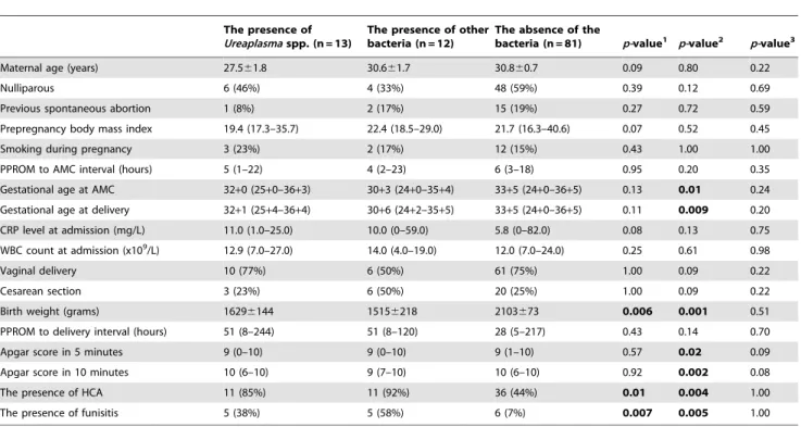
A total of 145 women with PPROM were recruited during the study period. Of these women, only 106 (73%) were included in the analyses, because aliquots of amniotic fluid samples were not available for 39 (20%) women. Of the 106 women, 25 (24%) presented with positive 16S rRNA in the amniotic fluid. Sequencing revealedUreaplasmaspp. in 12% of the women (13/ 106). Other bacteria were detected in 12% (12/106). Polymicro-bial finding (Sneathia sanguinegens and Leptotrichia amnionii) was detected in the amniotic fluid of one woman. Table 1 presents the microorganisms identified in the amniotic fluid from women included in the analyses. Table 2 shows the demographic and clinical characteristics of women withUreaplasmaspp., of women with other bacteria, and of women without any bacteria in the amniotic fluid. Women with either Ureaplasma spp. or other bacteria had lower birth weight of newborn, and a higher rate of histological chorioamnionitis and funisitis. In addition, women with other bacteria in the amniotic fluid had lower gestational age at sampling and delivery. No differences were found between the groups withUreaplasmaspp. and with other bacteria. All women in the study were Caucasian, as defined by self-report.
Table 1.The bacteria found in the amniotic fluid using 16S rRNA gene sequencing.
The microorganisms Number of cases
Ureaplasmaspp. 13
Granulicatella elegans 1
Haemophilus influenzae 2
Lactobacillus crispatus 1
Lactobacillus gallinarum 1
Leptotrichia amnionii 2
Peptostreptococcus anaerobicus 1
Sneathia sanguinegens 1
Streptococcus agalactiae 2
Streptococcus pneumoniae 1
Staphylococcusspp. 1
The variables are presented as number.
Two microorganisms in the amniotic fluid were detected in one woman (Sneathia sanguinegens+Leptotrichia amnionii).
doi:10.1371/journal.pone.0060399.t001
Table 2.Maternal and newborn characteristics based on the presence ofUreaplasmaspp. and other bacteria, and absence of the bacteria in the amniotic fluid.
The presence of Ureaplasmaspp. (n = 13)
The presence of other bacteria (n = 12)
The absence of the
bacteria (n = 81) p-value1 p-value2 p-value3
Maternal age (years) 27.561.8 30.661.7 30.860.7 0.09 0.80 0.22
Nulliparous 6 (46%) 4 (33%) 48 (59%) 0.39 0.12 0.69
Previous spontaneous abortion 1 (8%) 2 (17%) 15 (19%) 0.27 0.72 0.59
Prepregnancy body mass index 19.4 (17.3–35.7) 22.4 (18.5–29.0) 21.7 (16.3–40.6) 0.07 0.52 0.45
Smoking during pregnancy 3 (23%) 2 (17%) 12 (15%) 0.43 1.00 1.00
PPROM to AMC interval (hours) 5 (1–22) 4 (2–23) 6 (3–18) 0.95 0.20 0.35
Gestational age at AMC 32+0 (25+0–36+3) 30+3 (24+0–35+4) 33+5 (24+0–36+5) 0.13 0.01 0.24
Gestational age at delivery 32+1 (25+4–36+4) 30+6 (24+2–35+5) 33+5 (24+0–36+5) 0.11 0.009 0.20
CRP level at admission (mg/L) 11.0 (1.0–25.0) 10.0 (0–59.0) 5.8 (0–82.0) 0.08 0.13 0.75
WBC count at admission (x109/L) 12.9 (7.0–27.0) 14.0 (4.0–19.0) 12.0 (7.0–24.0) 0.25 0.61 0.98
Vaginal delivery 10 (77%) 6 (50%) 61 (75%) 1.00 0.09 0.22
Cesarean section 3 (23%) 6 (50%) 20 (25%) 1.00 0.09 0.22
Birth weight (grams) 16296144 15156218 2103673 0.006 0.001 0.51
PPROM to delivery interval (hours) 51 (8–244) 51 (8–120) 28 (5–217) 0.43 0.14 0.70
Apgar score in 5 minutes 9 (0–10) 9 (0–10) 9 (1–10) 0.57 0.02 0.09
Apgar score in 10 minutes 10 (6–10) 9 (7–10) 10 (6–10) 0.92 0.002 0.08
The presence of HCA 11 (85%) 11 (92%) 36 (44%) 0.01 0.004 1.00
The presence of funisitis 5 (38%) 5 (58%) 6 (7%) 0.007 0.005 1.00
Abbreviations: PPROM-preterm prelabor rupture of membranes; AMC-amniocentesis; CRP–C-reactive protein; WBC–white blood cells; HCA–histological chorioamnionitis.
p-value1the comparison between groups with the presence ofUreaplasmaspp. and without bacteria in amniotic fluid. p-value2the comparison between groups with the presence of other bacteria and without bacteria in amniotic fluid. p-value3the comparison between groups with the presence of
Ureaplasmaspp. and other bacteria in amniotic fluid.
Continuous variables were compared using parametric t-test (presented as mean6SEM) or a nonparametric Mann-WhitneyUtest [presented as median (range)]. Categorical variables were compared using Fisher exact test and presented as number (%).
Amniotic fluid protein profile of women withUreaplasma
spp.
The presence of Ureaplasma spp. in the amniotic fluid was associated with changes in 10 of 18 measured amniotic fluid proteins. Amniotic fluid levels of IL-6, IL-8, IL-10, BDNF, GM-CSF, MCP-1, MIP-1a, MMP-9, NT-3, and TREM-1 were higher in women with Ureaplasma spp. in the amniotic fluid compared with women with no bacteria in the amniotic fluid (see Table 3). The results are not adjusted for gestational age at sampling because no difference was found.
Amniotic fluid protein profile of women with other bacteria
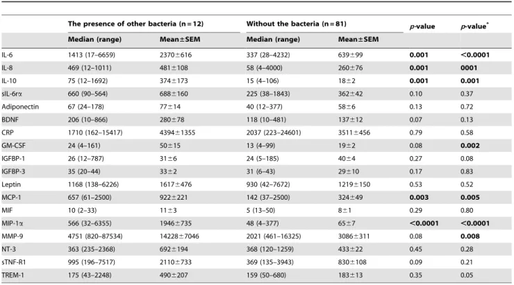
The presence of other bacteria in the amniotic fluid was related to changes in the amniotic fluid levels of five and seven proteins in crude analysis and adjusted analysis for gestational age at sampling, respectively (see Table 4). The amniotic fluid levels of IL-6, IL-8, IL-10, GM-CSF, MCP-1, MIP-1a, and MMP-9 were higher in women with other bacteria compared with those without bacteria in the amniotic fluid after adjustment for gestational age at sampling.
Intensity between intraamniotic and maternal
inflammatory response evoked byUreaplasma spp. and other bacteria
Only those proteins that showed changes in the presence of eitherUreaplasmaspp. or other bacteria in the amniotic fluid (IL-6, IL-8, IL-10, BDNF, GM-CSF, MCP-1, MIP-1a, MMP-9, NT-3, and TREM-1) were included in the analysis. No differences in
their levels were observed between women withUreaplasmaspp. and those with other bacteria (see Table 5). Furthermore, we did not find any difference between maternal serum CRP level and white blood cell count (see Table 2).
Discussion
A recent study showed that different types of bacteria evoke varied inflammatory responses in an explant model with fetal membranes [9]. Therefore, we hypothesized that the inflamma-tory response to different types of bacteria could be associated with diverse protein profiles that play a role in this process. The diversity of bacteria found in the amniotic fluid from pregnancies complicated by PPROM prevented us from creating more than two main cohorts of women with bacteria in their amniotic fluid. These two main cohorts consisted of women withUreaplasmaspp. and women with bacteria other thanUreaplasmaspp.
The following are the key findings from this study: i) the presence ofUreaplasmaspp. in the amniotic fluid is associated with a slightly different, more extensive protein profile of inflammatory response; ii) the changes in amniotic fluid IL-6, IL-8, IL-10, GM-CSF, MCP-1, MIP-1a, and MMP-9 are common for both
Ureaplasma spp. and other bacteria; and iii) the intensity of inflammatory response toUreaplasmaspp. is fully comparable with that to bacteria other thanUreaplasmaspp.
Different types of bacteria have been found to produce varied inflammatory responses in an explant model with fetal membranes [9,10,11,12]. Menon et al. found an association between the stimulation of fetal membranes by Ureaplasma parvum and an increase in TNF-a and IL-10, but not in IL-1b, IL-6, IL-8, and interferon-c levels in tissue explant model. On the other hand, Table 3.Amniotic fluid levels of selected proteins in women with the presence ofUreaplasmaspp. and without the bacteria in the amniotic fluid.
The presence ofUreaplasmaspp. (n = 13) Without bacteria (n = 81) p-value
Median (range) Mean±SEM Median (range) Mean±SEM
IL-6 1567 (61–5090) 21936463 337 (28–4232) 639699 ,0.0001
IL-8 456 (4–4000) 9366385 58 (4–4000) 260676 0.002
IL-10 183 (4–2199) 4696190 15 (4–106) 1862 ,0.0001
sIL-6ra 523 (65–2987) 8876262 225 (38–1843) 362642 0.09
Adiponectin 56 (4–357) 116631 40 (12–377) 5866 0.14
BDNF 269 (35–930) 308667 118 (10–481) 137612 0.002
CRP 3357 (1278–17786) 465161214 2037 (223–24601) 35116456 0.06
GM-CSF 38 (4–97) 3967 13 (4–99) 1962 0.002
IGFBP-1 33 (12–173) 48614 24 (5–185) 4064 0.79
IGFBP-3 30 (22–39) 3262 31 (6–43) 29610 0.49
Leptin 818 (234–4564) 12106332 930 (42–7672) 12196150 0.81
MCP-1 846 (38–2500) 10236261 142 (37–2500) 324649 0.004
MIF 7 (2–50) 1163 5 (13–50) 861 0.55
MIP-1a 718 (96–2567) 10596264 48 (4–377) 6567 ,0.0001
MMP-9 4193 (956–68255) 1496665425 2021 (461–16325) 30866311 0.004
NT-3 551 (217–957) 571671 368 (120–1259) 423622 0.04
sTNF-R1 1090 (249–4717) 14506388 369 (135–3943) 8306108 0.05
TREM-1 354 (114–2790) 5206195 159 (50–680) 183613 0.001
Continuous variables were compared using a nonparametric Mann-WhitneyUtest [presented as median (range) and mean6SEM]. Adiponectin, IGFB-1, IGFB-3, and MIF are presented in ng/mL; other mediators are showed in pg/mL.
stimulations by other bacteria, such asEscherichia coli, Streptococcus agalactiae, andGardanerella vaginalis, were related to higher levels of IL-1b, IL-6, and IL-10[9]. With this knowledge, we hypothesized that the presence of bacteria with a higher virulence potential in the amniotic fluid would be associated with a more pronounced
and extensive protein profile of intraamniotic inflammatory response.
Innate immunity provides prompt protection against the threat of invading bacteria and other microorganisms. Specific motifs on bacterial surfaces are recognized by pattern-recognition receptors, Table 4.Amniotic fluid levels of selected proteins in women with other bacteria and without bacteria in the amniotic fluid.
The presence of other bacteria (n = 12) Without the bacteria (n = 81) p-value p-value*
Median (range) Mean±SEM Median (range) Mean±SEM
IL-6 1413 (17–6659) 23706616 337 (28–4232) 639699 0.001 ,0.0001
IL-8 469 (12–1011) 4816108 58 (4–4000) 260676 0.001 0001
IL-10 75 (12–1692) 3746173 15 (4–106) 1862 0.001 0.001
sIL-6ra 660 (90–564) 6886160 225 (38–1843) 362642 0.10 0.37
Adiponectin 67 (24–178) 77614 40 (12–377) 5866 0.13 0.72
BDNF 206 (10–866) 280678 118 (10–481) 137612 0.07 0.13
CRP 1710 (162–15417) 439461355 2037 (223–24601) 35116456 0.79 0.58
GM-CSF 24 (4–161) 50615 13 (4–99) 1962 0.08 0.002
IGFBP-1 26 (12–787) 3166 24 (5–185) 4064 0.27 0.08
IGFBP-3 35 (20–44) 3362 31 (6–43) 29610 0.17 0.83
Leptin 1168 (138–6226) 16176476 930 (42–7672) 12196150 0.53 0.52
MCP-1 657 (61–2500) 9226221 142 (37–2500) 324649 0.003 0.005
MIF 10 (2–33) 1163 5 (13–50) 861 0.29 0.80
MIP-1a 566 (32–6355) 19466735 48 (4–377) 6567 ,0.0001 ,0.0001
MMP-9 4751 (820–87534) 1422867046 2021 (461–16325) 30866311 0.08 0.008
NT-3 363 (235–2368) 6926194 368 (120–1259) 433622 0.45 0.28
sTNF-R1 995 (196–7517) 21106733 369 (135–3943) 8306108 0.09 0.21
TREM-1 175 (43–2248) 4906207 159 (50–680) 183613 0.35 0.05
Continuous variables were compared using a nonparametric Mann-WhitneyUtest [presented as median (range) and mean6SEM].
p-value the comparison between groups with the presence of other bacteria and without bacteria in amniotic fluid.
p-value* Spearman partial correlation was performed to adjust by gestational age at sampling. Adiponectin, IGFB-1, IGFB-3, and MIF are presented in ng/mL; other mediators are showed in pg/mL. Statistically significant differences are marked in bold.
doi:10.1371/journal.pone.0060399.t004
Table 5.Amniotic fluid levels of proteins involved in intraamniotic inflammatory responses toUreaplasmaspp. and other bacteria.
The presence ofUreaplasmaspp. (n = 13) The presence of other bacteria (n = 12) p-value
Median (range) Mean±SEM Median (range) Mean±SEM
IL-6 1567 (61–5090) 21936463 1413 (17–6659) 23716616 1.00
IL-8 456 (4–4000) 9366349 469 (12–1011) 4826108 0.98
IL-10 183 (4–2199) 4696190 75 (12–1692) 1862 0.66
sIL-6ra 523 (65–2987) 8876262 660 (90–564) 6886160 1.00
BDNF 269 (35–930) 308667 206 (10–866) 280678 0.45
GM-CSF 38 (4–97) 3967 24 (4–161) 50615 0.94
MCP-1 846 (38–2500) 10236261 657 (61–2500) 9226221 0.98
MIP-1a 718 (96–2567) 10596264 566 (32–6355) 19466735 0.83
MMP-9 4193 (956–68255) 1496665425 4751 (820–87534) 1422867046 0.62
NT-3 551 (217–957) 571671 363 (235–2368) 6926194 0.45
TREM-1 354 (114–2790) 5206195 175 (43–2248) 4906207 0.21
Continuous variables were compared using a nonparametric Mann-WhitneyUtest [presented as median (range) and mean6SEM].
p-value the comparison between groups with the presence ofUreaplasmaspp. and other bacteria in amniotic fluid. Levels of mediators are showed in pg/mL.
and the activation of these receptors leads to the release of cytokines, chemokines, and other inflammatory mediators. Our results demonstrate that the protein profiles of intraamniotic inflammatory response toUreaplasmaspp. and to other bacteria are very similar, involving cytokines (IL-6, IL-10, and GM-CSF), chemokines [IL-8 (CXCL8 chemokine), MCP-1 (CCL2 chemo-kine), and MIP-1a(CCL3 chemokine)], and proteins involved in the breakdown of the extracellular matrix (MMP-9). A solid body of evidence indicates that IL-6, IL-8, and IL-10, along with IL-1b
and TNF-a(unfortunately, the levels of the latter two were below the detection limit in more than 50% of the cases), play a central role in the mechanism of intraamniotic inflammation [25,26]. GM-CSF is a cytokine with the ability to generate monocytes, neutrophils, and other granulocytes from precursor cells. There-fore, its higher levels could be one explanation for the observed local accumulation of these cells at sites of inflammation [27]. We found chemokines to be responsible for recruitment of first-line (IL-8, MIP-1a) and second-line (MCP-1) defense cells. Neutrophils that belong to the first line of the defense mechanism against invading microorganisms are thought to play an important role in the secretion of MMP-9, which is released following stimulation by IL-8 and other inflammatory mediators [28,29]. Moreover, MMP-9 plays an important role in inflammation due to the degradation of matrix protein and further activation of cytokines and chemokines [30]. Two specific proteins, NT-3 and TREM-1, were involved only in the intraamniotic inflammatory response to
Ureaplasma spp. in the amniotic fluid. Their exact roles in this pathway remain unclear.
In a study by Oh et al., it was proposed that genital mycoplasmas (Ureaplasma spp. and Mycoplasma hominis) exhibit more intense intraamniotic and maternal inflammatory responses than other microorganisms. We did not find any difference between the intensity of intraamniotic inflammatory response, as determined by the levels of 10 specific proteins, toUreaplasmaspp. and other bacteria. Moreover, no difference was revealed in maternal inflammatory response as well. We assume that the employment of different approaches for the detection of bacteria (cultivation vs. non-cultivation) may explain this discrepancy, making comparison of the results difficult.
In our study, MIAC was revealed in 24% women with PPROM. This rate is lower compare to data presented in the recent study by DiGiulio et al., where bacteria were identified in 45% of PPROM pregnancies [31,32]. Nevertheless, the results cannot be directly compared between their and our study.
DiGiulio et al. included women with PPROM with gestational age between 15+0236+6 weeks and they also included women with different ethnic background [31]. In our study, we only included Caucasian women with gestational age between 24+0236+6 weeks. Lower gestational age is associated with higher prevalence of MIAC in PPROM pregnancies. We think that differences in gestational age and ethnicity can partly explain the differences in rates of MIAC.
The strength of this study is that it provides new information concerning the amniotic fluid protein profile based on the levels of 18 mediators in response toUreaplasmaspp. and other bacteria in the amniotic cavity. Another important strength is that microbial invasion of the amniotic cavity was evaluated using non-cultivation techniques for bothUreaplasmaspp. and other bacteria. A unique aspect of this study is the precise preanalytic preparation of the amniotic fluid samples, which were collected using the same technique and examiners in a single institution. However, this study has some limitations. First, the sample size of the subgroups was relatively small. Second, we are aware the fact that our approach for revealing bacteria in the amniotic fluid is not optimal for the description of polymicrobial finding. The rate of polymicrobial finding identified in our study is lower than published previously [31,32]. However, the unraveling of amniotic fluid microbiome of pregnancies complicated by PPROM is beyond the scope of this study. Third, molecular methods can reveal the presence of bacterial DNA in the amniotic fluid regardless of its abundance; we did not quantify the amounts of bacteria in the amniotic fluid because the using of non-specific PCR approach. Fourth, we did not take into consideration the presence of viruses in the amniotic fluid.
In conclusion, Ureaplasma spp. elicit a protein profile of intraamniotic inflammatory response (determined by the amniotic fluid levels of 18 cytokines and mediators) that is slightly different from that of other bacteria. Nevertheless, the intensity of inflammatory response betweenUreaplasmaspp. and other bacteria seems to be very similar, when bacteria are detected using non-cultivation techniques.
Author Contributions
Conceived and designed the experiments: MK PC BV KS DH TC BJ. Performed the experiments: PC BV KS DH. Analyzed the data: MK TC BJ. Contributed reagents/materials/analysis tools: MK PC BV KS DH. Wrote the paper: MK.
References
1. Jacobsson B, Mattsby-Baltzer I, Andersch B, Bokstrom H, Holst RM, et al. (2003) Microbial invasion and cytokine response in amniotic fluid in a Swedish population of women with preterm prelabor rupture of membranes. Acta Obstet Gynecol Scand 82: 423–431.
2. Kacerovsky M, Pliskova L, Bolehovska R, Musilova I, Hornychova H, et al. (2011) The microbial load with genital mycolasmas correlated with the degree of histologic chorioamnionitis in preterm PPROM. Am J Obstet Gynecol. 2011; 205: 213.e1–e7.
3. Jacobsson B, Mattsby-Baltzer I, Andersch B, Bokstrom H, Holst RM, et al. (2003) Microbial invasion and cytokine response in amniotic fluid in a Swedish population of women in preterm labor. Acta Obstet Gynecol Scand 82: 120– 128.
4. Aaltonen R, Heikkinen J, Vahlberg T, Jensen J, Alanen A (2007) Local inflammatory response in choriodecidua induced by Ureaplasma urealyticum. BJOG 114: 1432–1435.
5. Oh KJ, Lee KA, Sohn YK, Park CW, Hong JS, et al. (2010) Intraamniotic infection with genital mycoplasmas exhibits a more intense inflammatory response than intraamniotic infection with other microorganisms in patients with preterm premature rupture of membranes. Am J Obstet Gynecol 203: 211 e211–218.
6. Kacerovsky M, Musilova I, Khatibi A, Skogstrand K, Hougaard DM, et al. (2012) Intraamniotic inflammatory response to bacteria: analysis of multiple
amniotic fluid proteins in women with preterm prelabor rupture of membranes. J Matern Fetal Neonatal Med 2012, ; doi: 10.3109/14767058.2012.671873. 7. Cobo T, Kacerovsky M, Holst RM, Hougaard DM, Skogstrand K, et al. (2012)
Intra-amniotic inflammation predicts microbial invasion of the amniotic cavity but not spontaneous preterm delivery in preterm prelabor membrane rupture. Acta Obstet Gynecol Scand 2012, ; doi: 10.1111/j.1600–0412.2012.01427.x. 8. Holst RM, Hagberg H, Wennerholm UB, Skogstrand K, Thorsen P, et al.
(2011) Prediction of microbial invasion of the amniotic cavity in women with preterm labour: analysis of multiple proteins in amniotic and cervical fluids. BJOG 118: 240–249.
9. Menon R, Peltier MR, Eckardt J, Fortunato SJ (2009) Diversity in cytokine response to bacteria associated with preterm birth by fetal membranes. Am J Obstet Gynecol 201: 306 e301–306.
10. Zaga-Clavellina V, Garcia-Lopez G, Flores-Herrera H, Espejel-Nunez A, Flores-Pliego A, et al. (2007) In vitro secretion profiles of interleukin (IL)-1beta, IL-6, IL-8, IL-10, and TNF alpha after selective infection with Escherichia coli in human fetal membranes. Reprod Biol Endocrinol 5:46.
12. Zaga-Clavellina V, Garcia-Lopez G, Flores-Espinosa P (2012) Evidence of in vitro differential secretion of human beta-defensins-1, -2, and -3 after selective exposure to Streptococcus agalactiae in human fetal membranes. J Matern Fetal Neonatal Med 25: 358–363.
13. Holst RM, Hagberg H, Wennerholm UB, Skogstrand K, Thorsen P, et al. (2011) Prediction of microbial invasion of the amniotic cavity in women with preterm labour: analysis of multiple proteins in amniotic and cervical fluids. BJOG 118: 240–249.
14. Holst RM, Hagberg H, Wennerholm UB, Skogstrand K, Thorsen P, et al. (2009) Prediction of spontaneous preterm delivery in women with preterm labor: analysis of multiple proteins in amniotic and cervical fluids. Obstet Gynecol 114: 268–277.
15. Vogel I, Goepfert AR, Thorsen P, Skogstrand K, Hougaard DM, et al. (2007) Early second-trimester inflammatory markers and short cervical length and the risk of recurrent preterm birth. J Reprod Immunol 75: 133–140.
16. Lemon KP, Klepac-Ceraj V, Schiffer HK, Brodie EL, Lynch SV, et al. (2010) Comparative analyses of the bacterial microbiota of the human nostril and oropharynx. J Mol Biol 215: 403–410.
17. He X, Hu W, He J, Guo L, Lux R, et al. (2011) Community-based interference against integration of Pseudomonas aeruginosa into human salivary microbial biofilm. Mol Oral Microbiol 26: 337–352.
18. Maldonado-Contreras A, Goldfarb KC, Godoy-Vitorino F, Karaoz U, Contreras M, et al. (2011) Structure of the human gastric bacterial community in relation to Helicobacter pylori status. ISME J 5: 574–579.
19. Shiga H, Kajiura T, Shinozaki J, Takagi S, Kinouchi Y, et al. (2012) Changes of faecal microbiota in patients with Crohn’s disease treated with an elemental diet and total parenteral nutrition. Dig Liver Dis 44: 736–742.
20. Nemoto H, Kataoka K, Ishikawa H, Ikata K, Arimochi H, et al. (2012) Reduced diversity and imbalance of fecal microbiota in patients with ulcerative colitis. Dig Dis Sci 57: 2955–2964.
21. Sakamoto M, Huang Y, Ohnishi M, Umeda M, Ishikawa I, et al. (2004) Changes in oral microbial profiles after periodontal treatment as determined by molecular analysis of 16S rRNA genes. J Med Microbil 53: 563–571. 22. Sakamoto M, Takeuchi Y, Umeda M, Ishikawa I, Benno Y (2003) Application of
terminal RFLP analysis to characterize oral bacterial flora in saliva of healthy subjects and patients with periodontitis. J Med Microbiol 52: 79–89. 23. Polgarova K, Behuliak M, Celec P (2010) Effect of saliva processing on bacterial
DNA extraction. New Microbiol 33: 373–379.
24. Altschul SF, Gish W, Miller W, Myers EW, Lipman DJ (1990) Basic local alignment search tool. J Mol Biol 215: 403–410.
25. Tashima LS, Millar LK, Bryant-Greenwood GD (1999) Genes upregulated in human fetal membranes by infection or labor. Obstet Gynecol 94: 441–449. 26. Romero R, Espinoza J, Goncalves LF, Kusanovic JP, Friel L, et al. (2007) The
role of inflammation and infection in preterm birth. Semin Reprod Med 25: 21– 39.
27. Hamilton JA (2002) GM-CSF in inflammation and autoimmunity. Trends Immunol 23: 403–408.
28. Xu X, Jackson PL, Tanner S, Hardison MT, Abdul Roda M, et al. (2011) A self-propagating matrix metalloprotease-9 (MMP-9) dependent cycle of chronic neutrophilic inflammation. PLoS One 6: e15781.
29. Chakrabarti S, Patel KD (2005) Regulation of matrix metalloproteinase-9 release from IL-8-stimulated human neutrophils. J Leukoc Biol 78: 279-288. 30. Van den Steen PE, Dubois B, Nelissen I, Rudd PM, Dwek RA, et al. (2002)
Biochemistry and molecular biology of gelatinase B or matrix metalloproteinase-9 (MMP-metalloproteinase-9). Crit Rev Biochem Mol Biol 37: 375–536.
31. DiGiulio DB, Romero R, Kusanovic JP, Gomez R, Kim CJ, et al. (2010) Prevalence and diversity of microbes in the amniotic fluid, the fetal inflammatory response, and pregnancy outcome in women with preterm pre-labor rupture of membranes. Am J Reprod Immunol 64: 38–57.