CLINICAL SCIENCE
Pleural tuberculosis: is radiological evidence of
pulmonary-associated disease related to the
exacerbation of the inflammatory response?
Leila Antonangelo,I,IIFrancisco S. Vargas,IJuliana Puka,IMa´rcia Seiscento,IMilena M. P. Acencio,ILisete R. Teixeira,I Ricardo M. Terra,III Roberta K. B. SalesI
IHospital das Clı´nicas da Faculdade de Medicina da Universidade de Sa˜o Paulo, Heart Institute (InCor), Pulmonary Division, Pleural Laboratory, Sa˜o Paulo/
SP, Brazil.IIHospital das Clı´nicas da Faculdade de Medicina da Universidade de Sa˜o Paulo, Department of Pathology, Clinical Laboratory and LIM III, Sa˜o Paulo/SP, Brazil.IIIHospital das Clı´nicas da Faculdade de Medicina da Universidade de Sa˜o Paulo, Heart Institute (InCor), Thoracic Surgery Division, Sa˜o Paulo/SP, Brazil.
OBJECTIVE:Pleural tuberculosis is the most frequently occurring form of extra pulmonary disease in adults. In up to 40% of cases, the lung parenchyma is concomitantly involved, which can have an epidemiological impact. This study aims to evaluate the pleural and systemic inflammatory response of patients with pleural or pleuropulmonary tuberculosis.
METHODS:A prospective study of 39 patients with confirmed pleural tuberculosis. After thoracentesis, a high resolution chest tomography was performed to evaluate the pulmonary involvement. Of the 39 patients, 20 exhibited only pleural effusion, and high resolution chest tomography revealed active associated-pulmonary disease in 19 patients. The total protein, lactic dehydrogenase, adenosine deaminase, vascular endothelial growth factor, interleukin-8, tumor necrosis factor-a, and transforming growth factor-b1levels were quantified in the patient serum and pleural fluid.
RESULTS: All of the effusions were exudates with high levels of adenosine deaminase. The levels of vascular endothelial growth factor and transforming growth factor-b1were increased in the blood and pleural fluid of all of
the patients with pleural tuberculosis, with no differences between the two forms of tuberculosis. The tumor necrosis factor-alevels were significantly higher in the pleural fluid of the patients with the pleuropulmonary form of tuberculosis. The interleukin-8 levels were high in the pleural fluid of all of the patients, without any differences between the forms of tuberculosis.
CONCLUSION:Tumor necrosis factor-awas the single cytokine that significantly increased in the pleural fluid of the patients with pulmonary involvement. However, an overlap in the results does not permit us to suggest that cytokine is a biological marker of concomitant parenchymal involvement. Although high resolution chest tomography can be useful in identifying these patients, the investigation of fast acid bacilli and cultures for M. tuberculosisin the sputum is recommended for all patients who are diagnosed with pleural tuberculosis.
KEYWORDS: Cytokines; Inflammation; Pleural Diseases; Tuberculosis.
Antonangelo L, Vargas FS, Puka J, Seiscento M, Acencio MM, Teixeira LR, Terra RM, Sales RK. Pleural tuberculosis: is radiological evidence of pulmonary-associated disease related to the exacerbation of the inflammatory response? Clinics. 2012;67(11):1259-1263.
Received for publication onApril 25, 2012;First review completed onMay 13, 2012;Accepted for publication onJuly 12, 2012
E-mail: [email protected]
Tel.: 55 11 2661-5695
INTRODUCTION
Pleural tuberculosis is the most common extra pulmonary form of tuberculosis in adults, particularly in countries where the prevalence of the disease is high or moderate (1-3). Although pulmonary tuberculosis can be a manifestation of primary infection, it is most commonly associated with the reactivation of pre-existing foci (1,2,4). It is generally
accepted that pleural tuberculosis results from a late hypersensitive reaction to the antigens of M. tuberculosis subsequent to the rupture of a subpleural caseous focus (1,2,4). The release of even a small number of bacilli from the lungs to the pleural space triggers a series of immune reactions that are mediated by T lymphocytes, which produce cytokines and stimulate macrophages to form a granuloma (4-7). These events trigger an inflammatory process in the pleural cavity: vascular permeability increases and an influx of leukocytes enter the pleural space, resulting in the accumulation of fluid and cells, which is a characteristic of the pleural exudates (1,2,6).
Cytokines are a group of polypeptides with multiple biological functions that act with other inflammatory mediators to coordinate the interaction between sensitized
Copyrightß2012CLINICS– This is an Open Access article distributed under
the terms of the Creative Commons Attribution Non-Commercial License (http:// creativecommons.org/licenses/by-nc/3.0/) which permits unrestricted non-commercial use, distribution, and reproduction in any medium, provided the original work is properly cited.
lymphocytes and macrophages to form granulomas (5-11). Among the involved mediators, we highlight the impor-tance of interleukin-8 (IL-8) (8-15), vascular endothelium growth factor (VEGF) (8,9,16-18), tumor necrosis factor-a
(TNF-a) (8,9,11,17-20), and transforming growth factor-b1
(TGF-b1) (7,9,20,21), all of which are considered to be key effectors in the inflammatory response of the pleural space (4-6).
Although pleural and pulmonary lesions were previously considered to be independent events, the advent of computed tomography has demonstrated concomitant lesions in more than 40% of cases (1,21-24). This concomitance may have an epidemiological impact, given that many patients with pleural tuberculosis are not adequately evaluated for intra-thoracic involvement. This finding enables us to speculate whether serum or pleural fluid inflammatory markers in pleural tuberculosis could suggest associated active pulmon-ary disease. The answer to this question is particularly relevant if we consider the potential risk of Mycobacterium tuberculosistransmission from these patients to contacts.
Therefore, this study compares the serum and pleural fluid expression of inflammatory mediators in pleural tuberculosis patients to evaluate whether pulmonary-asso-ciated involvement influences the magnitude of the inflam-matory response. We hypothesized that the more extensive the injury, the greater the inflammatory response. If this hypothesis is true, the inflammatory cytokines could be increased both locally (pleural fluid) and in the serum as a result of the pulmonary involvement.
METHODS
After approval by the local ethics committee, informed consent was obtained from 39 pleural tuberculosis patients. The patients were prospectively selected from the out-patient clinic of Pulmonary Diseases (InCor/FMUSP), Sa˜o Paulo, Brazil.
The diagnosis of pleural tuberculosis was based on the presence of a granuloma (upon pleural biopsy) associated with exudative effusion with increased adenosine deaminase (ADA. 40 IU/L) and/or a culture of the pleural fluid or fragment positive forMycobacterium tuberculosis(1,2,25). After thoracentesis, the patients underwent high resolution chest tomography (HRCT) and were subdivided into two groups based on the HRCT findings: pleuropulmonary (n = 19) or only pleural involvement (n = 20). The tomographic abnorm-alities that were suggestive of active pulmonary disease included the following: multi-segmental consolidation over the upper lung zones (homogenous opacity that reflected granulomatous inflammation of the parenchyma), thick-walled cavities (resulting from the coalescence of multiple inflammatory foci that necrotized and drained into the airways), centrilobular or confluent nodules, and the presence of the tree-in-bud pattern, which reflects the endobronchial dissemination of caseous necrosis and the granulomatous inflammation that fills and surrounds the alveolar ducts and respiratory bronchioli. Patients who had nonspecific tomo-graphic findings or images of residual scarring were not included in this series (23-25). HIV patients, patients previously treated for tuberculosis, and patients undergoing immunosuppressant therapy were also excluded.
Concomitant with thoracentesis, all of the patients under-went peripheral venous puncture and tuberculin skin tests. Pleural fluid samples were processed for cytology,
bacilloscopy,M. tuberculosiscultures, and to determine the adenosine deaminase levels (Giusti modified method) (26). Proteins (Biuret method) and lactic dehydrogenase (enzy-matic method) were also quantified in the blood and pleural fluid. Aliquots from both samples (serum and pleural fluid) were immediately centrifuged (1,500 rpm for 10 minutes at 4
˚
C), and the supernatant was stored at -80˚
C for posterior cytokine analyses.VEGF, IL-8, TNF-a, and TGF-b1were quantified using an
immunoenzymatic method (enzyme-linked immunosorbent assay, ELISA) according to the manufacturer’s instructions (R&D Systems Inc., Minneapolis, USA). The assays were performed in triplicate, and the results are expressed as means. The results were quantified by comparing the optic density (450 nm filter) in the ELISA reader (Powerwave, Biotek, USA) against a pre-established standard curve. The minimal detection values for IL-8, VEGF, and TGF-b1were
31 pg/mL, and the minimum detection value for TNF-awas 16 pg/mL. To evaluate the pleural inflammatory response, the results were compared with those obtained from the serum and pleural fluid of the patients with transudates that were caused by heart failure (27).
Statistical analysis
The results of the inflammatory marker analysis were compared using Student’s T-test or the Mann Whitney U-test according to the distribution of the variables. The SigmaStat 3.5 (SSI2006, California, USA) program was used for the statistical analyses, and the results are presented as medians (IQ 25th – 75th); p#0.05 was considered to be statistically significant.
RESULTS
Thirty-nine patients (35¡15 years) presented with pleural
effusions caused by tuberculosis, and 19 patients exhibited concomitant parenchyma involvement. In the patients with pleural effusion (n = 20), the detection of fast acid bacilli in the sputum was negative. In the pleuropulmonary group (n = 19), three patients exhibited positive fast acid bacilli in sputum. In two patients in the pleural group and six patients in the pleuropulmonary group, the response to the skin test exhibited indurations that were greater than 5 mm. Thus, the microbiological detection and the skin test contributed little toward diagnosing active pulmonary tuberculosis. The patients were not submitted to bronch-oalveolar lavage because they had previously been diag-nosed with pleural tuberculosis.
The HRCT findings that were considered to be suggestive of active pulmonary disease met the following criteria: the presence of centrilobular nodules (n = 14), confluent nodules (n = 11), tree-in-bud pattern (n = 15), pulmonary consolida-tion in the upper lung zones (n = 8), and thick-walled cavities (n = 3). Some patients exhibited more than one of these radiological changes.
represented the single parameter that was capable of differentiating between the two forms. IL-8 was increased in the pleural fluid of all the TB patients, and no differences in the levels of IL-8 were observed between the two forms. As expected, the protein and lactic dehydrogenase levels were higher in the serum and pleural fluid, respectively (Table 1; Figures 1 and 2).
DISCUSSION
The high expression of inflammatory mediators in the pleural fluid of pleural tuberculosis patients allows us to recognize the exudative inflammatory response at the site of active disease. The increased levels of VEGF and TGF-bin the serum of these patients reflect the systemic response
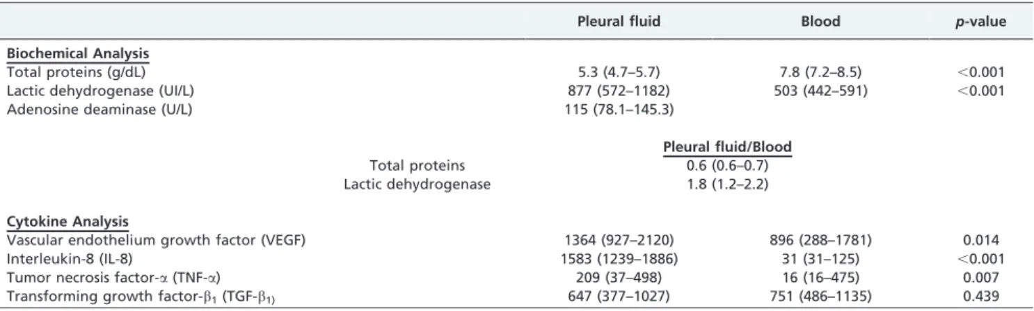
Table 1 -Results obtained in the patients with pleural effusion secondary to tuberculosis (n = 39). The data are expressed as medians (25thand 75thpercentiles).
Pleural fluid Blood p-value
Biochemical Analysis
Total proteins (g/dL) 5.3 (4.7–5.7) 7.8 (7.2–8.5) ,0.001
Lactic dehydrogenase (UI/L) 877 (572–1182) 503 (442–591) ,0.001
Adenosine deaminase (U/L) 115 (78.1–145.3)
Pleural fluid/Blood
Total proteins 0.6 (0.6–0.7)
Lactic dehydrogenase 1.8 (1.2–2.2)
Cytokine Analysis
Vascular endothelium growth factor (VEGF) 1364 (927–2120) 896 (288–1781) 0.014
Interleukin-8 (IL-8) 1583 (1239–1886) 31 (31–125) ,0.001
Tumor necrosis factor-a(TNF-a) 209 (37–498) 16 (16–475) 0.007
Transforming growth factor-b1(TGF-b1) 647 (377–1027) 751 (486–1135) 0.439
Figure 1 -Biochemical analysis of the fluid and blood of the patients with tuberculous pleural effusions. The data are expressed as medians: 25thand 75thpercentiles (*p
observed in cases of pleural tuberculosis. Although elevated TNF-a levels in the pleural fluid was the single parameter that was capable of differentiating between the two forms of the disease, the overlapping results do not suggest that this cytokine is a marker of pulmonary involvement.
Tuberculosis pleurisy is an acute and symptomatic disease that invariably evolves with exudative pleural effusions that are rich in cells and inflammatory mediators (1,2,4,7-10). Although tuberculosis pleurisy can indicate a manifestation of primary infection, it is most commonly associated with the reactivation of preexisting foci (1,2,4). Currently available laboratory methods frequently fail to demonstrate a possible concomitance with active pulmon-ary disease (1). In this regard, high resolution chest tomography has been helpful in identifying pulmonary lesions that are consistent with active disease in approxi-mately 40% of pleural tuberculosis patients (20-24). This finding has epidemiological implications, particularly if we consider that these patients are potential sources of infection and that their contacts are often not clinically or radiologi-cally evaluated. Therefore, we hypothesized that the patients with the pleuropulmonary form of tuberculosis could exhibit a greater inflammatory response because of the involvement of more than one anatomic site. This fact presumably determines systemic repercussions that could be detected by evaluating the serum and pleural inflamma-tory mediators.
Our findings corroborate previously described reports of increased inflammatory mediators in the pleural fluid of patients with pleural tuberculosis, thereby providing evi-dence of the compartmentalization of the inflammatory response at the active disease site (10,13,20,27). During the early phases of infection, after antigenic stimulation is triggered by the mycobacteria, cytokines such as VEGF, TNF-a, and IL-8 are transiently producedin vivo and alter the pleura permeability; this event precedes the exudative phase (20,28). However, the elevated serum levels of TNF-a
and TGF-bare thought to be related to prolonged antigenic stimulation, similar to observations in patients with pul-monary tuberculosis or late-diagnosed pleural tuberculosis (28-30).
In pleural tuberculosis, many cytokines are produced intracavitarially and modulate the inflammatory response that is triggered by the mycobacteria or its antigens to limit or aggravate the disease (7,19). Previous studies have related the findings of high serum or PF levels of TNF-a, TGF-b, and IL-8 to residual pleural thickening (19,20).
TGF-bis a mediator that is expressed in active infection and plays a fundamental role in the fibrotic scaring process. TGF-bis a key mediator in the immunopathogenesis of tuberculosis because it is able to modify the production and function of other cytokines, such as IL-1b and TNF-a, in addition to modulating the functions of T lymphocytes and macro-phages (7,20,29,30). In pleural tuberculosis, the excessive
production of TGF-bis believed to be related to the clinical progression of the disease, particularly in the physiopathol-ogy of pleural thickening (29). TGF-bpossesses proinflam-matory activity in low concentrations (pleural tuberculosis and healthy contacts of tuberculosis carries) and anti-inflammatory activity in high concentrations (pulmonary tuberculosis). We observed increased levels of TGF-bin the pleural fluid and blood of tuberculosis patients. Although higher TGF-blevels were observed in the pleuropulmonary form, there was no statistical significance when compared to the levels in patients with pleural disease.
Siawaya et al. (8) studied the cytokine and chemokine profiles of patients with different forms of tuberculosis to better understand the immunopathology of the disease and identify the biological markers that differentiate the various clinical forms of the disease. The authors concluded that systemic inflammatory markers, such as IL-8, TNF-a, and VEGF, are associated with pleural tuberculosis, whereas elevated levels of the factors involved in cell-mediated immunity, such as IL-12p40 and sCD40L, characterize pulmonary tuberculosis.
In conclusion, the present study demonstrates an intense intracavitary inflammatory response in the patients with pleural tuberculosis independent of parenchymal involve-ment. Although all of the cytokines were overexpressed in the pleural fluid, only TNF-awas significantly increased in the pleuropulmonary tuberculosis patients. This finding could suggest a more pronounced inflammatory response because of the concomitance of the anatomic sites involved. However, TNF-ashould not be considered to be a biological marker of pulmonary-associated disease. Although HRCT can be useful in identifying these patients, the identification of fast acid bacilli and/or the culture forM. tuberculosisin the sputum is recommended for all pleural tuberculosis patients.
ACKNOWLEDGMENTS
The study was supported by grants from the Fundac¸a˜o de Amparo a` Pesquisa do Estado de Sa˜o Paulo (FAPESP).
AUTHOR CONTRIBUTIONS
Antonangelo L, Vargas FS, Seiscento M, and Sales RK participated in the study design and conduction. Puka J, Acencio MM analyzed the patient files. Teixeira LR and Terra RM performed the statistical analysis. Antonangelo L, Vargas FS, Puka J, and Sales RK drafted and reviewed the manuscript. All of the authors reviewed and approved the manuscript.
REFERENCES
1. Light RW. Update on tuberculous pleural effusion. Respirology. 2010;15(3):451-8, http://dx.doi.org/10.1111/j.1440-1843.2010.01723.x. 2. Porcel JM. Tuberculous pleural effusion. Lung. 2009;187(5):263-70,
http://dx.doi.org/10.1007/s00408-009-9165-3.
3. Baumann MH, Nolan R, Petrini M, Lee YC, Light RW, Schneider E. Pleural tuberculosis in the United States: incidence and drug resistance. Chest. 2007;131(4):1125-32, http://dx.doi.org/10.1378/chest.06-2352. 4. Valdes L, Pose A, San Jose E, Martinez Vazquez JM. Tuberculous pleural
effusions. European journal of internal medicine. 2003;14(2):77-88, http://dx.doi.org/10.1016/S0953-6205(03)00018-9.
5. Cooper AM, Khader SA. The role of cytokines in the initiation, expansion, and control of cellular immunity to tuberculosis. Immunol Rev 2008;226:191-204, http://dx.doi.org/10.1111/j.1600-065X.2008. 00702.x.
6. Jantz MA, Antony VB. Pathophysiology of the pleura. Respiration. 2008;75(2):121-33, http://dx.doi.org/10.1159/000113629.
7. Antony VB. Immunological mechanisms in pleural disease. Eur Respir J. 2003;21(3):539-44, http://dx.doi.org/10.1183/09031936.03.00403902. 8. Siawaya JFD, Chegou NN, van den Heuvel MM, Diacon AH, Beyers N,
van Helden P, et al. Differential cytokine/chemokines and KL-6 profiles
in patients with different forms of tuberculosis. Cytokine. 2009;47(2):132-6, http://dx.doi.org/10.1016/j.cyto.2009.05.016.
9. Seiscento M, Vargas FS, Acencio MM, Teixeira LR, Capelozzi VL, Sales RK, et al. Pleural fluid cytokines correlate with tissue inflammatory expression in tuberculosis. Int J Tuberc Lung Dis. 2010;14(9):1153-8. 10. Akarsu S, Kurt ANC, Dogan Y, Yilmaz E, Godekmerdan A, Aygun AD.
The differential diagnostic values of cytokine levels in pleural effusions. Mediators Inflamm. 2005;2005(1):2-8, http://dx.doi.org/10.1155/ MI.2005.2.
11. Pokkali S, Das SD, Logamurthy R. Expression of CXC and CC type of chemokines and its receptors in tuberculous and non-tuberculous effusions. Cytokine. 2008;(3):307-14, http://dx.doi.org/10.1016/ j.cyto.2007.12.009.
12. Yang CS, Lee JS, Lee HM, Shim TS, Son JW, Jung SS, et al. Differential cytokine levels and immunoreactivities againstMycobacterium tubercu-losisantigens between tuberculous and malignant effusions. Respir Med. 2008;102(6)280-6, http://dx.doi.org/10.1016/j.rmed.2007.08.016. 13. Hoheisel G, Izbicki G, Roth M, Chan CHS, Leung JCK, Reichenberger F,
et al. Compartmentalization of pro-inflammatory cytokines in tubercu-lous pleurisy. Respir Med. 1998;92(1):14-7, http://dx.doi.org/10.1016/ S0954-6111(98)90025-7.
14. Saunders BM, Britton WJ. Life and death in the granuloma: immuno-pathology of tuberculosis. Immunology and cell biology. 2007;85(2):103-11, http://dx.doi.org/10.1038/sj.icb.7100027.
15. Park JS, Kim YS, Jee YK, Myong NH, Lee KY. Interleukin-8 production in tuberculous pleurisy: role of mesothelial cells stimulated by cytokine network involving tumour necrosis factor-alpha and interleukin-1 beta. Scandinavian journal of immunology. 2003;57(5):463-9, http:// dx.doi.org/10.1046/j.1365-3083.2003.01201.x.
16. Lim SC, Jung SI, Kim YC, Park KO. Vascular endothelial growth factor in malignant and tuberculous pleural effusions. J Korean Med Sci. 2000;15(3):279-83.
17. Momi H, Matsuyama W, Inoue K, Kawabata M, Arimura K, Fukunaga H, et al. Vascular endothelial growth factor and proinflammatory cytokines in pleural effusions. Respir Med. 2002;96(10):817-22, http:// dx.doi.org/10.1053/rmed.2002.1364.
18. Hamed EA, El-Noweihl AM, Mohamed AZ, Mahmoud A. Vasoactive mediators (VEGF and TNF-a) in patients with malignant and tubercu-lous pleural effusions. Respirology. 2004;9(1):81-6, http://dx.doi.org/ 10.1111/j.1440-1843.2003.00529.x.
19. Hua CC, Chang LC, Chen YC, Chang SC. Proinflammatory cytokines and fibrinolytic enzymes in tuberculous and malignant pleural effusions. Chest. 1999;116(5):1292-6, http://dx.doi.org/10.1378/chest.116.5.1292. 20. Olobo JO, Geletu M, Demissie A, Eguale T, Hiwot K, Aderaye G, et al.
Circulating TNF-a, TGF-b, and IL-10 in tuberculosis patients and healthy contacts. Scandinavian journal of immunology. 2001;53(1):85-91, http:// dx.doi.org/10.1046/j.1365-3083.2001.00844.x.
21. Valde´s L, Alvarez D, San Jose´ E, Penela P, Valle JM, Garcı´a-Pazos JM, et al. Tuberculous pleurisy: a study of 254 patients. Arch Intern Med. 1998;158(18):2017-21, http://dx.doi.org/10.1001/archinte.158.18.2017. 22. Kim HJ, Lee HJ, Kwon SY, Yoon HI, Chung HS, Lee CT, et al. The
prevalence of pulmonary parenchymal tuberculosis in patients with tuberculous pleuritis. Chest. 2006;129(5):1253-8, http://dx.doi.org/ 10.1378/chest.129.5.1253.
23. Nakanishi M, Demura Y, Ameshima S, Kosaka N, Chiba Y, Nishikawa S, et al. Utility of high-resolution computed tomography for predicting risk of sputum smear-negative pulmonary tuberculosis. Eur J Radiol. 2010;73(3):545-50, http://dx.doi.org/10.1016/j.ejrad.2008.12.009. 24. Seiscento M, Vargas FS, Bombarda S, Sales RK, Terra RM, Uezumi K,
et al. Pulmonary involvement in pleural tuberculosis: how often does it mean disease activity? Respir Med. 2011;105(7):1079-83, http:// dx.doi.org/10.1016/j.rmed.2011.02.014.
25. Chung KP, Chen JY, Lee CH, Wu HD, Wang JY, Lee LN, Yu CJ, Yang PC; TAMI Group. Trends and predictors of changes in pulmonary function after treatment for pulmonary tuberculosis. Clinics. 2011; 66(4):549-56, http://dx.doi.org/10.1590/S1807-59322011000400005. 26. Giusti G. Adenosine deaminase. In: Bergmeyer HU, editor. Methods of
Enzymatic Analysis. 1th ed. New York: Academic Press 1974.p.1092-9. 27. Marchi E, Acencio M, Antonangelo L, Sales RK, Vargas FS, Teixeira LR.
Citokines in pleural effusions. Am J Respir and Critical Care Medicine. 2002;v.168:p.A605.
28. Ruiz E, Alema´n C, Alegre J, Monasterio J, Segura RM, Armadans L, et al. Angiogenic Factors and angiogenesis inhibitors in exudative pleural effusions. Lung. 2005;183(3):185-95, http://dx.doi.org/10.1007/s00408-004-2533-0.
29. Ceyhan BB, Demiralp E, Karakurt ZL, Karakurt S, Sungur M. Transforming growth factor beta-1 level in pleural effusion. Respirology. 2003;8(3):321-5, http://dx.doi.org/10.1046/j.1440-1843.2003.00474.x.