Hawaiian Archipelago: Response, Outbreak
Status, Virulence, and a Method of
Treatment
Greta S. Aeby1,3*, Thierry M. Work2, Christina M. Runyon1,3, Amanda Shore-Maggio1,4, Blake Ushijima1,4, Patrick Videau4, Silvia Beurmann1,4, Sean M. Callahan1,3,4
1Hawai‘i Institute of Marine Biology, Kāne‘ohe, Hawaii, United States of America,2U.S. Geological Survey, National Wildlife Health Center, Honolulu Field Station, Honolulu, Hawaii, United States of America,3Marine Biology Graduate Program, University of Hawai‘i, Honolulu, Hawaii, United States of America,4Microbiology Department, University of Hawai‘i, Honolulu, Hawaii, United States of America
Abstract
A high number of coral colonies,Montiporaspp., with progressive tissue loss were reported
from the north shore of Kaua‘i by a member of the Eyes of the Reef volunteer reporting net-work. The disease has a distinct lesion (semi-circular pattern of tissue loss with an adjacent dark band) that was first observed in Hanalei Bay, Kaua‘i in 2004. The disease, initially termedMontiporabanded tissue loss, appeared grossly similar to black band disease
(BBD), which affects corals worldwide. Following the initial report, a rapid response was ini-tiated as outlined in Hawai‘i’s rapid response contingency plan to determine outbreak status and investigate the disease. Our study identified the three dominant bacterial constituents indicative of BBD (filamentous cyanobacteria, sulfate-reducing bacteria, sulfide-oxidizing bacteria) in coral disease lesions from Kaua‘i, which provided the first evidence of BBD in the Hawaiian archipelago. A rapid survey at the alleged outbreak site found disease to af-fect 6-7% of the montiporids, which is higher than a prior prevalence of less than 1% mea-sured on Kaua‘i in 2004, indicative of an epizootic. Tagged colonies with BBD had an average rate of tissue loss of 5.7 cm2/day over a two-month period. Treatment of diseased colonies with a double band of marine epoxy, mixed with chlorine powder, effectively re-duced colony mortality. Within two months, treated colonies lost an average of 30% less tis-sue compared to untreated controls.
Introduction
In the Caribbean, declines in live coral began in the late 1970s when disease outbreaks that af-fected the major reef-forming corals,Acropora palmataandA.cervicornis, were first reported [1]. Since that time, coral cover has declined precipitously from an average of 50% in the 1970s
OPEN ACCESS
Citation:Aeby GS, Work TM, Runyon CM, Shore-Maggio A, Ushijima B, Videau P, et al. (2015) First Record of Black Band Disease in the Hawaiian Archipelago: Response, Outbreak Status, Virulence, and a Method of Treatment. PLoS ONE 10(3): e0120853. doi:10.1371/journal.pone.0120853
Academic Editor:Christian R Voolstra, King Abdullah University of Science and Technology, SAUDI ARABIA
Received:October 30, 2014
Accepted:January 27, 2015
Published:March 16, 2015
Copyright:This is an open access article, free of all copyright, and may be freely reproduced, distributed, transmitted, modified, built upon, or otherwise used by anyone for any lawful purpose. The work is made available under theCreative Commons CC0public domain dedication.
Data Availability Statement:All 16S rRNA gene sequences and dsrA sequences identified in this study were deposited in the GenBank database under accession numbers KM924158-160, KJ914890-91, KM258122-127, KM924161-163, 165. All other relevant data are within the paper and its Supporting Information files.
to an average of 10% in 2002 [2] and coral diseases have contributed significantly to that de-cline [3]. Coral diseases are now degrading reefs of the Indo-Pacific [4–6]; numerous disease outbreaks have been reported from Australia [7,8], Philippines [9], Marshall Islands and Palau [10], and even the remote reefs of Palmyra Atoll [11]. The reefs of the Indo-Pacific appear to be starting down the same pathway of disease-induced destruction experienced by the reefs in the Caribbean and Florida Keys.
Within Hawai‘i, baseline information on coral diseases has been established; disease is wide-spread on reefs but occurs at a low prevalence [5,12,13]. However, disease outbreaks are starting to occur with increasing frequency. In 2003, an outbreak ofAcroporawhite syndrome caused mas-sive mortality of the table corals (Acropora cytherea) at French Frigate Shoals in the northwestern Hawaiian Islands (Papahānaumokuākea Marine National Monument) [14,15]. Additionally,
outbreaks ofMontiporawhite syndrome occurred in Kāne‘ohe Bay, O‘ahu in 2006 [16] and
‘Āhihi-Kīna‘u, Maui in 2008 [17]. Since coral diseases are an emerging issue in Hawai‘i, the State
developed a rapid response contingency plan (RRCP) to give managers the capacity to respond to bleaching or disease events in a timely and efficient manner [18]. Protocols were established to in-vestigate bleaching or disease events and aid in determining the significance, epizootiology, and potential causal linkages of outbreaks. A citizen science program, the Eyes of the Reef Network (http://eorhawaii.org/), was also developed to train community members in identifying and alert-ing managers to the occurrence of unusual disease events on Hawai‘i’s coral reefs.
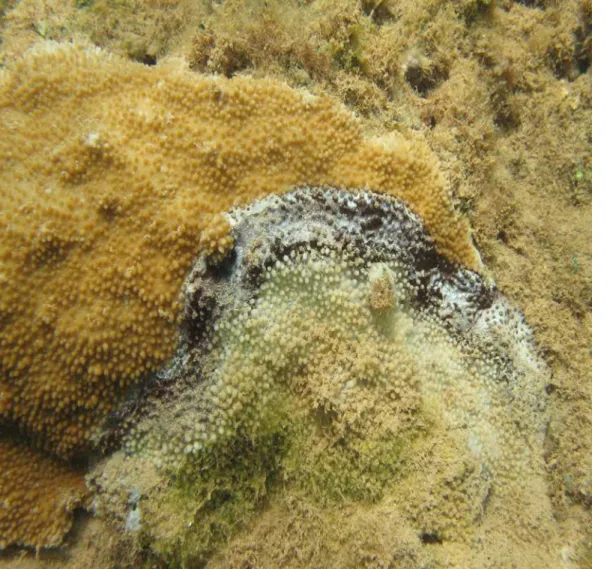
In the summer of 2012, a member of the Eyes of the Reef (EOR) network reported a high number of coral colonies,Montiporaspp. (M.capitata,M.patula,M.flabellata), on the north shore of Kaua‘i with progressive tissue loss. The corals had distinct disease lesions (semi-circular pattern of tissue loss with an adjacent dark band) (Fig. 1) that were first observed in Hanalei Bay, Kaua‘i in 2004 [19]. The lesion, initially termedMontiporabanded tissue loss, ap-peared grossly similar to black band disease (BBD), which affects numerous coral genera world-wide [3]. BBD is caused by a microbial consortium, visually dominated by filamentous
cyanobacteria, that creates the characteristic black band [20]. Other constituents of the BBD le-sion include sulfide-oxidizing bacteria (Beggiatoaspp.), sulfate-reducing bacteria that include members of theDesulfovibriogenus, and numerous heterotrophic bacteria [21–23]. The cyano-bacteria and sulfide-oxidizing cyano-bacteria exhibit vertical migration within the microbial mat under changing diel light conditions, which results in dynamic vertical microgradients in oxy-gen and sulfide [24,25]. The sulfate-reducing bacteria are responsible for the highly concentrat-ed sulfide and anoxic conditions underneath the BBD mat that is lethal to coral tissue [25,26]. Following the initial report by an EOR member, a rapid response was initiated, as outlined in Hawai‘i’s RRCP, to determine outbreak status and collect data for follow-up studies. The objec-tives of this study were to 1) conduct rapid surveys to determine outbreak status of the disease, 2) determine disease virulence by measuring rate of tissue loss on marked colonies, 3) test lesion occlusion as a method of disease treatment, and 4) determine whether the dominant microbial constituents of the disease lesion were consistent with BBD reported from other regions of the world. Our results indicate the outbreak status of the first report of BBD in the Hawaiian archi-pelago and show a successful method arresting progression of lesions. The ability to identify and respond quickly to this disease outbreak was facilitated by the prior development of pro-grams such as the Eyes of the Reef network and Hawai‘i’s Rapid Response Contingency Plan.
Methods
Ethics statement
This research was conducted under Special Activity Permit No. 2013–16 issued by the Hawai‘i Division of Aquatic Resources.
The funders had no role in study design, data collection and analysis, decision to publish, or preparation of the manuscript.
Rapid surveys
To determine disease prevalence (number of infected colonies/total colonies examined) at Anini, which is the reported outbreak site (22.227°N, 159.456°W), a diver swam from shore out to the reef crest and allMontiporaspp. colonies along an approximately 6 meter wide swath were examined and enumerated. All diseased colonies were photographed and enumer-ated. Surveys were conducted in October 2012.
Disease virulence and lesion occlusion for disease treatment
The rate of tissue loss on diseased colonies (n = 8) and colonies treated using the lesion occlu-sion method (n = 8) was measured by tagging and photographing colonies (M.capitata) though time. To measure disease progression, a band of marine epoxy (ZSPAR Splash Zone) was applied to the eight control colonies on bare skeleton roughly two centimeters behind the leading edge of the lesion. For the eight treatment colonies, the bacterial mat associated with disease lesions was loosened from the coral surface using the flat edge of a dive knife and re-moved by suction with a sterile 50cc syringe and used for identification of select bacteria as Fig 1.Montipora capitatacolony with signs of black band disease.
described below. To increase the probability of killing any potential bacterial pathogens, the marine epoxy was mixed with chlorine powder (calcium hypochlorite) (~15mL/ 50 mL epoxy), and then spread over the border of live tissue and bare skeleton (primary band). Another band of marine epoxy was applied to an area of healthy coral ca. two to five centimeters beyond the edge of the primary band as a“firebreak”or a second attempt to block disease progression if the primary band failed to halt disease progression (secondary band) (Fig. 2).
We conducted a preliminary trial using this double—band method for a tissue loss disease elsewhere and found that treatment was more effective if the epoxy was mixed with chlorine powder. There is the possibility that the chlorine can diffuse out of the marine epoxy and affect adjacent coral tissue. However, there was little mortality of coral tissue beyond the 2nd treat-ment bands suggesting, chlorine-impregnated epoxy caused minimal damage to the colony. This was confirmed after several months, whereupon corals were observed re-growing back over the epoxy, that itself, was colonized by filamentous and crustose coralline algae (Fig. 2) suggesting minimal longer-term toxicity.
All colonies (control and experimental) were photographed before and after marking or treatment and re-photographed two months later. All of the tagged colonies were flat, encrust-ing or platencrust-ing colonies, thereby allowencrust-ing rate of tissue loss per day to be measured digitally (Image J software, v1.46 NIH). The perimeter of the healthy tissue was digitally outlined and the encircled area calculated in cm2. Digital measurements were conducted in triplicate for each photograph and the averages were used for analysis.
Fig 2. The lesion occlusion method of disease treatment.A) InfectedM.capitatabefore treatment. B) Colony with marine epoxy over lesion and with a band of epoxy placed as a“firebreak”approximately 5 cm away. C) Same colony two months post-treatment. Note that the marine epoxy is overgrown with algae and the edge of the lesion is starting to grow over the epoxy. D) Control infectedM.capitatabefore marking with marine epoxy. E) Colony with marine epoxy approximately 5 cm behind lesion. F) Control colony after two months.
Identification of bacteria indicative of BBD
To determine if the dominant microbial constituents in BBD were present in the lesions char-acteristic ofMontiporabanded tissue loss, microscopy, culture, and PCR were used to identify representative filamentous cyanobacteria, sulfide-oxidizing bacteria, and sulfate-reducing bac-teria from lesion mabac-terial.
Isolation and growth of cyanobacteria.. Filamentous cyanobacteria identified under 10X
magnification were removed from disease samples using sterile forceps, rinsed thrice in auto-claved filtered seawater (FSW), plated onto the center of a plate of solid ASW:BG-11 medium [27], and incubated at 28°C under 2.0% CO2and 30 to 40μE/m2/sec cool white fluorescent
lighting. A subpopulation of filaments that had migrated to the periphery of the plate was transferred to fresh medium weekly. Bacteria were passaged five times before identification. Standard microscopy of filamentous bacteria was conducted [28].
Isolation and growth of sulfide-oxidizing bacteria.. White filamentous bacteria
contain-ing large inclusion bodies, grossly similar toBeggiatoaspp., were removed from disease sam-ples using a micropipettor and placed at the surface of J3 media deeps [29] within sterile polyethylene conicals, and buffered to pH 8.4 with Tris base, a medium designed to enrich sulfide-oxidizing bacteria. Inoculated J3 deeps were incubated at 28°C. Bacteria that were able to migrate into the J3 deep would indicate a motile bacterium capable of living in a micro-oxic, sulfide-rich environment, such asBeggiatoaspp.. Bacteria that migrated into the deep were re-isolated with a syringe penetrating through the side of the conical and re-inoculated onto the surface of fresh medium. Bacteria were passaged seven times before identification.
Molecular identification of bacteria.. To identify marine cyanobacteria, filaments either
removed directly from sampled lesions or strains cultured on ASW:BG-11, were boiled in sterile Milli-Q water for five minutes, centrifuged at 5000 x g for two minutes, and the supernatant was used as template DNA for PCR amplification of the16S rRNA genes using the cyanobacteria-specific primers CYA106F and CYA781R(a) [30]. PCR products from cyanobacterial filaments were sequenced directly using primer CYA106F.
To identify filamentous sulfide-oxidizing bacteria that were cultured as described above or taken directly from lesions, filaments were boiled in Milli-Q water for five minutes, centrifuged at 5000 x g for two minutes, and the supernatant was used as template DNA for PCR amplifica-tion of the 16S rRNA gene using universal bacterial primers 8F and 1513R [31].
ThedsrAgene encodes subunit A of dissimilatory sulfite reductase and has been used to in-dicate the presence of sulfate-reducing bacteria in environmental samples, including material from BBD [23,32–34]. Total DNA was extracted from coral lesion material using the Power-Soil DNA Extraction kit (MoBio) as per the manufacturer’s instructions. DNA from lesions was used as a template for PCR amplification of an approximately 200 bp region of thedsrA
gene using primers DSR1-F and DSR-R [32], which target a subset ofdsrAgene sequences pre-viously identified from sulfate-reducing bacteria [35] and fromDesulfovibrionacaeof BBD mi-crobial mats [23,32–34].
PCR products derived from the 16S rRNA gene of filamentous sulfide-oxidizing bacteria or
dsrAgenes of sulfur-reducing bacteria were cloned into theSmaI site of pBlueScript SK+ as previously described and sequenced using the primers M13F and M13R [36].
Data analyses
was healthy tissue at time zero (T0) and after two months (T1) was compared for treated and control corals.
Phylogenetic analysis of DNA sequences was conducted as previously described [37]. Briefly, sequences were aligned using BioEdit [38] and a maximum likelihood tree was constructed using the generalized time-reversible (GTR) algorithm [39] and 1000 bootstrap replicates were performed using MEGA5 [40]. All positions containing gaps and missing data were eliminated using the Maximum Likelihood method [41]. AlldsrAsequences were checked manually for chimeras using alignment in MEGA5 and BLAST before analysis. All 16S rRNA gene sequences anddsrAsequences identified in this study were deposited in the GenBank database under ac-cession numbers KM924158–60, KJ914890–91, KM258122–127, KM924161–163, 165.
Results
Rapid surveys
At Anini site one, 133 montiporid colonies were examined and 8 showed signs of the disease (prevalence = 6.0%). At Anini site two, 170 montiporids were examined and 13 were found in-fected (prevalence = 7.6%).
Rate of tissue loss in control and treated colonies
M.capitatacolonies treated with a double band of marine epoxy lost significantly less tissue as compared to the untreated control colonies.(Mann-Whitney test, W = 97.0, p = 0.003). Aver-age tissue loss was 1.1 (SE± 0.4) cm2/day for treated colonies compared to untreated colonies averaging 5.7 (SE±1.1) cm2of tissue loss per day.
Colony sizes were similar between groups with control colonies having an average total sur-face area (healthy and dead) of 692.7 cm2(range 148.6–2881.1 cm2) and treated colonies had an average of 703.4 cm2(range 72–1556.2 cm2). The average proportion of dead area on colonies at the beginning of the study was also similar in both control and treatment groups (9.7 ± 2.5% and 10.4 ± 1.2% respectively, Mann-Whitney, W = 60.0, p = 0.431). After two months, however, untreated control colonies exhibited significantly more mortality (avg. reduction in area of healthy tissue = 69.7 ± 10.6%) compared to treated colonies (avg. = 23.4 ± 7.1%) (Mann-Whitney test, W = 94.0, p = 0.007) (Fig. 2) and two control colonies experienced 100% mortality (case fatality rate = 25%).
For treated colonies, one colony had no tissue loss beyond the primary band and four addi-tional colonies showed no tissue loss beyond the secondary band during the observation peri-od. The remaining three out of eight treatment colonies experienced minimal tissue loss past the secondary band (avg. 0.52 cm2/day).
Identification of the pathogens indicative of BBD
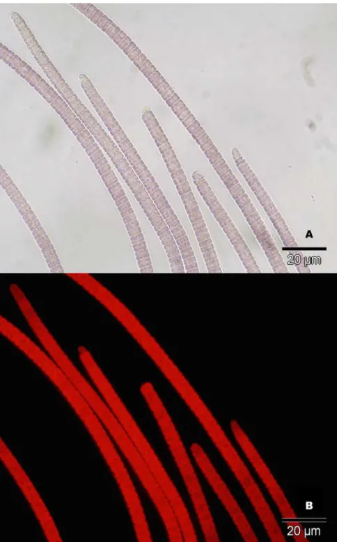
Cyanobacteria associated with disease lesions.. The visually dominant filamentous
(previously described asPhormidium corallyticum), the original BBD cyanobacterium isolated from the Florida Keys [44] (Fig. 4).
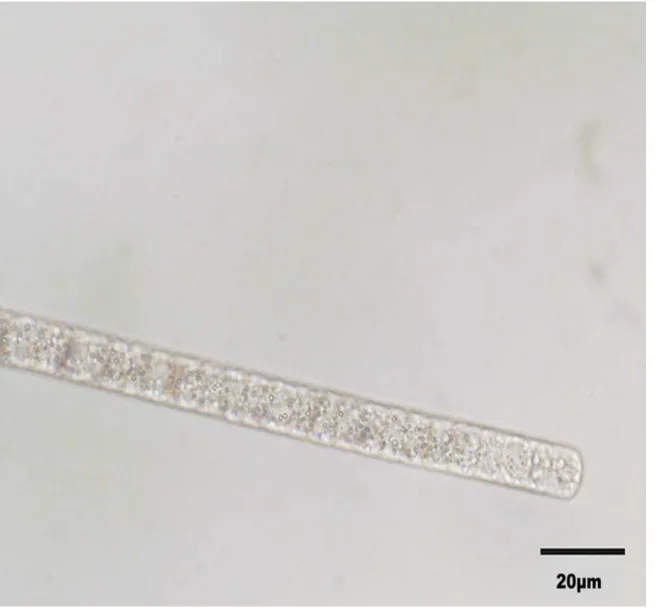
Beggiatoaassociated with disease lesions.. White filamentous microorganisms that
con-tained apparent sulfur granules and exhibited gliding motility, consistent withBeggiatoa, were Fig 3. Microscopy images of cyanobacteria isolated from disease lesions.A) Cyanobacteria under light microscope. B) Cyanobacteria under fluorescence microscope. Red autofluorescence is indicative of photosynthetic pigments. Images were taken at 60x magnification. Black bar represents 20μm.
observed from samples of coral lesions (Fig. 5). Inoculation of J3 medium, which is rich in hy-drogen sulfide, resulted in the migration of filaments two to three centimeters down into the deep, consistent with the motile behavior of sulfide-oxidizingBeggiatoa[28]. Sequences of 16S rRNA genes from filaments cultured on J3 deeps (KM924160, KM924158, KM924159) or taken directly from lesion material (KJ914891) clustered together (99 to 100% identical) and with otherBeggiatoaspecies (Fig. 6). Survival and behavior in hydrogen sulfide rich medium, the presence of large granules, gliding motility, and 16S rRNA gene sequence are all consistent with these isolates belonging to the genusBeggiatoa.
Sulfate-reducing bacteria associated with disease lesions.. A diverse group of
sulfate-reducing bacteria were present in the Kaua‘i coral lesions. ThedsrAgene was used as a proxy for the presence of sulfate-reducing bacteria [35,45] and partialdsrAsequences were recovered by PCR usingdsrA-specific primers from lesion material. Thirteen sequences were determined Fig 4. Phylogenetic relationship of cyanobacterium OCN074, isolated from disease lesions from Kaua‘i, to other cyanobacteria.All 16S rRNA gene sequences from the three Kaua‘i BBD cyanobacterial isolates were identical. A phylogenetic tree was generated using the Maximum Likelihood method. The ‘*’indicates cyanobacteria associated with BBD of Palau (*[42]) the Red Sea (**[41]), and the Florida Keys (***[43]). The tree with the highest log likelihood is shown, and 1000 bootstrap replicates were used. NCBI accession numbers are in brackets and bootstrap values are indicated at branch nodes. Scale bar represents two substitutions per 100 nucleotide positions.
and compared to those from other BBD samples and known sulfate-reducing bacteria (Fig. 7). Two general groups of sulfate-reducing bacteria were identified after phylogenetic analysis. Twelve sequences from the Kaua‘i BBD lesion (KM258124, KM258125, KM258127,
KM258122 (2 clones), KM258123, KM924162, KM924163, KM924161 (2 clones), KM924165 (2 clones)) clustered with sequences retrieved from the Red Sea BBD lesion material [23] and sequences from otherDesulfovibriostrains. One Kaua‘idsrAsequence (KM258126) clustered withdsrAsequences retrieved from the Great Barrier Reef BBD lesion material [34].
Discussion
This study establishes the presence of black band disease (BBD) on montiporids in the Hawai-ian archipelago. The three major microbial components indicative of BBD (filamentous cyano-bacteria, sulfide-oxidizing bacteria and sulfate-reducing bacteria) were identified in disease lesions from corals on Kaua‘i. A phototrophic, filamentous, motile cyanobacterium cultured from BBD lesions in Kaua‘i was genetically similar to bothPseudoscillatoriaandOscillatoria, Fig 5. Brightfield image of Beggiatoa sp. filaments removed from disease lesions.Image taken at 60x magnification. Black bar represents 20μm.
cyanobacteria found in BBD reported from other areas of the Indo-Pacific [43]. Motile, white filamentous bacteria from Kaua‘i BBD lesions were morphologically similar to other strains of
Beggiatoa[46], and corresponding 16S rRNA gene sequences were identified asBeggiatoa. Prior studies on BBD have exclusively used the distinct morphology ofBeggiatoato identify this bacterium [24,25]. Our study is the first to use morphology, behavior and molecular analy-sis for identification. Lastly, a range ofdsrAgene sequences were amplified from the Kaua‘i BBD lesions, indicative of a diverse group of bacteria that can reduce sulfate. No DNA se-quences were available forBeggiatoafound in BBD from other regions for our comparison. However, consistent with other studies, we found that although BBD is found on reefs world-wide, the specific bacterial species associated with this polymicrobial disease that have been ge-netically identified (cyanobacteria and sulfate-reducing bacteria) varies across regions [23,47, 48]. It has been suggested that the specific microbial communities found in BBD may be Fig 6. Phylogenetic relationship ofBeggiatoaspp. isolated from disease lesions from Kaua‘i to otherBeggiatoa.A phylogenetic tree was generated using the Maximum Likelihood method with sequences from 14 otherBeggiatoaand closely related sulfur-oxidizing strains including representative type strains from the Thiotrichaceae family. No 16S rRNA gene sequences fromBeggiatoafound in BBD from other regions were available for comparison. The tree with the highest log likelihood is shown, and 1000 bootstrap replicates were used. NCBI accession numbers are in brackets, and bootstrap values are indicated at branch nodes. Scale bar represents five substitutions per 100 nucleotide positions.
primarily derived from the local reef environment [48], which could explain the regional differ-ences in species found in BBD bacterial communities. More phylogenetic studies onBeggiatoa
are needed to further investigate this hypothesis.
Our study extends the known range of BBD within the Indo-Pacific, which has previously been documented in Palau [43], Great Barrier Reef (GBR) [4], Guam [49], American Samoa Fig 7. Phylogenetic relationship of sulfate-reducing bacteria from disease lesions from Kaua‘i to other sulfate-reducing bacteria.A phylogenetic tree was generated using the Maximum Likelihood method with a reference sequence library built from 25 knowndsrAgene sequences used as a proxy for the presence of sulfate-reducing bacteria [23,33]. The‘*’indicates gene sequences from BBD samples previously published from the Great Barrier Reef (*[33]), and the Red Sea (**[23]). The tree with the highest log likelihood is shown, and 1000 bootstrap replicates were used. NCBI accession numbers are in brackets, and bootstrap values are indicated at branch nodes. Scale bar represents two substitutions per 100
nucleotide positions.
[6], Indonesia [50], Philippines [51], Japan [52], Arabian Gulf [53], and the Red Sea [54]. Prior to this study, no occurrences of BBD were reported in Hawai‘i despite extensive disease surveys [5,12,13].
An Eyes of the Reef (EOR) member, who had been trained in disease identification, first re-ported the disease outbreak and the outbreak status of the disease was confirmed by rapid sur-veys as outlined in Hawai‘i’s rapid response contingency plan (RRCP) [18]. At the alleged outbreak site (Anini) on Kaua‘i’s north shore, disease prevalence was found to be 6 to 7%, which is higher than baseline levels of the disease, previously referred to asMontiporabanded tissue loss, recorded in nearby Hanalei Bay, Kaua‘i (prevalence<1%) [19]. Such high levels of disease compared to baseline levels constitute an epizootic. Since the disease was found to be at outbreak levels, a multi-agency research response was initiated. Having the ability to identify and respond quickly to disease outbreaks highlights the value of programs such as EOR and Hawai‘i’s RRCP. The need for standardized disease response protocols has been recognized elsewhere and resulted in the recent creation of multiple resources for biologists [55–57]. The importance of having prior knowledge of baseline disease levels cannot be overemphasized; it gives scientists and managers the capacity to identify and respond to changes in disease levels through time. Coral reefs are declining precipitously [58,59] and, in response to continued problems associated with anthropogenic overuse and global climate change, coral disease out-breaks are predicted to increase over time [60]. Numerous regions throughout the Indo-Pacific lack baseline coral disease surveys, which highlight the critical need for agencies, managers and scientists to work together to fill in these gaps in knowledge.
In other regions, BBD usually occurs at a low prevalence (<1%) but is considered a disease of concern because it is a chronic disease on reefs, often persisting for years and contributing to the long-term decline of susceptible coral species [61–63]. In Hawai‘i,Montiporabanded tissue loss, which is now known to be BBD, was first found in Hanalei Bay on Kaua‘i in 2004 [19], and found in subsequent surveys in 2007 and 2009 (S1 Table). Hence, we know the disease has been affecting montiporids on Kaua‘i for at least 10 years. We also found that BBD can cause significant colony mortality onMontipora capitatawith colonies losing an average of 68.7% of their live tissue within two months. In other regions, BBD prevalence and virulence is seasonal with higher prevalence and rate of tissue loss found during the warmer months [33,61]. Our study occurred from September to November; months that coincide with warmer water tem-peratures in Hawai‘i (http://www.nodc.noaa.gov/dsdt/cwtg/hawaii.html), which may partly ex-plain the high rate of tissue loss observed. BBD was also found to be more virulent than other tissue loss diseases in Hawai‘i. For example,Montiporawhite syndrome (MWS) is a tissue loss disease affectingM.capitatacolonies in Kāne‘ohe Bay, Oahu. MWS-affected colonies lost an
average of 3% of the colony per month [16], roughly one tenth the rate observed with colonies affected by BBD on Kaua‘i.
causes of disease, they may be useful for containing disease outbreaks, if caught early, or for re-ducing morbidity and mortality from outbreaks. As such, the different disease treatments being developed should be considered by resource managers as they continue to address the growing threats from coral disease events.
BBD is known to affect multiple host genera [3] but on Kaua‘i it was only observed on three species ofMontipora(M.capitata,M.flabellata,M.patula) even thoughPorites, known to be BBD susceptible in other regions [3], is also commonly found on the reefs surveyed. While fur-ther surveys on Kaua‘i may reveal ofur-ther infected genera, it initially appears that in Hawai‘i, montiporids are the primary coral genera affected by BBD. Similarly, acroporids consistently have the highest BBD levels on the GBR, although numerous genera are susceptible [4,63].
Interestingly,Montiporaspp. are abundant throughout the reefs of Hawai‘i [66,67] yet BBD has only been found on Kaua‘i despite extensive baseline disease surveys throughout the Ha-waiian archipelago [5,12,13]. What is different about the north shore of Kaua‘i that might be allowing BBD to persist and reach outbreak levels? Coastal coral reefs are increasingly exposed to excess nutrients, sediments, and pollutants discharged from land. Coastal development, agri-culture, and overgrazing have all contributed to increased terrestrial runoff and numerous studies have shown that sedimentation, turbidity, and nutrient enrichment can degrade local coral reefs [68]. The north shore of Kaua‘i has been plagued by chronic, impaired coastal waters having high levels of sedimentation [69], excess nutrient loading, and bacterial contamination [70,71]. At the sites we surveyed for coral disease, there were obvious signs of excessive sedi-mentation and sediment damage on corals. Increases in coral diseases have also been found as-sociated with reduced water quality from terrestrial run-off. In Australia, a 10-fold greater mean abundance of disease was found on reefs during the rainy summer months and it was concluded that rainfall and associated runoff were facilitating disease outbreaks [72]. Increased prevalence of BBD in the field has been associated with sewage effluent [73] and laboratory tests showed that the rate of tissue loss from BBD was increased with nutrient enrichment [74]. When experimentalin situnutrient enrichment of reefs in the Caribbean were conducted, cor-als exposed to chronic nutrient stress suffered a 3.5-fold increase in bleaching frequency and a two-fold increase in prevalence and severity of disease, compared to corals in control plots [75]. Additionally, nine months after removal of the nutrients, there were no differences in bleaching or disease levels in experimental versus control plots. This suggests that improve-ment in water quality may be an effective way to mitigate some coral diseases and improve overall coral reef integrity. It is likely that the chronically degraded water quality found on Kaua‘i’s north shore is contributing to the spread and severity of coral disease. Further studies are currently underway to examine this hypothesis.
Supporting Information
S1 Table. Disease surveys conducted within Hanalei Bay, Kauai in 2007 and 2009. (DOCX)
Acknowledgments
Author Contributions
Conceived and designed the experiments: GSA TMW CMR ASM BU PV SMC. Performed the experiments: GSA TMW CMR ASM BU PV SB SMC. Analyzed the data: GSA CMR ASM BU. Contributed reagents/materials/analysis tools: GSA TMW SMC. Wrote the paper: GSA TMW CMR ASM BU PV SB SMC.
References
1. Aronson RB, Precht WF. White-band disease and the changing face of Caribbean coral reefs. Hydro-biologia. 2001; 460(1–3): 25–38.
2. Gardner TA, Côté IM, Gill JA, Grant A, Watkinson AR. Long-term region-wide declines in Caribbean corals. Science. 2003; 301: 958–960. PMID:12869698
3. Sutherland KP, Porter JW, Torres C. Disease and immunity in Caribbean and Indo-Pacific
zooxanthel-late corals. Mar Ecol Prog Ser. 2004; 266: 265–272.
4. Willis BL, Page CA, Dinsdale EA. Coral Disease on the Great Barrier Reef. In: Rosenberg PE, Loya PY, editors. Coral Health and Disease. Springer, Berlin Heidelberg; 2004. p. 69–104.
5. Aeby GS. Baseline levels of coral disease in the Northwestern Hawaiian Islands. Atoll Res Bull. 2006; 543: 471–488.
6. Vargas-Ángel B, Wheeler B. Coral health and disease assessment in the US Pacific territories and affili-ated states. In: Kim K, Harvell D, Page C, editors. Diseases on coral reefs. Proceedings of the 11th In-ternational Coral Reef Symposium; 2008 July 7–11; Ft. Lauderdale, Florida. National Coral Reef Institute; 2009. p. 175–179.
7. Bruno JF, Selig ER, Casey KS, Page CA, Willis BL, Harvell CD, et al. Thermal stress and coral cover as drivers of coral disease outbreaks. PLoS Biol. 2007; 5: e124. PMID:17488183
8. Dalton SJ, Godwin S, Smith SDA, Pereg L. Australian subtropical white syndrome: a transmissible, temperature-dependent coral disease. Mar Freshw Res. 2010; 61: 342–350.
9. Raymundo LJ, Harvell CD, Reynolds TL.Poritesulcerative white spot disease: description, prevalence, and host range of a new coral disease affecting Indo-Pacific reefs. Dis Aquat Organ. 2003; 656(2): 95–104.
10. Sussman M, Willis BL, Victor S, Bourne DG. Coral pathogens identified for white syndrome (WS)
epizo-otics in the Indo-Pacific. PLoS One. 2008; 3: e2393. doi:10.1371/journal.pone.0002393PMID: 18560584
11. Williams GJ, Knapp IS, Aeby GS, Davy SK. Spatial and temporal patterns of scleractinian coral, soft
coral, and zoanthid disease on a remote, near-pristine coral reef (Palmyra Atoll, central Pacific). Dis Aquat Organ. 2011; 94: 89–100. doi:10.3354/dao02323PMID:21648237
12. Vargas-Ángel B. Coral Health and Disease Assessment in the U.S. Pacific Remote Island Areas. Bull
Mar Sci. 2009; 84: 211–227.
13. Aeby GS, Williams GJ, Franklin EC, Kenyon J, Cox EF, Work TM. Patterns of coral disease across the Hawaiian archipelago: relating disease to environment. PLoS One. 2011a; 6: e20370. doi:10.1371/ journal.pone.0020370PMID:21655248
14. Aeby GS. Outbreak of coral disease in the Northwestern Hawaiian Islands. Coral Reefs. 2006; 24: 481.
15. Aeby GS, Bourne DG, Wilson B, Work TM. Coral diversity and the severity of disease outbreaks: A cross-regional comparison ofAcroporaWhite Syndrome in a species-rich region (American Samoa) with a species-poor region (Northwestern Hawaiian Islands). J Mar Biol. 2011; 2011. PMID: 25505914
16. Aeby GS, Ross M, Williams GJ, Lewis TD, Work TM. Disease dynamics ofMontiporawhite syndrome within Kaneohe Bay, Oahu, Hawaii: distribution, seasonality, virulence, and transmissibility. Dis Aquat Organ. 2010; 91: 1–8. doi:10.3354/dao02247PMID:20853736
17. Ross M, Stender Y, Aeby GS. Outbreak of the coral disease,MontiporaWhite Syndrome in Maui, Hawai‘i. In: Yellowlees D, Hughes TP, editors. Proceedings of the 12th International Coral Reef Sym-posium; 2012 July 9–13; Queensland, Australia. James Cook University; 2012. 16B–3.
18. Aeby G, Hutchinson M, MacGowan P. Hawaii’s rapid response contingency plan for events of coral bleaching, disease or crown-of thorns starfish outbreaks Hawaii. Hawaii Division of Aquatic Resources; 2008.
20. Richardson LL. Black band disease. In: Rosenberg PE, Loya PY, editors. Coral Health and Disease. Springer, Berlin Heidelberg; 2004. pp. 325–336.
21. Cooney RP, Pantos O, Le Tissier MD, Barer MR, Bythell JC. Characterization of the bacterial consor-tium associated with black band disease in coral using molecular microbiological techniques. Environ Microbiol. 2002; 4: 401–413. PMID:12123476
22. Frias-Lopez J, Zerkle AL, Bonheyo GT, Fouke BW. Partitioning of bacterial communities between
sea-water and healthy, black band diseased, and dead coral surfaces. Appl Environ Microbiol. 2002; 68: 2214–2228. PMID:11976091
23. Barneah O, Ben-Dov E, Kramarsky-Winter E, Kushmaro A. Characterization of black band disease in Red Sea stony corals. Environ Microbiol. 2007; 9: 1995–2006. PMID:17635545
24. Carlton RG, Richardson LL. Oxygen and sulfide dynamics in a horizontally migrating cyanobacterial
mat: black band disease of corals. FEMS Microbiol Ecol. 1995; 18: 155–162.
25. Richardson LL. Horizontal and vertical migration patterns ofPhormidium corallyticumandBeggiatoa spp. associated with black-band disease of corals. Microb Ecol. 1996; 32: 323–335. PMID:8849426
26. Richardson LL, Miller AW, Broderick E, Kaczmarsky L, Gantar M, Sekar R. Sulfide, microcystein, and the etiology of black band disease. Dis Aquat Organ. 2009; 87(1–2): 79–90. doi:10.3354/dao02109 PMID:20099414
27. Allen MM. Simple conditions for growth of unicellular blue-green algae on plates 1, 2. J Phycol. 1968; 4: 1–4.
28. Borthakur PB, Orozco CC, Young-Robbins SS, Haselkorn R, Callahan SM. Inactivation of patS and hetN causes lethal levels of heterocyst differentiation in the filamentous cyanobacteriumAnabaenasp. PCC 7120. Mol Microbiol. 2005; 57: 111–123. PMID:15948953
29. Nelson DC, Jannasch HW. Chemoautotrophic growth of a marineBeggiatoain sulfide-gradient cul-tures. Arch Microbiol. 1983; 136: 262–269.
30. Nübel U, Garcia-Pichel F, Muyzer G. PCR primers to amplify 16S rRNA genes from cyanobacteria.
Appl Environ Microbiol. 1997; 63: 3327–3332. PMID:9251225
31. Aebischer T, Fischer A, Walduck A, Schlötelburg C, Lindig M, Schreiber S, et al. Vaccination prevents
Helicobacter pylori-induced alterations of the gastric flora in mice. FEMS Immunol Med Microbiol.
2006; 46: 221–229. PMID:16487303
32. Viehman S, Mills DK, Meichel GW, Richardson LL. Culture and identification ofDesulfovibriospp. from corals infected by black band disease on Dominican and Florida Keys reefs. Dis Aquat Organ. 2006; 69: 119–127. PMID:16703774
33. Sato Y, Bourne DG, Willis BL. Dynamics of seasonal outbreaks of black band disease in an assem-blage ofMontiporaspecies at Pelorus Island (Great Barrier Reef, Australia). Proc R Soc B Biol Sci. 2009; 276: 2795–2803. doi:10.1098/rspb.2009.0481PMID:19419991
34. Bourne DG, Muirhead A, Sato Y. Changes in sulfate-reducing bacterial populations during the onset of black band disease. ISME J. 2010; 5: 559–564. doi:10.1038/ismej.2010.143PMID:20811471
35. Leloup J, Loy A, Knab NJ, Borowski C, Wagner M, Jorgensen B. Diversity and abundance of sulfate-reducing microorganisms in the sulfate and methane zones of a marine sediment, Black Sea. Environ Microbiol. 2007; 9: 131–142. PMID:17227418
36. Messing J. New M13 vectors for cloning. Methods Enzymol. 1983; 101: 20–78. PMID:6310323
37. Ushijima B, Smith A, Aeby GS, Callahan SM.Vibrio owensiiinduces the tissue loss diseaseMontipora white syndrome in the Hawaiian reef coralMontipora capitata. PLoS One. 2012; 7: e46717. doi:10.
1371/journal.pone.0046717PMID:23056419
38. Hall TA. BioEdit: a user-friendly biological sequence alignment editor and analysis program for
Win-dows 95/98/NT. Nucleic acids symposium series. 1999. 41: 95–98.
39. Tavaré S. Some probabilistic and statistical problems in the analysis of DNA sequences. Lect Math Life Sci. 1986; 17: 57–86.
40. Tamura K, Peterson D, Peterson N, Stecher G, Nei M, Kumar S. MEGA5: molecular evolutionary genet-ics analysis using maximum likelihood, evolutionary distance, and maximum parsimony methods. Mol Biol Evol. 2011; 28: 2731–2739. doi:10.1093/molbev/msr121PMID:21546353
41. Guindon S, Gascuel O. A simple, fast, and accurate algorithm to estimate large phylogenies by maxi-mum likelihood. Syst Biol. 2003; 52: 696–704. PMID:14530136
42. Rasoulouniriana D, Siboni N, Ben-Dov E, Kramarsky-Winter E, Loya Y, Kushmaro A.Pseudoscillatoria
coraliigen. nov., sp. nov., a cyanobacterium associated with coral black band disease (BBD). Dis
Aquat Organ. 2009; 87: 91–96. doi:10.3354/dao02089PMID:20095244
44. Rützler K, Santavy DL. The black band disease of Atlantic reef corals. I. Description of the cyanophyte pathogen. PSZNI Mar Ecol. 1983; 4: 301–319.
45. Wagner M, Loy A, Klein M, Lee N, Ramsing NB, Stahl D, et al. Functional marker genes for identifica-tion of sulfate-reducing prokaryotes. Methods Enzymol. 2005; 397: 469–489. PMID:16260310
46. Ducklow HW, Mitchell R. Observations on naturally and artificially diseased tropical corals: a scanning
electron microscope study. Microb Ecol. 2005; 5: 215–223.
47. Frias-Lopez J, Bonheyo GT, Jin Q, Fouke BW. Cyanobacteria associated with coral black band disease in Caribbean and Indo-Pacific reefs. Appl Environ Microbiol. 2003; 69: 2409–2413. PMID:12676731
48. Miller AW, Richardson LL. A meta-analysis of 16S rRNA gene clone libraries from the polymicrobial black band disease of corals. FEMS Microbiol Ecol. 2011; 75: 231–241. doi:10.1111/j.1574-6941. 2010.00991.xPMID:21114503
49. Myers RL, Raymundo LJ. Coral disease in Micronesian reefs: a link between disease prevalence and host abundance. Dis Aquat Organ. 2009; 87: 97–104. doi:10.3354/dao02139PMID:20095245
50. Haapkylä J, Unsworth RK, Seymour AS, Melbourne-Thomas J, Flavell M, Willis B, et al. Spatio-temporal coral disease dynamics in the Wakatobi Marine National Park, South-East Sulawesi, Indone-sia. Dis Aquat Organ. 2009; 87: 105–115. doi:10.3354/dao02160PMID:20095246
51. Raymundo LJ, Halford AR, Maypa AP, Kerr AM. Functionally diverse reef-fish communities ameliorate coral disease. Proc Natl Acad Sci. 2009; 106: 17067–17070. doi:10.1073/pnas.0900365106PMID: 19805081
52. Weil E, Irikawa A, Casareto B, Suzuki Y. Extended geographic distribution of several Indo-Pacific coral reef diseases. Dis Aquat Organ. 2012; 98: 163–170. doi:10.3354/dao02433PMID:22436464
53. Riegl B. Effects of the 1996 and 1998 positive sea-surface temperature anomalies on coral, coral
dis-ease and fish in the Arabian Gulf (Dubai, UAE). Mar Bio. 2002; 140(1): 29–40.
54. Al-Moghrabi S. Unusual black band disease (BBD) outbreak in the northern tip of the Gulf of Aqaba (Jordan). Coral Reefs. 2001; 19: 330–331. doi:10.1007/s003380000127
55. Raymundo LJ, Couch CS, Harvell CD, Raymundo J, Bruckner AW, Work TM, et al. Coral Disease Handbook Guidelines for Assessment, Monitoring & Management. Coral Reef Targeted Research and Capacity Building for Management Program; 2008.
56. Woodley CM, Bruckner AW, McLenon AL, Higgins JL, Galloway SB, Nicholson J. Field manual for in-vestigating coral disease outbreaks. US Department of Commerce, National Oceanic and Atmospheric Administration, National Ocean Service; 2008.
57. Beeden R, Maynard JA, Marshall PA, Heron SF, Willis BL. A framework for responding to coral disease outbreaks that facilitates adaptive management. Environ Manage. 2012; 49: 1–13. doi:10.1007/ s00267-011-9770-9PMID:22042407
58. Hughes TP, Baird AH, Bellwood DR, Card M, Connolly SR, Folke C, et al. Climate change, human im-pacts, and the resilience of coral reefs. Science. 2003; 301: 929–933. PMID:12920289
59. Bellwood DR, Hughes TP, Folke C, Nyström M. Confronting the coral reef crisis. Nature. 2004; 429: 827–833. PMID:15215854
60. Sokolow S. Effects of a changing climate on the dynamics of coral infectious disease: a review of the
evidence. Dis Aquat Organ. 2009; 87: 5–18. doi:10.3354/dao02099PMID:20095237
61. Edmunds PJ. Extent and effect of black band disease on a Caribbean reef. Coral Reefs. 1991; 10: 161–165.
62. Bruckner AW, Bruckner RJ, Williams J, Ernest H. Spread of a black-band disease epizootic through the coral reef system in St. Ann's Bay, Jamaica. Bull Mar Sci. 2007; 61(3): 919–928.
63. Page C, Willis B. Distribution, host range and large-scale spatial variability in black band disease preva-lence on the Great Barrier Reef, Australia. Dis Aquat Organ. 2006; 69: 41–51. PMID:16703765
64. Hudson H. First aid for massive corals infected with black band disease: an underwater aspirator and
post-treatment sealant to curtail re-infection. Diving Sci 21st Century. 2000.
65. Williams GJ. Contrasting recovery following removal of growth anomalies in the coralsAcroporaand
Montipora. Dis Aquat Organ. 2013; 106: 181–185. doi:10.3354/dao02652PMID:24113251
66. Jokiel PL, Brown EK, Friedlander A, Rodgers SK, Smith WR. Hawai’i coral reef assessment and moni-toring program: Spatial patterns and temporal dynamics in reef coral communities. Pac Sci. 2004; 58: 159–174.
67. Maragos JE, Potts DC, Aeby GS, Gulko D, Kenyon J, Siciliano D, et al. 2000–2002 rapid ecological as-sessment of corals (Anthozoa) on shallow reefs of the Northwestern Hawaiian Islands. Part 1: Species and Distribution. Pac Sci. 2004; 58: 211–230.
69. Storlazzi CD, Field ME, Bothner MH, Presto MK, Draut AE. Sedimentation processes in a coral reef em-bayment: Hanalei Bay, Kauai. Mar Geol. 2009; 264: 140–151.
70. Knee KL, Layton BA, Street JH, Boehm AB, Paytan A. Sources of nutrients and fecal indicator bacteria to nearshore waters on the north shore of Kauai (Hawaii, USA). Estuaries Coasts. 2008; 31: 607–622.
71. Derse E, Knee KL, Wankel SD, Kendall C, Berg CJ, Paytan A. Identifying sources of nitrogen to Hanalei
Bay, Kauai, utilizing the nitrogen isotope signature of macroalgae. Environ Sci Technol. 2007; 41: 5217–5223. PMID:17822082
72. Haapkylä J, Unsworth R, Flavell M, Bourne DG, Schaffelke B, Willis BL. Seasonal rainfall and runoff
promote coral disease on an inshore reef. PLoS One. 2011; 6(2):e16893. doi:10.1371/journal.pone. 0016893PMID:21347317
73. Kaczmarsky LT, Draud M, Williams EH. Is there a relationship between proximity to sewage effluent
and the prevalence of coral disease. Caribb J Sci. 2005; 41: 124–137.
74. Voss JD, Richardson LL. Nutrient enrichment enhances black band disease progression in corals. Coral Reefs. 2006; 25: 569–576.