Resveratrol Causes Antiatherogenic Effects in an Animal Model of
Atherosclerosis
Rossane Serafim Matos, Liz Andréa Villela Baroncini, Leonardo Brandão Précoma, Guilherme Winter, Pedro
Henrique Lambach Caron, Flávia Kaiber, Dalton Bertolim Précoma
Pontifícia Universidade Católica do Paraná, Curitiba, PR, Brazil
Abstract
Background: Resveratrol protects the cardiovascular system by a number of mechanisms, including antioxidant and anti-platelet activities.
Objective: To assess the potential anti-inflammatory and antiatherogenic effects of resveratrol using rabbits fed a hypercholesterolemic diet (1% cholesterol).
Methods: Twenty white male rabbits were selected and divided into two groups: control group (CG), 10 rabbits; and resveratrol group (RG), 10 rabbits. The animals were fed a hypercholesterolemic diet for 56 days. For the RG diet, resveratrol (2mg/kg weight/day) was added from days 33 - 56.
Results: There was no significant difference in the total serum cholesterol, HDL-cholesterol, LDL-cholesterol, and triglycerides between the groups. Of the CG, 70% had advanced aortic atherosclerotic lesions (types III, IV, V, or VI). All animals from the RG had mild aortic atherosclerotic lesions (types I or II, or no lesions). The intima area and the intima/media layer area ratio was significantly lower in the RG as compared to the CG (p<0.001). Positive areas for VCAM-1 molecules were lower in the RG (p=0.007). The MCP-1 and IL-6 concentrations were lower in the RG than the CG (p=0.039 and p=0.015, respectively).
Conclusion: Resveratrol had significant anti-atherogenic and anti-inflammatory effects in an animal model with rabbits fed a hypercholesterolemic diet (1% cholesterol). (Arq Bras Cardiol 2012;98(2):136-142)
Keywords: Revesratrol; antioxidants; atherosclerosis; dyslipidemias.
Mailing Address: Liz Andréa Villela Baroncini •
Rua Buenos Aires, 764 / 601, Batel - 80250-070 – Curitiba, PR, Brazil E-mail: [email protected]; [email protected]
Manuscript received May 26, 2011, revised manuscript received July 20, 2011; accepted August 09, 2011.
Methods
Animals
Twenty white adult New Zealand male rabbits at a mean age of 30 days were selected. Animals were handled in compliance with the Guiding Principles in the Care and Use of Animals. Protocol approval was obtained from the Animal Research Committee of the Pontifícia Universidade Católica. The animals were divided into 2 groups, as follows: control group (CG), 10 rabbits; and resveratrol group (RG), 10 rabbits. During the 56 days of the study, the animals were fed a specific diet for the species (Nuvilab®) plus 1% cholesterol from lyophilized eggs. From days 29 – 56, resveratol (Resveratrol®; extracted from the Galena® Laboratory) was added to the RG diet (2mg/kg/day) and administered by oral gavage. On day 56, the animals underwent dissection of the aortic arch and descending aorta. Anesthesia was induced with ketamine (Vetanarcol®, 3.5 mg/kg; König) and intramuscular xylazine (Coopazine®, 5 mg/kg; Coopers). After the procedure, the rabbits were sacrificed by a lethal dose of barbiturate. The sample size was calculated based on the study by Zou et al6.
Blood chemistry
Blood samples were obtained on the first day of the experiment and immediately before sacrifice by cardiac puncture. Clinical
Introduction
laboratory assessment included total cholesterol (TC), high-density lipoprotein cholesterol (HDL-C), low-high-density lipoprotein cholesterol (LDL-C) and triglycerides (TGC). Measurements were taken using an automated system (Abbott Architect ci8200; Abbott Laboratories, Abbott Park, IL, USA).
Histologic analysis
The arteries were removed and washed with 10% formaldehyde buffered with phosphate (ph=7.6) and waxed. For evaluation of this experimental model, qualitative and quantitative histologic measurements were adopted. The hematoxylin and eosin (HE) stained slices were analyzed blindly in a microscopy jellyfish five-head Olympus® BX 40. The atherosclerotic lesions were graded from 0 - VI according to the qualitative criteria proposed by Stary et al7-9. The morphometric analysis was performed on stained orcein (elastic) to determine the area of the intima and the medial layers of the aortic arch and descending aorta, as well as to determine the intima/media layer area ratio (IMR - the area of the intimal layer divided by the area of the medial layer). For this assessment, the more injured segment or the segment with more advanced stage of atherosclerosis was previously selected in the HE-stained slice. The analyses were performed microscopically in conjunction with Image Pro-plus® 4.5 software (Media Cybernetics Inc., Silver Spring, MD, USA).
Immunohistochemistry
We evaluated the concentration of the vascular cell adhesion molecule (VCAM-1), monocyte chemotactic protein-1 (MCP-1), and interleucin-6 (IL-6) in the intima of the arteries using primary
monoclonal antibodies. The reading was performed with an Olympus® BX50 microscope, with an objective of 20 times.
Statistical analysis
Categoricalvariables were expressed as percentages and continuous variables were expressed as the mean ± SD and medians. The Shapiro-Wilks test was used for testing sample normality. For symmetry conditions, some variables were submitted to logarithmic transformation. For dichotomic nominal variables, the Fisher exact test was used to compare the groups. For quantitative parameters, Student’s t-test and Mann-Whitney non-parametric test were used for the comparison between CG and RG. Statistical significance was indicated by p < 0.05.
Results
Two animals from the CG died in the middle of the experiment and were removed from the final analysis. The initial mean body weight of animals in the CG (1.78 kg) and RG (1.82 kg) were similar (p=0.274). At the end of the study, CG animals exhibited a mean body weight of 2.58 kg, while RG animals had a mean body weight of 2.39 kg (p=0.537).
Lipid proiles
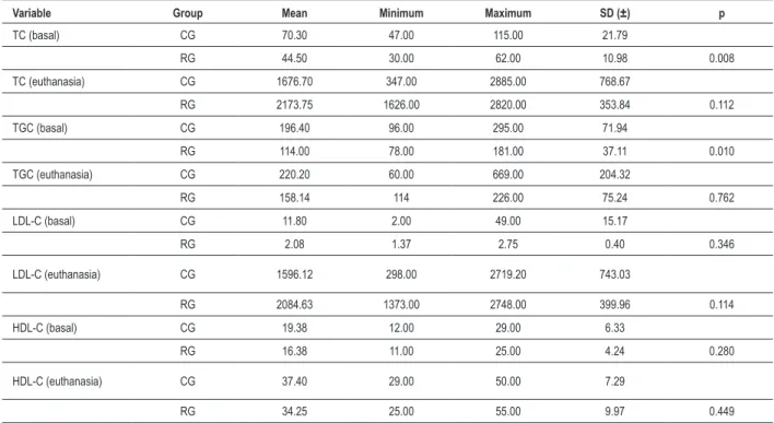
Baseline TC, HDL-C, LDL-C, and TGC levels were relatively equivalent in all groups before initiation of the diet. At the end of the experiment, an analysis of TC, HDL-C, LDL-C, and serum TG indicated no significant difference between the groups (Table 1).
Table 1 - Lipid proile between control group (CG) and Resveratrol group (RG)
Variable Group Mean Minimum Maximum SD (±) p
TC (basal) CG 70.30 47.00 115.00 21.79
RG 44.50 30.00 62.00 10.98 0.008
TC (euthanasia) CG 1676.70 347.00 2885.00 768.67
RG 2173.75 1626.00 2820.00 353.84 0.112
TGC (basal) CG 196.40 96.00 295.00 71.94
RG 114.00 78.00 181.00 37.11 0.010
TGC (euthanasia) CG 220.20 60.00 669.00 204.32
RG 158.14 114 226.00 75.24 0.762
LDL-C (basal) CG 11.80 2.00 49.00 15.17
RG 2.08 1.37 2.75 0.40 0.346
LDL-C (euthanasia) CG 1596.12 298.00 2719.20 743.03
RG 2084.63 1373.00 2748.00 399.96 0.114
HDL-C (basal) CG 19.38 12.00 29.00 6.33
RG 16.38 11.00 25.00 4.24 0.280
HDL-C (euthanasia) CG 37.40 29.00 50.00 7.29
RG 34.25 25.00 55.00 9.97 0.449
Histologic analysis
At the end of the experiments, it was shown that there was a statistically significant difference between the animal groups with respect to the probability of type I, II, III, IV, V, and VI lesions or absence of lesions. Of those animals in the CG, 70% had advanced atherosclerotic lesions (types III, IV, and V) in the aortic arch and descending aorta, while 100% of the RG animals had mild atherosclerotic lesions (types I and II, or no lesions) in the aortic arch and descending aorta (Figure 1).
Morphometric analysis
The intimal area was significantly lower in the RG than the CG (p = 0.001; Figure 1). There was a significantly greater reduction in the intima/media layer area ratio (IMR) in the RG than in the CG (p = 0.001). There was no statistically significant difference between the medial layer area between the groups in the aortic arch and descending aorta (Table 2).
Immunohistochemistry
An analysis of positive areas for VCAM-1 revealed a significant difference between groups, with a higher concentration in CG
(p=0.007). Analysis of MCP-1 and IL-6 showed significantly higher values in the CG compared with the RG (p=0.039 and p=0.015, respectively; Table 3; Figure 2).
Discussion
Resveratrol, present in the grape juice and wine, is considered the major polyphenol responsible for cardiovascular benefits because of its antioxidant and anti-platelet activities10,11. Resveratrol has anti-inflammatory properties which are manifested as inhibition of ICAM-1 and VCAM-1expression and the attachment of monocytes to endothelial cells, inhibition of lipopolysaccharide (LPS)-induced synthesis of TNF-a and IL-1-b, and release of IL-6 from monocytes12. Resveratrol also inhibits the migration and proliferation of vascular smooth muscle cells (SMCs) to the intima layer, which is considered the sine qua non of atherogenesis13,14. Collectively, these effects are thought to be responsible for decreased incidence of heart disease in the French population, and is thus termed the French Paradox (a very low mortality rate due to cardiovascular disease, despite a high-fat diet), because of the population’s
preference for red wine15. In this study, we induced atherosclerotic lesions with the addition of lyophilized egg to the normal diet of rabbits during 4 weeks. This experimental model of inducing atherosclerosis using lyophilized egg is rarely described in the literature, though it is proven to be efficient and less costly when compared to the use of industrialized cholesterol. Resveratrol was added in the diet of RG after 4 weeks to test its capacity to interfere in established atherosclerotic lesions. We demonstrated that resveratrol inhibits the progression of atherosclerotic lesions by reducing the intima area and the IMR, and showing no advanced lesions in the aortic arch and descending aorta in the RG. In a similar study using rabbits, Castro et al16 concluded that resveratrol worked as a preventive agent in the development of atherosclerotic lesions as they observed a low-degree of foam cell invasion in the tunica media of animals treated with resveratrol. The mechanisms by which resveratrol inhibits the formation of advanced atherosclerotic lesions appears to be through its inhibitory effect on LDL oxidation, platelet aggregation, and vascular proliferation of smooth muscle cells. LDL oxidation is a main cause of endothelial injury and induction of the expression of pro-inflammatory molecules in endothelial cells. Resveratrol has been shown to protect lipids from peroxidative degradation and to stop the uptake of oxidized LDLs in the vascular wall in a concentration-dependent manner17-19. However, there are conflicting results regarding
the effects of resveratrol on lipid levels, because some studies have failed to show a reduction in plasma lipids levels induced by such a substance20. In this study, we did not uncover any evidence of a significant difference in lipid profiles between groups, which may have reflected the short period of the experiment. Daugherty et al21, in a similar study in hypercholesterolemic rabbits, showed that the reduction in atherosclerotic lesions secondary to the reduction in LDL-C levels is a process that takes months, or even years. In contrast, in this study, we showed that a hyphercholesterolemic diet causes the development of atherosclerotic lesions associated with an inflammatory process, and resveratrol reduced the concentration of VCAM-1, MCP-1, and IL-6 in the intima layer of hypercholesterolemic rabbits. It is known that inflammation mediates all stages of atherosclerosis from initiation to progression, and eventually plaque rupture17. Indeed, VCAM-1, MCP-1, and IL-6 facilitate the adherence of monocytes to the endothelial surface and facilitate leukocyte transendothelial migration22-25. This process is exacerbated when blood LDL levels are high from poor dietary habits or an inherited genetic pre-disposition, and signals the endothelium to increase adhesion molecule expression and potentiate the inflammatory response26. Even though we did not make the correlation between blood lipid levels and the inflammatory process, we believe that such a possible correlation should be the subject of future studies. Also, we did not analyze the monocyte/macrophage concentration Table 2 - Quantitative histopathological parameters between control group (CG) and resveratrol group (RG) expressed in square micrometer.
Variable Group n Mean Median SD (±) p*
Intima
CG 10 75.10 68.25 51.66 < 0.001
RG 8 4.76 1.38 6.75
Media CG 10 176.59 196.99 52.69 0.315
RG 8 209.70 205.49 39.77
IMR CG 10 0.41 0.37 0.25 < 0.001
RG 8 0.03 0.01 0.04
IMR represents intima/media area ratio. *Mann- Whitney.
Table 3 - Immunohistochemical variables in descending aorta between control group (CG) and resveratrol group (RG) expressed in square micrometer
Variable Group n Mean Median SD (±) p*
VCAM-1 CG 10 5339.07 5209.03 3791 0.007
RG 8 1452.96 1035.59 1137
MCP-1 CG 10 11772 11003 7947 0.039
RG 8 5509 4781 2745
IL-6 CG 10 3852 3623 2694 0.015
RG 8 1273 1128 580
that appears to have a crucial role in the development of atherosclerosis. This cell appears to be involved in all stages of atherosclerotic plaque development and is increasingly regarded as a candidate for therapeutic intervention and as a potential biomarker of disease progression and response to therapy27-29.
Conclusions
Resveratrol had significant atherogenic and anti-inflammatory effects in an animal model of rabbits fed a hypercholesterolemic diet (1% cholesterol).
Figure 2 - Immunohistochemistry showing positive areas for MCP-1, VCAM-1, and IL-6. A – Control group. Black arrow indicates intima layer with positive areas for MCP-1 (brown-speckled areas). B – Resveratrol group. Black arrow indicates intima layer without positive areas for MCP-1. C – Control group. Positive areas for VCAM-1 (brown-speckled areas). D – Resveratrol group. Positive areas for VCAM-VCAM-1. E- Control group. Positive areas for IL-6 (brown-speckled areas). F- Resveratrol group. Positive areas for IL-6.
Potential Conflict of Interest
No potential conflict of interest relevant to this article was reported.
Sources of Funding
There were no external funding sources for this study.
Study Association
References
1. Turner RT, Evans GL, Zhang M, Maran A, Sibonga JD. Is resveratrol an estrogen agonist in growing rats? Endocrinology. 1999;140(1):50-4.
2. Pendurthi US, Rao LVM. Resveratrol suppresses agonist-induced monocyte adhesion to cultured human endothelial cells. Thromb Res. 2002;106(4-5):243-8.
3. Wilson T, Knight TJ, Beitz DC, Lewis DS, Engen RL. Resveratrol promotes atherosclerosis in hypercholesterolemic rabbits. Life Sci. 1996;59(1):PL15-21.
4. Pace-Asciak CR, Rounova O, Hahn SE, Diamandis EP, Goldberb DM. Wines and grape juices as modulators of platelet aggregation in healthy human subjects. Clin Chim Acta. 1996;246(1-2):163-82.
5. Pace-Asciak CR, Hahn S, Diamandis EP, Soleas G, Goldberg DM. The red wine phenolics trans-resveratrol and quercetin block human platelet aggregation and eicosanoid synthesis: implications for protection against coronary heart disease. Clin Chim Acta. 1995;235(2):207-19.
6. Zou JG, Wang ZR, Huanhg YZ, Cao KJ, Wu JM. Effect of red wine and wine polyphenol resveratrol on endothelial function in hypercholesterolemic rabbits. Int J Mol Med. 2003;11(3):317-20.
7. Stary HC, Blankenhorn DH, Chandler AB, Glagov S, Insull W Jr, Richardson M, et al. A definition of the intima of human arteries and of its atherosclerosis- prone regions: a report from the Committee on Vascular Lesions of the Council on Arteriosclerosis, American Heart Association. Circulation. 1992;85(1):391-405.
8. Stary HC, Chandler AB, Glagov S, Guyton JR, Insull W Jr, Rosenfeld ME, et al. A definition of initial, fatty streak, and intermediate lesions of atherosclerosis: a report from the Committee on Vascular Lesions of the Council on Arteriosclerosis, American Heart Association. Circulation. 1994;89(5):2462-78.
9. Stary HC, Chandler AB, Dinsmore RE, Fuster V, Glagov S, Insull W Jr, et al. A definition of advanced types of atherosclerotic lesions and a histological classification of atherosclerosis: a report from the Committee on Vascular Lesions of the Council on Arterisoclerosis, American Heart Association. Circulation. 1995;92(5):355-74.
10. Fulgenzi A, Bertelli AAE, Magni E, Ferrero E, Ferrero ME. In vivo inhibition of TNFa-induced vascular permeability by resveratrol. Transplant Proc. 2001;33(3):2341-3.
11. Olas B, Wachowicz B. Resveratrol and vitamin C as antioxidants in blood platelets. Thromb Res. 2002;106(2):143-8.
12. Slater SJ, Seiz JL, Cook AC, Stagliano BA, Buzas CJ. Inhibition of protein kinase C by resveratrol. Biochim Biophys Acta. 2003;1637(1):59-69.
13. Poussier B, Cordova AC, Becquemin JP, Sumpio BE. Resveratrol inhibits vascular smooth muscle cell proliferation and induces apoptosis. J Vasc Surg. 2005;42(6):1190-7.
14. Chao HH, Juan SH, Liu JC, Yang HY, Yang E, Cheng TH, et al. Resveratrol inhibits angiotensin II-induced endothelin-1 gene expression and subsequent proliferation in rat aortic smooth muscle cells. Eur J Pharmacol. 2005;515(1-3):1-9.
15. Rocha KK, Souza GA, Ebaid GX, Seiva FR, Cataneo AC, Novelli EL. Resveratrol toxicity: effects on risk factors for atherosclerosis and hepatic oxidative stress in standard and high-fat diets. Food Chem Toxicol. 2009;47(6):1362-7.
16. Castro M, Pacheco MMR, Machado MRF. Morphology of aortic arch in rabbits with atherosclerosis treated with resveratrol. Intern J Appl Res Vet Med. 2009;7(4):190-5.
17. Fan E, Zhang L, Jiang S, Bai Y. Beneficial effects of resveratrol on atherosclerosis. J Med Food. 2008;11(4):610-4.
18. Nigdikar SV, Williams NR, Griffin BA, Howard AN. Consumption of red wine polyphenols reduces the susceptibility of low-density lipoproteins to oxidation in vivo. Am J Clin Nutr. 1998;68(2):258-65.
19. Shanmuganayagam D, Warner TF, Krueger CG, Reed JD, Folts JD. Concord grape juice attenuates platelet aggregation, serum cholesterol and development of atheroma in hypercholesterolemic rabbits. Atherosclerosis. 2007;190(1):135-42.
20. Castro M, Veiga APM, Pacheco MR. Plasma lipid profile of experimentally induced hyperlipidemic New Zealand white rabbits is not affected by resveratrol. J Appl Res. 2009;9(1):18-22.
21. Daugherty A, Schonfeld G, Sobel BE, Lange LG. Metabolism of very low density lipoproteins after cessation of cholesterol feeding in rabbits: a factor potentially contributing to the slow regression of atheromatous plaques. J Clin Invest. 1986;77(4):1108-15.
22. Tang W, Pankow JS, Carr JJ, Tracy RP, Bielinski SJ, North KE, et al. Association of sICAM-1 and MCP-1 with coronary artery calcification in families enriched for coronary heart disease or hypertension: the NHLBI Family Heart Study. BMC Cardiovasc Disord. 2007;7:30.
23. Ferrero ME, Bertelli AE, Fulgenzi A, Pellegatta F, Corsi MM, Bonfrate M, et al. Activity in vitro of resveratrol on granulocyte and monocyte adhesion to endothelium Am J Clin Nutr. 1998;68(6):1208-14.
24. Carluccio MA, Siculella L, Ancora MA, Massaro M, Scoditti E, Storelli C, et al. Olive oil and red wine antioxidant polyphenols inhibit endothelial activation: antiatherogenic properties of mediterranean diet phytochemicals. Arterioscler Thromb Vasc Biol. 2003;23(4):622-9.
25. Wung BS, Hsu MC, Wu CC, Hsieh CW. Resveratrol suppreses IL-6-induced ICAM-1 gene expression in endothelial cells: effects on the inhibition of STAT3 phosphorylation. Life Sci. 2005;78(4):389-97.
26. McLaren JE, Ramji DP. Interferon gamma: a master regulator of atherosclerosis. Cytokine Growth Factor Rev. 2009;20(2):125-35.
27. Saha P, Modarai B, Humphries J, Mattock K, Waltham M, Burnand KG, et al. The monocyte/macrophage as a therapeutic target in atherosclerosis. Curr Opin Pharmacol. 2009;9(2):109-18.
28. Ghosh S, Zhao B, Bie J, Song J. Macrophage cholesteryl ester mobilization and atherosclerosis. Vascul Pharmacol. 2010;52(1-2):1-10.