Asymmetric chromosome segregation in
Xanthomonas citri
ssp.
citri
Amanda P. Ucci1, Paula M. M. Martins2, Ivy F. Lau1, Maurı´cio Bacci Jr2, Jose Belasque Jr3& Henrique Ferreira1,2
1
Depto. de Cieˆncias Biologicas, Faculdade de Cieˆncias Farmaceˆuticas, Universidade Estadual Paulista, Rodovia Araraquara/Jau´ Km 1, CP 502, Araraquara, Sa˜o Paulo 14801-902, Brazil
2Depto. de Bioquı´mica e Microbiologia, Instituto de Biocieˆncias, Universidade Estadual Paulista, Av. 24A, 1515, Rio Claro, Sa˜o Paulo 13506-900, Brazil 3Fundo de Defesa da Citricultura, Av. Dr. Adhemar P. de Barros, 201, Araraquara, Sa˜o Paulo 14807-040, Brazil
Keywords
Cell division arrest, chromosome segregation, citrus canker.
Correspondence
Henrique Ferreira, Depto. de Cieˆncias Biolo´gicas, Faculdade de Cieˆncias Farmaceˆuticas, Universidade Estadual Paulista, Rodovia Araraquara/Jau´ Km 1, CP 502, Araraquara, Sa˜o Paulo 14801-902 Brazil. Tel: +55 19 3526 4187; Fax: +55 16 3301 6940;
E-mail: henrique.ferreira@linacre.oxon.org
Funding Information
A. P. U. and P. M. M. M. received scholarships from FAPESP (2010/02041-8) and CNPq (142293/2009-1), respectively. This work was funded by FAPESP (grants 2004/ 09173-6; 2010/05099-7). Beneficia´rio de auxı´lio financeiro da CAPES, Brasil.
Received: 21 August 2013; Revised: 24 October 2013; Accepted: 4 November 2013
MicrobiologyOpen2014; 3(1): 29–41
doi: 10.1002/mbo3.145
Abstract
This study was intended to characterize the chromosome segregation process of
Xanthomonas citrissp. citri(Xac) by investigating the functionality of the ParB factor encoded on its chromosome, and its requirement for cell viability and virulence. Using TAP tagging we show that ParB is expressed in Xac. Disrup-tion ofparB increased the cell doubling time and precluded the ability of Xac to colonize the host citrus. Moreover, Xac mutant cells expressing only trun-cated forms of ParB exhibited the classical phenotype of aberrant chromosome organization, and seemed affected in cell division judged by their reduced growth rate and the propensity to form filaments. The ParB-GFP localization pattern in Xac was suggestive of an asymmetric mode of replicon partitioning, which together with the filamentation phenotype support the idea that Xac may control septum placement using mechanisms probably analogous toCaulobacter crescentus, and perhaps Vibrio cholerae, and Corynebacterium glutamicum. Xac exhibits asymmetric chromosome segregation, and the perturbation of this pro-cess leads to an inability to colonize the host plant.
Introduction
Citrus canker is a serious disease present in the major cit-rus-producing areas around the world. Currently, there are no effective curative measures to safeguard the orch-ards, where the eradication of affected trees is the only reliable method to prevent the spread of the causal agent Xac to regions where it has not been detected (Gottwald et al. 2002). The eradication of plants is a highly contro-versial, costly, and difficult method to control citrus can-ker. In addition, its effectiveness is only possible in areas
fruits, symptomatic fruit drop, defoliation, and stem die-back (Behlau et al. 2010).
Citrus canker has an endemic status in some of the main citrus-producing areas in the world, the state of Florida, USA, and in the southern portion of South America. In Florida, the eradication program was aban-doned in January 2006, and as a consequence, citrus can-ker is spreading throughout the region. In both areas, growers adopt the disease management system. On the other hand, the state of Sa˜o Paulo, Brazil, still keeps an eradication program with mandatory elimination of foci of the disease; however, due to a diminished effort to detect and eradicate infected trees, citrus canker is becom-ing an epidemic (see references within Ferreira and Belas-que [2011]). As a result, in some years growers may have to manage citrus canker in the state of Sa˜o Paulo as it is already done in the southern states of Brazil. For these reasons in depth knowledge of the biology of Xac may help in the development of strategies to control and mini-mize the losses caused by this plant pathogen.
Recently, we characterized some esters of gallic acid that interfere with the subcellular localization of ParB-green fluorescent protein (GFP) and also GFP-ZapA in Xac (Silva et al. 2013). As ParB and ZapA are compo-nents of the bacterial chromosome segregation (segro-some) and cell division (divisome) machineries (Gueiros-Filho and Losick 2002; Mierzejewska and Jagu-ra-Burdzy 2012), respectively, alkyl gallates are believed to target components of the segrosome and/or the divi-some (Silva et al. 2013). ParB-like factors are DNA-binding proteins encoded on plasmids or on the chro-mosomes of many bacteria (Leonard et al. 2005b; Gerdes et al. 2010; Mierzejewska and Jagura-Burdzy 2012). They act together with cognate ATPases, ParA-like factors, to operate efficient replicon segregation in prokaryotes. Seg-regation of low copy number plasmids and chromo-somes requires the specific interaction of ParB with short cis-acting elements designated parS, which on bac-terial chromosomes are normally located around the ori-gins of replication (Livny et al. 2007). Homology searches showed that Xac possesses at least one of these
cis elements (TGTTCCACGTGGAACG; genomic coordi-nates c2753..2768), which is located right at the 3′-end of the dnaN gene on the bacterial chromosome (Livny et al. 2007). Active segregation of ParB–parS complexes (also known as the bacterial centromeres), and conse-quently the origins of replication, occurs with the help of ParA filaments that help to orient newly replicated chromosomes to opposite positions within the cells, a mechanism elegantly demonstrated for the models
Vibrio cholerae (V. cholerae) and Caulobacter crescentus
(C. crescentus) (Fogel and Waldor 2006; Ptacin et al. 2010). In the case of ZapA, this protein is well
con-served among Bacteria, and shows septal localization dependent on FtsZ (Gueiros-Filho and Losick 2002). ZapA stimulates FtsZ polymer bundling, as well as the cross-linking of FtsZ filaments, which is believed to sta-bilize the Z-ring during cytokinesis (Gueiros-Filho and Losick 2002; Low et al. 2004; Mohammadi et al. 2009; Dajkovic et al. 2010).
The fact that alkyl gallates are able to target at once chro-mosome segregation and cell division in Xac is supported by demonstrations that these processes are interlinked in other bacteria, for example, C. crescentus, Streptomyces coelicolor (S. coelicolor), Mycobacterium smegmatis (M. smegmatis), andCorynebacterium glutamicum(C. glutami-cum) (Easter and Gober 2002; Thanbichler and Shapiro 2006a; Jakimowicz et al. 2007; Donovan et al. 2010; Ginda et al. 2013). InS. coelicolor,for instance, ParAB is required for proper distribution of chromosomal copies within the aerial hyphae during sporulation (Jakimowicz et al. 2005, 2007). ParB bound to theoriCregion organizes segregation complexes, while ParA coordinates their spatial distribution for a concomitant assembly of multiple septa. Therefore, lack of ParA interferes not only with chromosome segrega-tion but also with septum placement in S. coelicolor
(Jakimowicz et al. 2007). InC. crescentus, MipZ, an inhibi-tor of FtsZ polymerization, follows ParB/parS precluding the formation of the Z-ring at sites other than in the middle of the cells (Thanbichler and Shapiro 2006a; Kiekebusch et al. 2012). In a recent model, ParB/DNA was proposed to catalyze the conversion of MipZ monomers into the active/ dimeric inhibitor of FtsZ, and given the polar localization of ParB/DNA in C. crescentus, a cloud of active inhibitor is formed and surrounds the polar regions of the cells (Kie-kebusch et al. 2012). In the case ofM. smegmatis, deletion ofparAproduced aberrant septum placement (Ginda et al. 2013); this phenotype was attributed to overall chromo-some disorganization, and it was suggested that another factor, maybe analogous to MipZ, could link both processes. Finally, C. glutamicum possesses an orphan
parA-like gene that codes for a protein designated PldP (ParA-like division protein) (Donovan et al. 2010). Muta-tion ofpldPhad just a mild effect on segregation producing a slight increase in the number of anucleate cells, and over-expression led to cell elongation; however, a fluorescent form of PldP localized close to the septum, and protein interaction analysis showed that PldP associated with both ParB and ParA, making it a possible candidate for a septum inhibitor.
planta. Subsequent analyses of the ParB-GFP subcellular localization pattern coupled to the observation that chro-mosome segregation and cell division may be interlinked in this plant pathogen raised the hypothesis that the strat-egies used by Xac to deal with these tasks resembles those that have been described for the model organism
C. crescentus(Thanbichler and Shapiro 2006b).
Materials and Methods
Bacterial strains and plasmids
The strains and plasmids used are listed in Table 1; oligo-nucleotides are listed in Table S1.E. coliwas cultivated at 37°C in luria broth (Sambrook et al. 1989); Xac was cultivated at 30°C in nutrient yeast glycerol (NYG) (Daniels et al. 1984). The TAP-tag expression vector pHF5Ca (FJ562210) is a variant of pPM2a (Martins et al. 2010). To construct pHF5Ca, we substituted thegfpmut1 cassette of pPM2a by thetap1479, which was extracted from pBS1479 (Puig et al. 2001) using PCR with primers PBS1479F/ PBS1479R;gfpmut1was released from pPM2a by aBamHI/
XbaI digest, whereas tap1479 was treated with the same enzymes prior to ligation. The ParB-TAP expression vector, pAPU1, was constructed by the ligation of parB (PCR amplified from the chromosome of Xac with primers Par-BF20070822/ParBR20080530) into the BamHI site of pHF5Ca. pAPU2 was generated by the ligation of parB124-769(PCR amplified using primers parBintF/parBintR), into the cloning vector pCR-2.1-TOPO (Invitrogen, Grand Island, NY). The ParB-GFP expression plasmid, pAPU3, is a derivative of pPM7g, which carries theparB gene (PCR
amplified with primers ParBF20070822/ParB20100309) ligated betweenBamHI/XhoI restriction sites.
General methods
Electrotransformation of Xac was followed (Ferreira et al. 1995). Southern and Western blot analyses were carried out following the instructions contained into the DIG (Roche, Indianapolis, IN) and the Amersham (GE, Fair-field, CT) ECL Western blotting kits, respectively. To detect ParB-TAP we used a horseradish peroxidase-conjugated anti-horse (IgG) raised in rabbits as an unique antibody (Sigma-A6917, Sigma-Aldrich, St. Louis, MO).
Pathogenicity tests
The host plant used was Rangpur lime (C. limonia
Osbeck). Citrus plants were cultivated under greenhouse conditions at 25–35°C. For the infiltration tests, Xac cells were cultivated in NYG medium until the OD600 nm of ~1. Cells were subsequently diluted to 105colony forming unit (CFU)/mL in PBS 1X, and infiltrated on the abaxial surface of leaves using hypodermic syringes without nee-dles. Symptoms were observed during the course of 60 days. All the tests were performed in triplicates.
Microscopy
Wild-type and mutant strains of Xac were immobilized onto agarose-covered slides for microscope observations as previously described (Martins et al. 2010). Cells were visualized using an Olympus BX-61 microscope and
doc-Table 1. Strains and plasmids.
Relevant characteristics1 References
Strain
Xanthomonas citrissp.citri IBSBF 1594; formerly known asXanthomonas axonopodis
pv. citri strain 306 (Xac); ApR
da Silva et al. (2002), Schaad et al. (2005, 2006)
E. coliDH10B Cloning strain Invitrogen
E. coliBL21(DE3) Protein expression strain (pET system) Novagen Xacamy::pAPU1 pHF5Ca-parBintegrated into theamylocus of Xac; ApRKmR This work XacparB::pAPU1 pHF5Ca-parBintegrated inparB; ApRKmR
XacparB::pAPU2 pCR-2.1-TOPO-parB124-769integrated inparB; ApRKmR XacparB::pAPU3 pPM7g-parBintegrated inparB; ApRKmR
Plasmids
pPM2a and pPM7g GFP expression vectors;xylR pxyl gfpmut1 bla neo Martins et al. (2010)
pHF5Ca TAP-tag expression vector;xylR pxyl tap1479 bla neo This work
pAPU1 pHF5Ca-parB:xylR pxyl parB-tap1479 bla neo
pAPU2 Derivative of pCR-2.1-TOPO (Invitrogen) carryingparB124-769;bla neo
pAPU3 pPM7g-parB:xylR pxyl parB-gfpmut1 bla neo
1
umented with a monochromatic XM-10 camera. Image processing and analyses were conducted using the soft-ware Cell^F (Olympus, Center Valley, PA).
Statistics
Statistical analysis was performed using one-way analysis of variance (ANOVA) followed by Tukey posttest (P<0.05).
Results
ParB is expressed in Xac
The annotation of the genome sequence of Xac showed the presence of at least fiveparA-like open reading frames (ORFs) with the following designations: XAC0192, XAC1907, XAC2205, XAC2433, and XAC3905. The last one, XAC3905, was found in a small operon (parAB) with XAC3906, which was identified as a chromosomal parB
homologue (da Silva et al. 2002). In order to verify if
parB was expressed in Xac, we monitored the activity of the parAB promoter (pparAB) by the ability of a Xac mutant strain to express a TAP-tagged version of ParB (ParB-TAP). This mutant was prepared by the integration of the suicide plasmid pAPU1 (carrying the fusion parB-tap1479) by a single crossover event into the parB locus of Xac (Xac parB::pPAU1) using an established protocol (Martins et al. 2010). Following pAPU1 integration, parB
was duplicated, where the native copy of the gene was adjacent to the xylose promoter (pxyl) carried by the plas-mid, whereas the parB-tap1479 fusion had its expression governed by the native promoter of the parAB operon (Fig. 1B). The genomic structure of two selected Xac
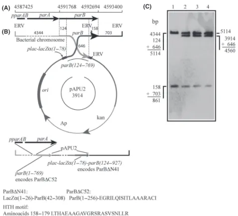
parB::pPAU1 mutants was evaluated by Southern blot using theparBgene as a probe (1C), and the detection of the hybridization bands of 1.031 bp and 5.615 bp con-firmed the integration of pAPU1 into the parB locus (compare lanes 1–3 and 2–3). To detect the production of ParB-TAP by XacparB::pPAU1, cells were cultivated in NYG medium until the mid-log phase, and subsequently processed for the immune detection of the TAP-tagged protein by Western blot (Fig. 1D). A signal of ~54 kDa was observed for the two mutant strains analyzed, which is consistent with the size expected for ParB-TAP (lanes 5–6). No bands could be detected for the wild-type strain (lane 7), whereas Xac transformed with the empty vector (Xacamy::pHF5Ca) produced a signal relative to the TAP tag of~21 kDa (lane 8). We also included in these analy-ses two mutants in which pAPU1 had integrated into the
amy locus through recombination between the amy106-912 fragment carried by pAPU1 and the a-amylase gene of Xac (Xac amy::pPAU1), hence, they express ParB-TAP
(A)
(B)
(C) (D)
Figure 1. Activity of the XacparABoperon. The activity of theparAB
operon was demonstrated by the ability of XacparB::pPAU1 mutants to express ParB-TAP under the control of the nativeparABpromoter. The ParB-TAP expression vector was integrated into theparBlocus of Xac by a single crossover event (B) generating the genomic structure depicted at the bottom of (B). (A) Genomic coordinates of theparAB
operon. (B) Schematics of the integration of pPAU1 (carrying the
parB-tap fusion) into the parB locus. Numbers above the map and around the circle indicate the sizes in base-pairs of the DNA fragments delimited; ERV, EcoRV restriction sites. (C) Southern blot analysis of Xac parB::pPAU1 mutants: genomic DNA was digested with EcoRV and subsequently probed with a DIG-labeledparB. The sizes of the DNA fragments detected correspond to those illustrated in (B). Lanes 1 and 2, XacparB::pPAU1 mutants 1 and 2, respectively; lane 3, wild-type Xac. (D) Western blot was used to detect ParB-TAP (54 kDa) in the protein extracts of Xac parB::pPAU1 and Xacamy:: pPAU1 mutants. Lanes: 1–2, Xac amy::pPAU1 mutant 1; 3–4, Xac
ectopically to serve as a control. Note that both Xacamy:: pPAU1 mutants also express ParB-TAP, but at higher lev-els (compare lanes 1 and 3 with 2 and 4 for noninduced and xylose-induced cultures, respectively, with lanes 5–6). The lower expression of ParB-TAP by the Xac parB:: pPAU1 mutants is probably due to the fact that these cells haveparB-tap1479 under the control ofpparAB, and therefore, they express ParB-TAP at physiological/natural levels. In summary, the stable production of ParB-TAP from the nativeparABpromoter confirmed the activity of theparABoperon in Xac.
Xac parB mutant is unable to induce citrus canker symptoms
Several unsuccessful attempts were made to delete the
parBgene of Xac. To circumvent this problem and to be able to extend our functional characterization of the
parAB operon of Xac, we decided to construct a mutant strain that can only express truncated forms of ParB. This was achieved by transforming Xac with the suicide vector pAPU2 that carried a DNA fragment corresponding to
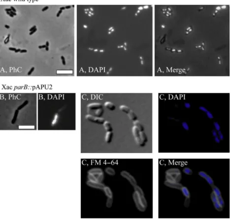
parB124-769. Upon integration into the parB locus (Fig. 2B) two new forms of parBshould arise: one under the control of pparAB, which lacks the coding sequence for the 52 C-terminal residues of ParB (codes for an equivalent of ParB∆C52, and that has a DNA addition from the vector coding for extra 18 residues not related to LacZ [Fig. 2B]), and another that can only be expressed by the activity of theplac promoter of pPAU2, and that codes for a LacZa-ParB chimera (LacZa 1-26-ParB42-308, which corresponds to a ParB deleted for the first 41 N-terminal aminoacids, ParB∆N41). Although ParB∆C52 carries a predicted HTH motif (Fig. 2B; resi-dues 158–179 identified using GYM2.0; Narasimhan et al. 2002), this protein is not expected to form dimers and
(A)
(B)
(C)
Figure 2. The strategy used to disruptparBin Xac. The suicide vector pPAU2, which carries theparB124–769fragment, was integrated into the
parBlocus of Xac by a single crossover event; integration generated the genomic structure depicted underneath the vector in which ParB∆52 can be expressed under the control of the native promoter of ParB (pparAB). (A) Genomic coordinates ofparAB. (B) Schematics of integration: upon plasmid insertion, two new forms ofparBwill be generated: one coding for ParB∆N41, a fusion between the first 26 amino acids of LacZa(coded by the expression vector) plus residues 42–308 of ParB, and another that codes for ParB∆C52, which lacks the 52 C-terminal residues of ParB and has an addition of the 18 new amino acids shown. Both truncated ParB proteins still carry the helix-turn-helix (HTH) motif shown. The numbers above the map and around the circle indicate the sizes of the DNA fragments delimited; the predicted helix-turn-helix motif of ParB is depicted below the drawings; ERV,EcoRV restriction sites. (C) Southern blot analysis: total DNA of the three XacparB::pAPU2 mutants was digested with
should be impaired in DNA binding (by analogy to data from the biochemical characterization of Spo0J∆20, and also of a C-terminal deletion form of ParB from Thermus thermophilus (Leonard et al. 2004; Murray et al. 2006); and H. Ferreira and J. Errington, unpublished data). On the other hand, ParB∆N41, if correctly expressed and folded, would be compromised in its ability to interact and/or stimulate the ParA partner as demonstrated for the ParAB system of T. thermophilus (Leonard et al. 2005a).
Upon transformation of Xac with pAPU2, and the expected integration of this plasmid into the bacterium chromosome, the genomic context surrounding the parB
locus of three independently selected Xac parB::pAPU2 mutants, which were derived from the same disruption event, was checked by Southern blot (Fig. 2C), where the observation of the diagnostic hybridization band of 4.560 bp confirmed the alterations. The three mutant strains exhibited a slower growth rate when compared with the wild-type strain. Growth curves of these mutants displayed a slower doubling time on the first 24 h, being only able to reach the usual culture limit for Xac (OD600 nm ~2) after 30 h of growth. This contrasts with the 18 h normally taken by the wild-type strain to approach the growth peak (Figure S1). Note that it took twice as much for the mutant to get to the same culture limit of OD600 nm~2. Furthermore, the recovery of kana-mycin-resistant mutants after transformation took almost 5 days (120 h), whereas wild-type colonies become visible in up to 48 h.
As a plant pathogen, the ability to colonize its host is a vital process for Xac. Since the disruption ofparBaltered the growth pattern of the Xac parB::pAPU2 mutants on rich medium, we tested whether these cells would still be able to colonize citrus plants. The three Xac parB::pAPU2 mutants were infiltrated in leaves of Rangpur lime along-side the wild-type strain. While the region inoculated with the wild-type strain evolved erumpent–corky and brownish lesions surrounding the infiltration point, none of the mutant strains was capable of inducing the typical symptoms of citrus canker (Fig. 3), even after the long incubation periods of 60 days. We conclude that disrup-tion of parB in Xac retarded the cell doubling time and promoted a loss of the ability to colonize the host citrus.
Alteration of the normal ParB function disrupts cell division in Xac
Disruption, deletion, and/or the overexpression of ParB-like proteins in several microorganisms, includingC. cres-centus, C. glutamicum, and Pseudomonas aeruginosa (P. aeruginosa), may lead to pleiotropic effects such as cell filamentation, loss of motility, and perturbations in the
basic processes of chromosome segregation and cell divi-sion (Mohl and Gober 1997; Bartosik et al. 2009; Dono-van et al. 2010). In this work, the inability to colonize citrus observed for Xac parB::pAPU2 could be a conse-quence of at least two other deficiencies beyond the per-turbation of the segrosome: (1) metabolic and/or physiological alterations, and (2) the perturbation of pathogenicity systems/factors indirectly triggered by the disruption of parB. To eliminate such possibilities, we began by examining the mutants under the microscope in order to detect any morphological changes in the cells that could support a segregation defect.
In general, cultures of the parB mutant Xac parB:: pAPU2 had a mix of filaments and rods (Figs. 4, 5). Per-centages of the entire cell types documented are shown in Table 2. Filaments and/or irregular chains were approxi-mately 5% of the cell types in a culture (n=400) (Fig. 5). Close inspection of these filamented cells showed the presence of septal constrictions (arrows in Fig. 5), in which the division pattern displayed seems as if a particu-lar rod started to grow and divide normally, losing the ability to form septa in successive cellular cycles; thus, fil-amentation seems to happen with elder rods attached to their tips. Despite the propensity to grow in long fila-ments, the division machinery seems operative in these mutants.
Considering the nonfilamented cells of Xac parB:: pAPU2, they looked apparently longer than the wild-type strain (compare Fig. 4A and B; Table 2). In order to quantitatively evaluate this, we measured 200 individuals
of each culture (wild-type and Xac parB::pAPU2), and calculated the average cell length. We observed significant differences between them: the average wild-type cell
length was 1.440.31lm, whereas the Xac parB:: pAPU2 mutants measured 2.560.42lm.
To subsequently estimate the effects ofparBdisruption on the chromosome organization of Xac parB::pAPU2, cells were labeled with 40,6-diamidino-2-phenylindole (DAPI) and visualized by fluorescence microscopy (Figs. 4, 5). Cultures of Xac parB::pAPU2 exhibited an increase in the number of anucleate rods (~30% of the cell types, Fig. 4B and C), which contrasts with an absence of DNA-free cells in cultures of wild-type Xac (Table 2). When we looked at filamented cells of the Xac
parBmutant, we observed a considerable accumulation of chromosomal mass, sometimes adopting the form of a continuum in which the nucleoid fills up the whole of a cell compartment extending through and preventing the closure of a septum (Fig. 5; arrow on the right). Minicells
Figure 4. Morphological analysis of XacparB:: pPAU2 mutant cells. The XacparB::pPAU2 mutant, which expresses only truncated forms of ParB, was compared with Xac wild type using various combinations of phase contrast (PhC), differential interference contrast (DIC), and fluorescence microscopy; the membrane and nucleoid stains FM 4–64 and DAPI, respectively, were used as indicated. Wild-type and mutant cells were cultivated in NYG medium at 30°C, and inspected around the OD600 nm~0.3; cells were immobilized onto 1% agarose-covered slides. (A) Xac wild type. (B–C) XacparB::pPAU2. Scale bar corresponds to 4lm.
Figure 5. The filamentation phenotype of XacparBmutant. The Xac
parB mutant (Xac parB::pPAU2) was investigated under phase contrast (PhC) and fluorescence microscopy of DAPI-stained cells (DAPI) in order to evaluate their chromosome organization. Here, we show the picture of a representative filament from cells cultivated in NYG medium at 30°C, and visualized around the OD600 nm ~0.3; arrows show the closure of septa; on the right (DAPI), septum is closing without chromosome clearance. Scale bar corresponds to 4lm.
Table 2. Morphological analysis of XacparB::pAPU2. Cell length
(n=200),lm
Filaments
(n=400) Anucleate Minicells
Xac wild type 1.440.31 0 0 0
XacparB::pAPU2 2.560.42* 5% 30% 0
were not detected neither in cultures of Xac wild-type nor in the parBmutant (Table 2). Altogether, data show that the disruption of parB in Xac leads to a severe impair-ment of the chromosome segregation process, which also culminated in an unexpected alteration of its ability to stimulate citrus canker symptoms.
Subcellular localization of ParB-GFP
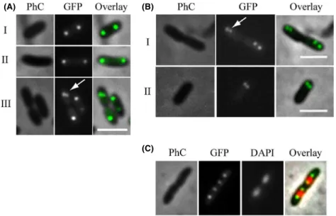
The localization patterns of several chromosomally encoded ParB-like proteins have been documented in vivo and it constitutes an excellent indicative of function in chromosome segregation (Glaser et al. 1997; Mohl and Gober 1997; Jakimowicz et al. 2005; Fogel and Waldor 2006; Bartosik et al. 2009; Maloney et al. 2009; Donovan et al. 2010). To further extend our characterization of Xac ParB, we prepared a mutant strain, XacparB::pAPU3, which expresses ParB-GFP under the control ofpparABat physiological levels (Fig. 6) (Xac parB::pAPU3 was con-structed using the same strategy depicted above for the expression of ParB-TAP). In Figure 6A (I and II), we show the localization of ParB-GFP in cells without a clear sign of septal constriction. Note the presence of two foci per compartment, each occupying one of the cellular poles. This localization pattern was very similar to the one documented for the ParB homologue of C. crescentus
(Mohl and Gober 1997). Inspection of several fields revealed another localization pattern in which the ParB-GFP focus is splitting in two in just one of the cellular compartments (Fig. 6A-III, arrow), whereas in the other it apparently remains as a single entity. The pattern seen in Figure 6A-III is probably a resultant of either the initi-ation of a new repliciniti-ation event in only one of the cell compartments, at a late stage of septation (which implies
an asynchrony in the process of initiation of chromosome replication), or the brighter individual focus would in fact be two ParB-GFP foci superimposed. We favor the latter possibility as we also observed localization patterns in which the cells seemed to be in synchrony with regard to the initiation of replication (Fig. 6B). In Figure 6B-I, we see dividing cells and two closely positioned ParB-GFP foci occupying in each compartment just one of the polar regions. Here, new replication events that would separate the origin regions of the chromosomes are taking place at the poles; hence, the replisomes should be nearby. The cellular type shown in Figure 6B-II (a nondividing rod) seems to support the idea that the origins in Xac initiate replication toward one of the cellular poles, which is simi-lar to the replication patterns exhibited by C. crescentus,
V. cholerae, and C. glutamicum (Mohl and Gober 1997; Fogel and Waldor 2006; Donovan et al. 2010). Finally, we performed DAPI staining of cells and captured individuals in late stages of septum closure judged by the placement of the ParB-GFP foci within the cells (Fig. 6C). We observed four ParB-GFP foci evenly distributed inside the two linked cell compartments, where ParB-GFP colocaliz-es with the edgcolocaliz-es of the nucleoids. If we consider that these edges comprise the origins of replication, similar to
B. subtilis and many other systems (Leonard et al. 2005b; Mierzejewska and Jagura-Burdzy 2012), parS in Xac is located in this region (at ~3 kb from the origin in the chromosome of Xac (Livny et al. 2007)) and ParB-GFP is expected to be bound to it.
In Figure 6B-II, we showed evidence that Xac initiates chromosome replication close to a cellular pole. This sug-gests that chromosome segregation is asymmetric in this plant pathogen as it was also proposed for C. crescentus,
V. cholerae, and C. glutamicum (Fogel and Waldor 2006;
(A) (B)
(C)
Figure 6. Localization of ParB-GFP. The localization pattern of ParB-GFP was analyzed in different cell types of a XacparB::pAPU3 expressing ParB-GFP from the nativepparAB
promoter. (A, B, C) represent cells
photographed during different moments of the cell cycle; arrows mark recently divided origins of replication (see text). Cells were cultivated in NYG medium at 30°C until the OD600nmof
Donovan et al. 2010; Ptacin et al. 2010). In order to fur-ther characterize the asymmetric pattern of chromosome segregation in Xac, we performed time-lapse microscopy using Xac parB::pAPU3 (Fig. 7). From t=0 to
t=20 min the leftmost dividing rods started new chro-mosomal replication events that can be seen by the divi-sion of the ParB-GFP foci (arrows in t =20 min). Note that at the beginning of the process (from t= 0 until
t=20), the origins of replication, and consequently the ParB-GFP foci associated with them, were situated near the cellular poles and opposite to the septum. From
t=20 to t=40, we see the innermost origins/ParB-GFP foci moving toward the septum until they reach the
region that will become the new pole after cytokinesis. Therefore, we conclude that Xac exhibits asymmetric chromosome segregation.
Discussion
We demonstrated recently that esters of gallic acid per-turbed the cell division and/or the chromosome segrega-tion apparatuses of Xac. Thus, these compounds constitute promising antimicrobial agents against citrus canker and probably other bacterial diseases (Silva et al. 2013). Here, we further characterized the Xac mutant strain used by Silva et al. (2013) that is labeled for the centromere (XacparB::pAPU3), and extended the studies with ParB encoded by this plant pathogen. First, we showed that Xac ParB is stably expressed and operates on chromosome segregation. Chromosomally encoded ParB-like proteins have been characterized in several bacteria (recently reviewed by (Mierzejewska and Jagura-Burdzy 2012)), where the perturbation of their normal function and/or their cellular dosage with respect to their associ-ated ParA partners often lead to an increase in the num-ber of anucleate cells, cell filamentation, and nucleoid disorganization. Disruption of parB in Xac produced these classical phenotypes (Figs. 4, 5). In addition, in the absence of full-length ParB, Xac was not able to colonize the host citrus (Fig. 3). Second, the subcellular localiza-tion of ParB-GFP suggests an asymmetric mode of repli-con segregation in Xac, which resembles the chromosome segregation patterns observed for C. crescentus, V. chole-rae, and C. glutamicum (Fogel and Waldor 2006; Dono-van et al. 2010; Ptacin et al. 2010). This, along with the fact that lack of ParB produced a remarkable cell filamen-tation phenotype suggests that ParB may be involved with cell division in Xac.
The inability to produce disease symptoms in planta
documented for the Xac parB mutant is a novel effect, which reinforced our previous observations that alkyl gal-lates that were able to perturb the subcellular localization of ParB-GFP also precluded the ability of Xac to colonize citrus (Silva et al. 2013). In the past decade, several stud-ies gradually unveiled the participation of ParAB proteins with various cellular processes other than chromosome segregation. In P. aeruginosa, for example, absence of ParB altered bacterial growth and affected swarming and swimming motilities (Lasocki et al. 2007; Bartosik et al. 2009). Slower growth rates were also detected in our experiments for theparBmutant of Xac. In C. crescentus, it was demonstrated that ParB operates on cell division by sequestering the FtsZ inhibitor MipZ to areas away from the cell center where the divisome should assemble (Thanbichler and Shapiro 2006b; Kiekebusch et al. 2012). In the Gram-positive bacterium B. subtilis the ParB-like
protein Spo0J recruits structural maintenance of chromo-somes to the origin of replication where it was proposed to assist in the organization of the region to enable more efficient chromosome segregation (Sullivan et al. 2009). Although Spo0J may help in replicon partition, it is required for sporulation in B. subtilis, and recently it has also been implicated with the control of initiation of DNA replication by modulating the action of the ParA-like factor Soj (Ireton et al. 1994; Scholefield et al. 2011). The involvement of ParAB proteins with control of DNA replication was also reported for V. cholerae (Kadoya et al. 2011). We are not certain yet whether the pathoge-nicity shutdown following parBdisruption in Xac has any association with pathogenicity systems/routes encoded by the bacterium. We have employed yeast two-hybrid analy-ses in an attempt to identify interactions of ParB with factors belonging to known pathogenicity systems in Xac (A. Ucci, S. C. Farah, and H. Ferreira, unpubl. results); but so far, nothing was detected. Therefore, we believe that the loss of virulence may well be simply a response to the perturbation of the vital cellular processes of chro-mosome segregation and/or cell division.
In this work, we showed evidence for asymmetric chro-mosome segregation in Xac (Figs. 6, 7). First, we see newly duplicated origins of replication labeled with ParB-GFP occupying just one of the cellular poles (Figs. 6B-I, 7). Later, the two ParB-GFP foci are well separated from each other within the same cellular compartment (Figs. 6C, 7), which suggests that segregation occurs with the internally located origins migrating from the point of duplication toward the opposite pole (Fig. 7). Asymmet-ric chromosome segregation was documented for C. cres-centus, V. cholerae, and C. glutamicumas well (Fogel and Waldor 2006; Donovan et al. 2010; Ptacin et al. 2010). For the most extensively characterized system, C. crescen-tus, the chromosome is oriented such that the origin of replication (designated cori) is close to what is called the old pole (C. crescentus is known for exhibiting a strict polar asymmetry, in which diverse protein factors/systems are arranged in one but not at both cellular poles at once). Before DNA replication starts, ParB is bound to
parS located around cori; ParB/parS organizes a centro-mere (Toro et al. 2008) that is kept anchored to the old pole by PopZ (Bowman et al. 2008; Ebersbach et al. 2008). When replication fires, the centromere is dupli-cated, and one copy of this region is captured by ParA fil-aments that come from the opposite cell pole, where they are anchored to TipN (a factor that marks the new pole) (Ptacin et al. 2010; Schofield et al. 2010). ParB/parS then interacts with ParA, ParB stimulates ParA depolymeriza-tion that shortens the filament, which is followed by sub-sequent recapture of ParB/parS; orientation of the centromeric region to the new pole operates chromosome
partitioning. A related segregation mechanism was also proposed for V. cholerae(Fogel and Waldor 2006), which presents some differences with that of C. crescentus, one of which is the use of a distinct polar anchor designated HubP, which interacts with ParA (Yamaichi et al. 2012). As for C. glutamicum, DivIVA was demonstrated to be the factor that tethers the ParB/DNA complex at the cell pole (Donovan et al. 2012). Considering the similarities between systems, in particular the ParB-GFP subcellular localization patterns documented for C. crescentus, V. cholerae, C. glutamicum, and Xac, we expect to identify protein factors of equivalent functions in Xac in the near future.
The localization pattern of ParB-GFP in Xac as well as the observation that chromosome segregation and cell division may be interlinked raised the idea that Xac could control septum placement in a manner similar to C. cres-centus (Thanbichler and Shapiro 2006b). Curiously, simi-lar findings and suggestions were recently reported forM. smegmatis (Ginda et al. 2013), where deletion of parA
produced aberrant phenotypes of chromosome segrega-tion and septum posisegrega-tioning leading to cell elongasegrega-tion. The association of chromosome segregation and cell divi-sion in Xac was first proposed by Silva et al. (2013), who demonstrated that alkyl gallates able to perturb the locali-zation of ParB-GFP in this bacterium apparently acted on septum assembly as well. In C. crescentus, septum site selection depends on the centromere migration dynamics (Thanbichler and Shapiro 2006a), where MipZ, an inhibi-tor of FtsZ polymerization, interacts and localizes with ParB bound to parS (Thanbichler and Shapiro 2006a; Kiekebusch et al. 2012). As the centromeric complexes occupy the poles, the FtsZ inhibitor MipZ is kept away from the cell center where the septum is assembled. Therefore, disruption of ParB in C. crescentus perturbs the control of septation and leads to cell filamentation. Xac does not have an obvious mipZ homologue on its genome. MipZ is conserved in a-proteobacteria, and belongs to the Mrp/MinD family of P-loop ATPases, being structurally related to the ParA superfamily mem-bers Soj and MinD (Kiekebusch et al. 2012). However, Xac carries at least four parA-like ORFs (XAC0192, XAC1907, XAC2205, and XAC2433) that could have a
MinCD. Ultimately, the filamentation observed for the Xac parBmutant strain could be caused by the action of a nucleoid occlusion system, analogous to the Noc/SlmA systems ofB. subtilisandE. coli(Wu and Errington 2004; Bernhardt and de Boer 2005). As illustrated in Fig. 5, chromosome replication in the XacparBmutant, followed by inadequate segregation, apparently produces cells with several chromosomal copies that if associated with factors able to inhibit septum assembly/closure would lead to fil-amentation. However, Xac does not seem to encode any Noc/SlmA-like protein that could fulfill the function of a nucleoid occlusion factor (Wu and Errington 2004; Bern-hardt and de Boer 2005).
Acknowledgments
A. P. U. and P. M. M. M. received scholarships from FAPESP (2010/02041-8) and CNPq (142293/2009-1), respectively. We thank C. S. Farah for helping us with the YTH experiments and valuable discussion. This work was funded by FAPESP (Grants: 2004/09173-6; 2010/05099-7), PROPe-UNESP, and PADC-FCF-UNESP. Beneficia´rio de auxı´lio financeiro da CAPES, Brasil.
Conflict of Interest
None declared.
References
Bartosik, A. A., J. Mierzejewska, C. M. Thomas, and G.
Jagura-Burdzy. 2009. ParB deficiency inPseudomonas aeruginosa
destabilizes the partner protein ParA and affects a variety of physiological parameters. Microbiology 155:1080–1092. Behlau, F., L. Amorim, J. Jr Belasque, A. Bergamin Filho,
R. P. Jr Leite, J. H. Graham, et al. 2010. Annual and polyetic progression of citrus canker on trees protected with copper sprays. Plant Pathol. 59:1031–1036.
Belasque, J. Jr, J. C. Barbosa, A. Bergamin Filho, and
C. A. Massari. 2010. Provaveis consequeˆncias do abrandamento da metodologia de erradicacßa˜o do cancro cı´trico no Estado de Sa˜o Paulo. Trop. Plant Pathol. 35:314–317.
Bernhardt, T. G., and P. A. de Boer. 2005. SlmA, a nucleoid-associated, FtsZ binding protein required for blocking septal ring assembly over chromosomes inE. coli. Mol. Cell 18:555–564.
Bowman, G. R., L. R. Comolli, J. Zhu, M. Eckart, M. Koenig, K. H. Downing, et al. 2008. A polymeric protein anchors the chromosomal origin/ParB complex at a bacterial cell pole. Cell 134:945–955.
Dajkovic, A., S. Pichoff, J. Lutkenhaus, and D. Wirtz. 2010. Cross-linking FtsZ polymers into coherent Z rings. Mol. Microbiol. 78:651–668.
Daniels, M. J., C. E. Barber, P. C. Turner, M. K. Sawczyc, R. J. Byrde, and A. H. Fielding. 1984. Cloning of genes involved in pathogenicity ofXanthomonas campestrispv. campestris using the broad host range cosmid pLAFR1. EMBO J. 3:3323–3328.
Donovan, C., A. Schwaiger, R. Kramer, and M. Bramkamp. 2010. Subcellular localization and characterization of the ParAB system fromCorynebacterium glutamicum. J. Bacteriol. 192:3441–3451.
Donovan, C., B. Sieger, R. Kramer, and M. Bramkamp. 2012. A syntheticEscherichia colisystem identifies a conserved origin tethering factor in Actinobacteria. Mol. Microbiol. 84:105–116.
Easter, J. Jr, and J. W. Gober. 2002. ParB-stimulated nucleotide exchange regulates a switch in functionally distinct ParA activities. Mol. Cell 10:427–434.
Ebersbach, G., A. Briegel, G. J. Jensen, and C. Jacobs-Wagner. 2008. A self-associating protein critical for chromosome attachment, division, and polar organization in caulobacter. Cell 134:956–968.
Ferreira, H., and J. Jr Belasque. 2011. Proceedings of the international workshop on Xanthomonas citri/citrus canker. Ribeira˜o Preto, Brazil, ISBN: 978-85-64947-04-7, Available at (http://www.fcfar.unesp.br/wxc/download/
workshop_Xanthomonas.pdf) UNESP/FUNDECITRUS. Ferreira, H., F. J. A. Barrientos, R. L. Baldini, and Y. B.
Rosato. 1995. Electrotransformation in three pathovars of
Xanthomonas campestris. Appl. Microbiol. Biotechnol. 43:651–655.
Fogel, M. A., and M. K. Waldor. 2006. A dynamic, mitotic-like mechanism for bacterial chromosome segregation. Genes Dev. 20:3269–3282.
Gerdes, K., M. Howard, and F. Szardenings. 2010. Pushing and pulling in prokaryotic DNA segregation. Cell 141:927–942. Ginda, K., M. Bezulska, M. Ziolkiewicz, J. Dziadek, J.
Zakrzewska-Czerwinska, and D. Jakimowicz. 2013. ParA of
Mycobacterium smegmatisco-ordinates chromosome segregation with the cell cycle and interacts with the polar growth determinant DivIVA. Mol. Microbiol. 87:998–1012. Glaser, P., M. E. Sharpe, B. Raether, M. Perego, K. Ohlsen,
and J. Errington. 1997. Dynamic, mitotic-like behavior of a bacterial protein required for accurate chromosome partitioning. Genes Dev. 11:1160–1168.
Gottwald, T. R., J. H. Graham, and T. S. Schubert. 2002. Citrus canker: the pathogen and its impact. Online Plant Health Prog. doi:10.1094/PHP-2002-0812-01-RV. Gueiros-Filho, F. J., and R. Losick. 2002. A widely conserved
bacterial cell division protein that promotes assembly of the tubulin-like protein FtsZ. Genes Dev. 16:2544–2556. Ireton, K., N. W. Gunther, and A. D. Grossman. 1994. spo0J is
Jakimowicz, D., B. Gust, J. Zakrzewska-Czerwinska, and K. F. Chater. 2005. Developmental-stage-specific assembly of ParB complexes inStreptomyces coelicolorhyphae. J. Bacteriol. 187:3572–3580.
Jakimowicz, D., P. Zydek, A. Kois, J. Zakrzewska-Czerwinska, and K. F. Chater. 2007. Alignment of multiple chromosomes along helical ParA scaffolding in sporulatingStreptomyces
hyphae. Mol. Microbiol. 65:625–641.
Kadoya, R., J. H. Baek, A. Sarker, and D. K. Chattoraj. 2011. Participation of chromosome segregation protein ParAI of
Vibrio choleraein chromosome replication. J. Bacteriol. 193:1504–1514.
Kiekebusch, D., K. A. Michie, L. O. Essen, J. Lowe, and M. Thanbichler. 2012. Localized dimerization and nucleoid binding drive gradient formation by the bacterial cell division inhibitor MipZ. Mol. Cell 46:245–259.
Lasocki, K., A. A. Bartosik, J. Mierzejewska, C. M. Thomas, and G. Jagura-Burdzy. 2007. Deletion of the parA (soj) homologue inPseudomonas aeruginosacauses ParB instability and affects growth rate, chromosome segregation, and motility. J. Bacteriol. 189:5762–5772.
Leonard, T. A., P. J. Butler, and J. Lowe. 2004. Structural analysis of the chromosome segregation protein Spo0J from
Thermus thermophilus. Mol. Microbiol. 53:419–432. Leonard, T. A., P. J. Butler, and J. Lowe. 2005a. Bacterial
chromosome segregation: structure and DNA binding of the Soj dimer–a conserved biological switch. EMBO J. 24:270– 282.
Leonard, T. A., J. Moller-Jensen, and J. Lowe. 2005b. Towards understanding the molecular basis of bacterial DNA segregation. Philos. Trans. R. Soc. Lond. B Biol. Sci. 360:523–535.
Livny, J., Y. Yamaichi, and M. K. Waldor. 2007. Distribution of centromere-like parS sites in bacteria: insights from comparative genomics. J. Bacteriol. 189:8693–8703. Low, H. H., M. C. Moncrieffe, and J. Lowe. 2004. The crystal
structure of ZapA and its modulation of FtsZ polymerisation. J. Mol. Biol. 341:839–852.
Lutkenhaus, J. 2007. Assembly dynamics of the bacterial MinCDE system and spatial regulation of the Z ring. Annu. Rev. Biochem. 76:539–562.
Maloney, E., M. Madiraju, and M. Rajagopalan. 2009. Overproduction and localization ofMycobacterium tuberculosisParA and ParB proteins. Tuberculosis 89(Suppl. 1):S65–S69.
Martins, P. M., I. F. Lau, M. Bacci, J. Belasque, A. M. do Amaral, S. R. Taboga, et al. 2010. Subcellular localization of proteins labeled with GFP inXanthomonas citrissp.citri: targeting the division septum. FEMS Microbiol. Lett. 310:76–83.
Mierzejewska, J., and G. Jagura-Burdzy. 2012. Prokaryotic ParA-ParB-parS system links bacterial chromosome segregation with the cell cycle. Plasmid 67:1–14.
Mohammadi, T., G. E. Ploeger, J. Verheul, A. D. Comvalius, A. Martos, C. Alfonso, et al. 2009. The GTPase activity of
Escherichia coliFtsZ determines the magnitude of the FtsZ polymer bundling by ZapA in vitro. Biochemistry 48:11056– 11066.
Mohl, D. A., and J. W. Gober. 1997. Cell cycle-dependent polar localization of chromosome partitioning proteins in
Caulobacter crescentus. Cell 88:675–684.
Murray, H., H. Ferreira, and J. Errington. 2006. The bacterial chromosome segregation protein Spo0J spreads along DNA from parS nucleation sites. Mol. Microbiol. 61:1352–1361.
Narasimhan, G., C. Bu, Y. Gao, X. Wang, N. Xu, and K. Mathee. 2002. Mining protein sequences for motifs. J. Comput. Biol. 9:707–720.
Ptacin, J. L., S. F. Lee, E. C. Garner, E. Toro, M. Eckart, L. R. Comolli, et al. 2010. A spindle-like apparatus guides bacterial chromosome segregation. Nat. Cell Biol. 12:791– 798.
Puig, O., F. Caspary, G. Rigaut, B. Rutz, E. Bouveret, E. Bragado-Nilsson, et al. 2001. The tandem affinity purification (TAP) method: a general procedure of protein complex purification: a generic protein purification method for protein complex characterization and proteome exploration. Methods 24:218–229.
Sambrook, J., E. F. Fritsch, and T. Maniatis. 1989. Molecular cloning: a laboratory manual. Cold Spring Harbor Laboratory Press, Cold Spring Harbor, NY. Schaad, N. W., E. Postnikova, G. H. Lacy, A. Sechler,
I. Agarkova, P. E. Stromberg, et al. 2005. Reclassification of
Xanthomonas campestrispv. citri (ex Hasse 1915) Dye 1978 forms A, B/C/D, and E asX. smithiisubsp.citri(ex Hasse) sp. nov. nom. rev. comb. nov.,X. fuscanssubsp.aurantifolii
(ex Gabriel 1989) sp. nov. nom. rev. comb. nov., and
X. alfalfaesubsp.citrumelo(ex Riker and Jones) Gabriel et al., 1989 sp. nov. nom. rev. comb. nov.;X. campestrispv. malvacearum (ex smith 1901) Dye 1978 asX. smithiisubsp.
smithiinov. comb. nov. nom. nov.;X. campestrispv. alfalfae (ex Riker and Jones, 1935) dye 1978 asX. alfalfaesubsp.
alfalfae(ex Riker et al., 1935) sp. nov. nom. rev.; and “var. fuscans” ofX. campestrispv. phaseoli (ex Smith, 1987) Dye 1978 asX. fuscanssubsp.fuscanssp. nov. Syst. Appl. Microbiol. 28:494–518.
Schaad, N. W., E. Postnikova, G. Lacy, A. Sechler, I. Agarkova, P. E. Stromberg, et al. 2006. Emended classification of xanthomonad pathogens on citrus. Syst. Appl. Microbiol. 29:690–695.
Schofield, W. B., H. C. Lim, and C. Jacobs-Wagner. 2010. Cell cycle coordination and regulation of bacterial chromosome segregation dynamics by polarly localized proteins. EMBO J. 29:3068–3081.
switch from an activator to an inhibitor of DNA replication initiation. Mol. Microbiol. 79:1089–1100.
Silva, I. C., L. O. Regasini, M. S. Petronio, D. H. Silva, V. S. Bolzani, J. Jr Belasque, et al. 2013. Antibacterial Activity of Alkyl Gallates againstXanthomonas citrisubsp.citri. J. Bacteriol. 195:85–94.
da Silva, A. C., J. A. Ferro, F. C. Reinach, C. S. Farah, L. R. Furlan, R. B. Quaggio, et al. 2002. Comparison of the genomes of twoXanthomonaspathogens with differing host specificities. Nature 417:459–463.
Sullivan, N. L., K. A. Marquis, and D. Z. Rudner. 2009. Recruitment of SMC by ParB-parS organizes the origin region and promotes efficient chromosome segregation. Cell 137:697–707.
Thanbichler, M., and L. Shapiro. 2006a. Chromosome organization and segregation in bacteria. J. Struct. Biol. 156:292–303.
Thanbichler, M., and L. Shapiro. 2006b. MipZ, a spatial regulator coordinating chromosome segregation with cell division inCaulobacter. Cell 126:147–162.
Toro, E., S. H. Hong, H. H. McAdams, and L. Shapiro. 2008.
Caulobacterrequires a dedicated mechanism to initiate chromosome segregation. Proc. Natl. Acad. Sci. USA 105:15435–15440.
Wu, L. J., and J. Errington. 2004. Coordination of cell division and chromosome segregation by a nucleoid occlusion protein inBacillus subtilis. Cell 117:915–925.
Yamaichi, Y., R. Bruckner, S. Ringgaard, A. Moll, D. E. Cameron, A. Briegel, et al. 2012. A multidomain hub anchors the chromosome segregation and
chemotactic machinery to the bacterial pole. Genes Dev. 26:2348–2360.
Supporting Information
Additional Supporting Information may be found in the online version of this article:
Figure S1.Growth curve of the mutant XacparB::pAPU2. Bacteria were cultivated for 36 h in NYG medium at 30°C and 200 rpm. Each point in the graph corresponds to an average of optical densities (OD600nm) calculated from three independent experiments; vertical bars indicate standard deviation values calculated for each average. Here, we show the measurements done for mutant 3; however, mutants 1 and 2 exhibited similar growth pat-terns. Blue, wild-type Xac; red, XacparB::pAPU2.