LDL Receptor-Related Protein-1 (LRP1)
Regulates Cholesterol Accumulation in
Macrophages
Anna P. Lillis1,5, Selen Catania Muratoglu1,2, Dianaly T. Au1, Mary Migliorini1,3, Mi-Jeong Lee4, Susan K. Fried4, Irina Mikhailenko1,2, Dudley K. Strickland1,2,3*
1Center for Vascular and Inflammatory Diseases, University of Maryland School of Medicine, Baltimore, MD 21201, United States of America,2Department of Physiology, University of Maryland School of Medicine, Baltimore, MD 21201, United States of America,3Department of Surgery, University of Maryland School of Medicine, Baltimore, MD 21201, United States of America,4Department of Medicine, Section of
Endocrinology, Diabetes and Nutrition, Boston University School of Medicine, Boston, MA 02118, United States of America,5Department of Pathology, Duke University Medical Center, Durham, NC 27710, United States of America
*dstrickland@som.umaryland.edu
Abstract
Within the circulation, cholesterol is transported by lipoprotein particles and is taken up by cells when these particles associate with cellular receptors. In macrophages, excessive li-poprotein particle uptake leads to foam cell formation, which is an early event in the devel-opment of atherosclerosis. Currently, mechanisms responsible for foam cell formation are incompletely understood. To date, several macrophage receptors have been identified that contribute to the uptake of modified forms of lipoproteins leading to foam cell formation, but the contribution of the LDL receptor-related protein 1 (LRP1) to this process is not known. To investigate the role of LRP1 in cholesterol accumulation in macrophages, we generated mice with a selective deletion of LRP1 in macrophages on an LDL receptor (LDLR)-deficient background (macLRP1-/-). After feeding mice a high fat diet for 11 weeks, peritoneal macro-phages isolated fromLrp+/+mice contained significantly higher levels of total cholesterol than those from macLRP1-/- mice. Further analysis revealed that this was due to increased levels of cholesterol esters. Interestingly, macLRP1-/- mice displayed elevated plasma cho-lesterol and triglyceride levels resulting from accumulation of large, triglyceride-rich lipopro-tein particles in the circulation. This increase did not result from an increase in hepatic VLDL biosynthesis, but rather results from a defect in catabolism of triglyceride-rich lipoprotein particles in macLRP1-/- mice. These studies reveal an importantin vivocontribution of mac-rophage LRP1 to cholesterol homeostasis.
Introduction
Key events during the development of atherosclerosis include the accumulation of lipoprotein particles in the subendothelial arterial intima, where they are taken up by recruited monocytes/ a11111
OPEN ACCESS
Citation:Lillis AP, Muratoglu SC, Au DT, Migliorini M, Lee M-J, Fried SK, et al. (2015) LDL Receptor-Related Protein-1 (LRP1) Regulates Cholesterol Accumulation in Macrophages. PLoS ONE 10(6): e0128903. doi:10.1371/journal.pone.0128903
Academic Editor:Patricia Aspichueta, University of Basque Country, SPAIN
Received:August 19, 2014
Accepted:May 3, 2015
Published:June 10, 2015
Copyright:© 2015 Lillis et al. This is an open access article distributed under the terms of theCreative Commons Attribution License, which permits unrestricted use, distribution, and reproduction in any medium, provided the original author and source are credited.
Data Availability Statement:All relevant data are within the paper.
Funding:This research was funded by research grants R01 HL114379 (DKS), R01 HL120388 (DKS), T32 HL007698 (DKS), from the National Heart, Lung, and Blood Institute, of the National Institutes of Health, and P30 DK072488 (SKF) from the National Institute of Diabetes and Disgestive and Kidney Disease of the National Institutes of Health.
macrophages [1]. This process occurs via interaction of cholesterol ester-rich lipoprotein parti-cles with specific macrophage receptors that target the internalized lipoproteins to lysosomes. Here the lipoprotein particles are processed and the cholesterol esters are hydrolyzed to free cholesterol [2]. In cells, excess free cholesterol is subsequently re-esterified and stored in the cy-toplasm as lipid droplets, which under conditions of excessive uptake results in the morpholog-ic appearance known as foam cells [3].
There is considerable interest in identifying macrophage receptors that participate in the in-ternalization of lipoprotein particles leading to cholesterol accumulationin vivo. Early studies confirmed that native forms of LDL particles are not taken up by macrophages and therefore do not contribute to excessive cholesterol uptake in these cells [3–5]. It was subsequently dis-covered that modified forms of LDL are internalized by macrophages leading to unregulated cholesterol uptake [3,4,6]. Further investigation revealed that macrophages express a number of scavenger receptors that recognize oxidized forms of LDL. Cell based studies have identified two members of this receptor family, SR-A (Msr1) and CD36 (Cd36) that can mediate the cel-lular internalization of oxidized LDL leading to cholesterol ester accumulation and foam cell formation [7]. Peritoneal macrophages isolated fromCd36-/-,apoE-/-double knockout mice or
Msr1-/-,apoE-/-double knockout mice on a high fat diet show a marked reduction in free and esterified cholesterol [8] confirming thein vivoimportance of these receptors in cholesterol ac-cumulation. However, targeted deletion of both SR-A and CD36 on an apoE-deficient back-ground does not abrogate macrophage foam cell formation, revealing that other mechanisms exist in macrophages that contribute to lipid uptake [9]. More recently, the lectin-type oxidized LDL receptor (LOX1) has been identified in endothelial cells as well as macrophages [10] which binds oxidized forms of LDL, and enhances the development of atherogenesis in LDL re-ceptor-deficient mice [11].
Certain modifications of LDL, such as incubation with secreted sphingomyelinase, results in aggregation of the LDL particles which leads to enhanced macrophage uptake and cholesterol loading in these cells [12–14]. Various mechanisms have been suggested for the internalization of aggregated LDL particles by macrophages [15], including receptor-mediated uptake by the LDL receptor-related protein 1 (LRP1) [16–18]. LRP1 is a large endocytic receptor that was originally identified when Herz et al. [19] cloned a large protein containing multiple LDLa re-peats and when Ashcom et al. [20] and Moestrup et al. [21] isolated and sequenced the liver re-ceptor responsible for catabolism ofα2-macroglobulin (α2M)-proteinase complexes [22]. In addition to its ability to bindα2M-proteinase complexes, early cross-linking studies revealed that LRP1 can also bind apolipoprotein E-containing liposomes [23] suggesting that LRP1 might also function as a receptor for chylomicron and VLDL remnants rich in apoE. This was confirmed in studies revealing that genetic deletion of hepatic LRP1 in LDL receptor-deficient mice resulted in a substantial increase in remnant accumulation in the plasma [24].
LRP1 is abundant in several cells, including macrophages. Prior work has revealed that mice with a selective deletion of LRP1 in macrophages have more extensive atherosclerosis when crossed into an apoE/LDL receptor double knockout mouse [25] or when bone marrow from LRP1-/- mice are transplanted into irradiated LDL receptor-deficient mice [26–28]. Addition-ally, genetic deletion of LRP1 in macrophages also results in more extensive vascular remodel-ing upon injury [29]. The mechanisms by which macrophage LRP1 modulates the
employing mice with tissue-selective deletion of theLrp1gene in macrophages. The results re-veal an important contribution of macrophage LRP1 to cholesterol uptake in macrophages.
Materials and Methods
Animals
Mice with LRP1 deleted in macrophages were generated on an LDLR-deficient background by crossing LysMCre mice [31] (kindly provided by I. Förster, Munich) withLDLR-/-,LRP1flox/flox
mice [24] (kindly provided by J. Herz, Dallas) as described [32]. Littermate siblings of Cre-, termed LRP1+/+ (LDLR-/-,LRP1flox/flox,Cre-/-) or Cre+, termed macLRP1-/- (LDLR-/-,LRP1flox/
flox,Cre-/+) were used in all experiments. Mice were genotyped by polymerase chain reaction
(PCR) as described [29]. Mice were weaned at 3 weeks, maintained on a 12-hour light/12-hour dark cycle and fed standard rodent chow (4% wt/wt fat, Harlan Teklad) and waterad libitum. For lipoprotein studies, mice were placed on a“Western”diet (21% wt/wt fat, 0.2% wt/wt cho-lesterol, Harlan Teklad TD-88137) for the indicated times. All animal experiments were ap-proved by the University of Maryland School of Medicine Animal Care and Use Office.
Macrophage isolation
Peritoneal macrophages were harvested from macLRP1-/- and LRP1+/+ mice four days after intraperitoneal injection of 1 ml thioglycollate broth (Brewer modified; 5% wt/vol; Becton Dickinson) by flushing the peritoneum with 3x5 ml ice cold PBS. Macrophages were washed with ice cold PBS and incubated in DMEM containing 10% fetal calf serum and penicillin/ streptomycin in 10 cm cell culture plates at 37°C. Bone marrow derived macrophages were generated as described [32].
Antibodies and immunoblotting
Anti-LRP1 R2629 has been described [22]. Anti-apoE antibodies were purchased from AbCam. Rabbit polyclonal anti-apoC3 was obtained from Santa Cruz, while anti-Mac-2 IgG was purchased from Cedarlane laboratories. For immunoblotting, cell extracts were prepared as described [33]. 30μg of protein from each lysate was resolved by SDS-PAGE under
non-re-ducing conditions on 4–12% Tris-glycine SDS gels. Proteins were transferred to nitrocellulose, and membranes were blocked with 3% milk in Tris-buffered saline for 1 hour. Membranes were incubated for 1 hour at room temperature with the indicated antibody at 1μg/ml in
Tris-buffered saline with 3% milk and 0.05% Tween 20. Membranes were washed, incubated with HRP-conjugated goat-anti-rabbit secondary antibody (1:10,000) and then washed and devel-oped with ECL Reagents (Pierce), or by incubation with an appropriate IRDye (LI-COR Biosci-ences) conjugated secondary antibody. In this case, immunoreactive bands were detected using LI-COR's Odyssey Infrared Imaging System.
Lipoprotein analysis
Blood samples were drawn from mice following fasting by retro-orbital sinus bleeds. Serum was assayed for total cholesterol (Cholesterol E kit, Wako Diagnostics) and total triglycerides (Sigma) using commercially available spectrophotometric kits. Lipoproteins from pooled serum were separated on a Superose 6 FPLC column run at a flow rate of 0.5 ml/min. 100μl of
sinus bleeds was collected in EDTA-containing tubes (final concentration 4 mM) and plasma was separated by low speed centrifugation at 4°C. Samples of plasma from two LRP1+/+ and two macLRP1-/- mice were pooled. After the plasma was adjusted to a density of 1.019 g/ml with NaBr, aliquots of 1 ml were transferred to the bottoms of centrifuge tubes. Each was over-laid with 4 ml of a NaBr solution of density 1.019 g/ml. Following 20 h of ultracentrifugation in a SW 55TI rotor (Beckman) at 34,000 rpm and at 16°C, 0.5 ml fractions were collected from the top of the tubes (labeled VLDL fraction). Protein content of these fractions was determined using the BCA assay (Pierce). For SDS-PAGE analysis, 15μg was separated on a 4–20%
Tris-glycine gel under reducing conditions and stained with Coomassie blue. To measure the apoC3 content of the VLDL fraction, acetone precipitation was performed by adding 1 volume (40μg
of protein) to 4 volumes of ice cold acetone and incubating for 2 h at -20°C. All samples were centrifuged, and the pellets were dissolved in SDS reducing buffer and resolved on a 4–20% Tris-glycine gel. Proteins were transferred to nitrocellulose, and membranes were blocked with 3% milk in Tris-buffered saline for 1 h. Membranes were incubated overnight at 4°C with either rabbit polyclonal anti-apoC3 (1μg/ml in TBS with 3% milk and 0.05% Tween 20) or rabbit
polyclonal anti-apoE (1μg/ml in TBS with 3% milk and 0.05% Tween 20). Immunoreactive
bands were detected and analyzed as described above.
For the gavage experiments, 13 week old male mice who had been fed standard chow diet were fasted before olive oil gavage (10 ml/kg; Sigma-Aldrich) [35]. Blood samples were ob-tained by tail snipping at the indicated times. Serum was analyzed with Serum TG Determina-tion Kit (Sigma-Aldrich) according to the manufacturer’s protocol.
Binding and uptake of
125I-labeled
α
2M
*
and aggregated LDL by
macrophages
Freshly isolated thioglycollate-elicited peritoneal macrophages were plated on 12-well plates. Iodination of trypsin-activatedα2M (α2M) and uptake experiments were performed as
de-scribed [36]. Aggregated LDL was prepared by vortexing DiI-labeled LDL in PBS at room tem-perature as described [18]. Aggregated LDL was incubated with macrophages cultured in 5% lipoprotein-free serum for 24 h at 37°C. Following incubation, cells were washed and the amount of LDL internalized was measured by fluorescence microscopy.
Cholesterol assays in macrophages
Thioglycollate-elicited peritoneal macrophages (2x10⁶cells) isolated from macLRP1-/- and LRP1+/+ mice were plated in 100 mm dishes in serum free media for 3 h at 37°C. Cells were scraped off the plate into 4 ml PBS and centrifuged at 1000 rpm for 5 min. Cell pellets were ex-tracted with 300μl isopropanol with sonication (2 x 10 s, medium setting). The cell extract was
centrifuged at 12000 rpm for 5 min and the supernatant was removed and assayed for choles-terol. Total cholesterol and free cholesterol was determined using the Invitrogen Amplex Red Cholesterol Assay in the presence or absence of cholesterol esterase respectively. Samples were diluted 10- to 40-fold before assaying. The cell pellet was taken up in 50μl 4M guanidinium
hy-drochloride and the protein concentration determined using the Pierce BCA assay.
Cholesterol efflux assay
(Alfa Aesar) and 1μCi/mL [1,2-3H(N)]-cholesterol (PerkinElmer) in MGM for 24 hours at
37°C. Following incubation, the cells were washed twice with PBS, incubated in low serum growth medium (DMEM supplemented with 1% FBS, penicillin, and streptomycin), and treat-ed with or without 8-Br-cyclic AMP (Sigma) for 16–18 hours. To induce efflux, cells were washed twice with PBS and incubated in DMEM containing 50μg/mL HDL (Intracel). At the
indicated time points, the medium was collected and centrifuged at maximum speed for 10 minutes and cells were harvested by addition of NP-40 lysis buffer. Radioactivity in the medi-um and cell lysate was quantified by liquid scintillation counting, and percent cholesterol efflux was calculated as follows: (cpmMedia/(cpmMedia+ cpmCell)) x 100 after subtracting background efflux (efflux in the absence of HDL).
In vivo hepatic VLDL-triglyceride production
After a 4 hour fasting period, mice (macLRP1-/-, n = 11 and LRP1+/+, n = 10) were injected with 500 mg/kg body weight Triton WR1339 (Tyloxapol 0.15g/ml; Sigma) in 0.9% NaCl. At the indicated timepoints following injection, blood samples were obtained by tail snipping and the serum was assayed for triglycerides as described above.
Heparin releasable LPL and apoE from macrophages
Cultured bone marrow-derived or thioglycollate-elicited macrophages were kept in serum free media overnight and then incubated with heparin (50 U/ml) for 30 min at 37°C. Lipase activity was measured as described below. For apoE immunoblot analysis, StrataClean beads (Strata-gene) were added to the media. The samples were centrifuged, incubated with reducing SDS sample buffer, and heated at 95°C for 3 min. The samples were then resolved on 4–20% Tris-glycine gels.
Lipase assays
Fasted LRP1+/+ or macLRP1-/- mice were euthanized after 3 weeks on the Western diet or a chow diet. Livers, epididymal fat pads, gastrocnemius and soleus muscles were isolated, and were incubated with heparin (50 U/ml) on ice for 45 min to elute lipoprotein lipase bound to cell surface heparin-sulfate proteoglycans. Lipase activities were determined as previously de-scribed [38].
Gene expression analysis
Total RNA was extracted from thioglycollate-elicited peritoneal macrophages using Trizol (Invitrogen) reagent as directed by the manufacturer. 1μg total RNA was used to synthesize
cDNA by using the First Strand cDNA Synthesis Kit (SABiosciences). Quantitative real-time PCR was performed on an ABI 7900 HT instrument (Applied Biosystems) by using RT2 Real-Time SYBR Green/ROX PCR Master Mix (SABiosciences). Gene expressions were normalized to beta glucuronidase, (Gusb), hypoxanthine guanine phosphoribosyl transferase(Hprt1)and heat shock protein 90 alpha(Hsp90ab1)housekeeping genes. Data were analyzed based on ΔΔCtfold-change method.
Statistics
p-values less than 0.05 were considered significant. The Figure Legends indicate which test was performed.
Results
Effective tissue-specific Lrp1 gene deletion in Kupffer cells
In the current experiments, effective deletion of LRP1 in macrophages was confirmed by quan-titative real time PCR (Fig 1a). In addition, we measured the functional activity of LRP1 in elic-ited peritoneal macrophages by assessing their ability to mediate the internalization of125 I-labeledα2M. The results reveal a substantially diminished ability of thioglycollate-elicited
peritoneal macrophages from the macLRP1-/- mice to internalize this LRP1 ligand, confirming loss of functional activity in macrophages from macLRP1-/- mice (Fig 1b). These results
Fig 1. Effective deletion of theLrp1gene in elicited macrophages, bone marrow-derived macrophages, and Kupffer cells.(a) Quantitative real-time PCR was performed on bone marrow derived macrophages to determineLrp1levels (**p = 0.005, Student’s t-test comparing LRP1+/+ with macLRP1-/-, n = 3 independent experiments). (b) Elicited peritoneal-derived macrophages from LRP1+/+ and macLRP1-/- mice were measured for their ability to internalize activated forms of125I-labeled
α2-macroglobulin (*p = 0.003, Student’s t-test comparing LRP1+/+ with macLRP1-/-, n = 2 independent experiments). (c) macLRP1-/- (open symbols) and LRP1+/+ (closed symbols) mice were injected with125
I-α2M*alone or with excess RAP. Plasma was sampled over time and counted. Counts were normalized to the amount of radioactivity detected at 1 min following injection. (125I-α2M*, n = 6;125I-α2M* + RAP, n = 3). Error bars denote SEM. (d-i) Formalin-fixed paraffin-embedded liver sections from Western-fed LRP1+/+ (d-f) and macLRP1-/- mice (g-i) were stained with Alexa 488-anti-Mac-2 (d,g) and Alexa 546-anti-LRP1 (e,h) antibodies. Fluorescent channels merged with phase contrast images of the tissue (f, i) show yellow co-staining of LRP1 and Mac-2 in LRP1+/+ livers (f). No co-staining was observed in the macLRP1-/- livers (i). Livers from three sibling pairs of mice were stained. Representative fields from one pair are shown.
revealed an effective and reproducible ablation of LRP1 expression in macrophages isolated from macLRP1-/- mice consistent with our previous results [29,32].
Resident liver macrophages, known as Kupffer cells, also express LRP1. To evaluate Kupffer cell LRP1 expression in LRP1+/+ and macLRP1-/- mice, immunohistochemistry was per-formed on sections of formalin-fixed paraffin-embedded livers from mice fed a Western diet for four weeks. Tissues were co-stained for LRP1 and the Kupffer cell marker, Mac-2, using an Alexa 546-conjugated rabbit polyclonal anti-LRP1 IgG, and an Alexa 488-labeled rat monoclo-nal anti-Mac 2 antibody. The results (Fig1d–1i) revealed co-localization of Mac-2 and LRP1 staining in the LRP1+/+ mice, but no co-localization was detected in the macLRP1-/- mice. As anticipated, the results confirm that resident macrophages are depleted of LRP1 in macLRP1-/-mice. To ensure that hepatic LRP1 function was normal in these mice, we examined the clear-ance of125I-labeledα2Min LRP1+/+ and macLRP1-/- mice. This LRP1 ligand is known to be
taken up by hepatocytes [39]. The results confirm normal hepatocyte-mediated clearance of 125I-labeledα2Min macLRP1-/- mice that is effectively blocked by the receptor associated
protein, a potent LRP1 antagonist (Fig 1c). Together, these data reveal an effective ablation of LRP1 antigen and function in thioglycollate-elicited peritoneal macrophages and Kupffer cells in the macLRP1-/- mice, but normal LRP1 function in hepatocytes.
Deletion of LRP1 in macrophages reduces accumulation of cholesterol
esters in vivo
To determine if LRP1 influences cholesterol accumulation in macrophagesin vivo, we har-vested macrophages from LRP1+/+ and macLRP1-/- mice fed a Western diet for 11 weeks. Compared with macrophages expressing LRP1, macrophages lacking LRP1 demonstrated less Oil Red O staining (Fig2aand2b) and 34% less total cholesterol (Fig 2c). The increased choles-terol content in macrophages isolated from LRP1-expressing mice resulted from higher levels of cholesterol esters in these cells (Fig 2c). Even when mice were fed a Western diet for shorter periods of time (3 weeks), LRP1 expressing macrophages still accumulated increased amounts of total cholesterol, resulting from increased levels of cholesterol esters (Fig 2d).
LRP1-deficient macrophages are defective in internalizing aggregated
LDL
To define potential mechanisms by which macrophage LRP1 mediates the uptake of lipopro-tein particles, we examined the ability of peritoneal macrophages to mediate the uptake of cho-lesterol derived from lipoproteins. Initial experiments were performed in which
LRP1-expressing or LRP1-deficient macrophages were cultured in the presence of delipidated serum or delipidated serum containing LDL, oxidized LDL, VLDL, or chylomicrons. After 3 days, the cholesterol content of the macrophages was quantified (Fig 3a). The results reveal no differences between LRP1 expressing and LRP1-deficient macrophages. Several studies have re-ported the ability of LRP1 to mediate the uptake of aggregated LDL [16–18], which is generated by the action of secreted acid sphingomyelinase [12–14]. We examined the internalization of DiI-labeled aggregated LDL by peritoneal macrophages from LRP1+/+ and macLRP1-/- mice. The results reveal uptake of fluorescently labeled aggregated LDL particles in macrophages from LRP1+/+ mice (Fig 3b). In contrast, LRP1-deficient macrophages are not effective in in-ternalizing aggregated LDL (Fig 3c). Quantitative analysis of the data confirmed significantly more uptake of aggregated LDL particles in macrophages expressing LRP1 (Fig 3d).
regulators of ABCA1 [40]. Thus, we examined cholesterol efflux from peritoneal macrophages isolated from LRP1+/+ and macLRP1-/- mice. The rate of cholesterol efflux was linear up to 6 h, with no differences between macrophages isolated from macLRP1-/- and LRP1+/+ mice (Fig 3e). cAMP is known to induce cholesterol efflux [41], and we measured its effect on cho-lesterol efflux in macLRP1-/- macrophages. As shown inFig 3f, cAMP stimulated cholesterol efflux in both LRP1+/+ and macLRP1-/- macrophages, with no differences noted between them. We also measured protein and mRNA levels ofAbca1andapoEin elicited peritoneal macrophages isolated from LRP1-expressing and LRP1-deficient mice that were fed a Western diet for 11 weeks. Consistent with the cholesterol efflux results, we noted no significant differ-ences in ABCA1 and apoE protein (Fig4a–4c) and mRNA levels (Fig4dand4e) in these cells. In addition, we also found that the mRNA levels for the cholesterol transporter ATP-binding cassette transporter G1 (Abcg1) were similar in LRP1+/+ and LRP1-deficient macrophages (Fig 4f). Finally, we found no difference in mRNA levels of the ATP-binding cassette transport-er 2 (Abca2) gene (data not shown).
Fig 2. Peritoneal macrophages from macLRP1-/- mice are defective in cholesterol ester accumulation.(a) LRP1+/+ (upper panel) or macLRP1-/- mice (lower panel) were fed a Western diet for 11 weeks. After this period, thioglycollate-elicited peritoneal macrophages were analyzed by Oil red O staining. (b). Quantification of the percentage of cells containing Oil red O positive staining. Macrophages from LRP1+/+ (n = 3) and macLRP1-/- (n = 3) were pooled and stained with Oil red O. Four fields for each genotype were counted. (*p = 0.0016, Student’s t-test comparing LRP1+/+ with macLRP1-/-). (c,d) Thioglycollate-elicited peritoneal macrophages from LRP1+/+ and macLRP1-/- (n = 3 for both) were isolated from mice following 11 weeks (c) or 3 weeks (d) on a Western diet, and cholesterol levels were measured. (*p<0.05,**p<0.004, Student’s t-test comparing LRP1+/+ with macLRP1-/-, n = 7). TC, total cholesterol; FC,
free cholesterol; CE, cholesterol ester.
Lipoprotein lipase (LPL) is abundantly expressed in macrophages, and since macrophage-derived LPL plays a significant role in the regulation of serum triglycerides [42], we measured heparin-releasable lipase activity in culture media from thioglycollate-elicited macrophages in LRP1+/+ and macLRP1-/- mice. The results revealed a 21% decrease in the steady state levels of cell surface lipase activity from LRP1-deficient macrophages when compared to LRP1 +/+ macrophages (Fig 4g).
Elevated remnant lipoprotein levels in the plasma of macLRP1-/- mice
To determine if genetic deletion of LRP1 in macrophages alters plasma lipoprotein levels, we fed adult LRP1+/+ and macLRP1-/- mice either a chow or Western diet for 6 weeks and then collected the serum after fasting and measured total serum cholesterol and triglyceride levels. We noted a striking and statistically significant increase in plasma cholesterol (Fig 5a) and plas-ma triglyceride levels (Fig 5b) in macLRP1-/- mice when compared to LRP1+/+ mice after six weeks on the Western diet.
To identify the lipoprotein fraction(s) that are increased in macLRP1-/- mice, plasma sam-ples from fasted mice were subjected to fast protein liquid chromatography (FPLC) gel filtra-tion chromatography. LRP1+/+ or macLRP1-/- mice fed a chow diet displayed no difference in Fig 3. Macrophages from macLRP1-/- mice are defective in internalizing aggregated LDL. (a)Thioglycollate-elicited peritoneal macrophages from LRP1+/+ and macLRP1-/- mice were cultured in DMEM supplemented with 5% delipidated fetal calf serum containing 80μg/ml LDL, oxidized LDL, VLDL, or chlylomicrons for 3 days at 37°C. Lipids were then extracted and total cholesterol was measured and normalized to total cell protein. (b,c) Thioglycollate-elicited peritoneal macrophages from LRP1+/+ and macLRP1-/- mice were cultured in DMEM supplemented with 5% delipidated fetal calf serum. DiI-labeled aggregated LDL was then incubated with LRP1+/+ (b) or macLRP1-/- macrophages (c) for 24 h at 37°C and analyzed for lipoprotein internalization by fluorescence microscopy. (d) The extent of internalized fluorescence was quantified using Velocity Software (*p<0.001, Student’s t-test comparing LRP1 +/+ with macLRP1-/-) (e) Cholesterol efflux of peritoneal macrophages from LRP1+/+ and macLRP1-/- mice (n = 3). (f) Cholesterol efflux measured at 6 h in the presence or absence of cAMP (n = 3).
their lipoprotein profile, either measured by cholesterol content or triglyceride content. Mice fed a Western diet displayed significantly elevated VLDL/LDL levels in the macLRP1-/- mice (Fig5cand5d).
To further characterize the lipoproteins that accumulate in the macLRP1-/- mice, the frac-tion of lipoproteins with d<1.019 g/ml was isolated by density gradient centrifugation, and
analyzed by SDS-PAGE and immunoblotting. When normalized to protein loading, this analy-sis revealed no significant changes in the apoE content of the lipoprotein particles (Fig6aand
6b). In addition, the ratios of apoB100 and apoB48 to apoE were not significantly different in Fig 4. No changes in ABCA1 and ApoE protein and mRNA levels in macLRP1-/- macrophages.(a) Cell extracts from thioglycollate-elicited peritoneal macrophages isolated from mice fed a Western diet for 11 weeks were subjected to immunoblot analysis for ABCA1 or ApoE (n = 4). (b) ABCA1 protein levels and (c) ApoE protein levels normalized to GAPDH were quantified using NIH ImageJ software. (d,e) Quantitative RT-PCR was employed to measure levels ofAbca1(d) andapoE(e) andAbcg1(f) mRNA (n = 3). (g) Lipase activity was measured following heparin elution from LRP1+/+ and macLRP1-/-macrophages. (*p<0.001, n = 4 Student’s t-test comparing LRP1+/+ with macLRP1-/-).
the lipoprotein particles isolated from macLRP1-/- mice when compared to LRP1+/+ mice (Fig6cand6d). Studies have shown that excess apoC3 results in hypertriglyceridemia due to delayed catabolism of VLDL and chylomicron remnants [43,44], and thus we examined the levels of apoC3 associated with the d<1.019 g/ml fraction by immunoblot analysis. These
re-sults revealed variable amounts of apoC3 levels associated with these fractions from both LRP1 +/+ and macLRP1-/- mice with no significant difference between these groups of mice (Fig6e
and6f).
Elevated remnant lipoproteins result from a catabolism defect in
macLRP1-/- mice
Increased accumulation of triglyceride-rich particles in the plasma could result from either in-creased biosynthesis of VLDL particles from the liver or dein-creased catabolism. In order to dif-ferentiate between the two, we measured the triglyceride biosynthesis rate. This was
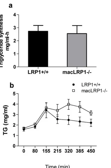
accomplished by measuring temporal increases in plasma triglycerides under conditions in which triglyceride hydrolysis by lipoprotein lipase is inhibited by injecting the nonionic deter-gent Triton WR-1339 [45]. The results (Fig 7a) demonstrate that there is no difference in the rate of triglyceride synthesis between LRP1+/+ and macLRP1-/- mice, suggesting that the Fig 5. Macrophage LRP1 deficiency leads to accumulation of triglyceride-rich lipoproteins in the plasma of LDLR-deficient mice.Mice were fed either a normal chow (a,left;b,left) or Western diet (a,right;b,right;c,d) for six weeks. Aliquots of serum from fasted mice were assayed for cholesterol (a) or triglyceride (b) (*p = 0.0004,**p = 0.003, n = 19 per group, Student’s t-test comparing LRP1+/+ with macLRP1-/-). (c,d) 100μl of pooled serum (n = 19/ group) from macLRP1-/- and LRP1+/+ mice was fractionated over a Superose 6 FPLC column. Plasma cholesterol (c) and triglyceride (d) levels were quantified. Three independent experiments were performed, each with a unique cohort of mice. Data from one representative experiment is shown.
increase in triglycerides in macLRP1-/- mice is likely due to an alteration in the processing (i.e. lipolysis) and/or the clearance of the particles.
To test this possibility, we measured the plasma triglyceride levels following an intragastric bolus of olive oil administration. The results, shown inFig 7b, confirm that at 2.5 hours follow-ing gavage, the level of triglyceride-rich lipoproteins in the plasma were comparable in both the LRP1+/+ and macLRP1-/- mice, confirming that the VLDL synthesis rates are identical in Fig 6. Normal ratios of apolipoproteins in the isolated d<1.019 lipoprotein fraction.The d<1.019
fraction from pooled plasma of LRP1+/+ and macLRP1-/- mice was isolated. (a) 15μg of protein from the d<1.019 fraction was analyzed by SDS-PAGE (n = 2) (b) ApoE area was quantified using NIH ImageJ. (c) Ratio of apoB100 to apoE and (d) ratio of apoB48 to apoE were determined using NIH ImageJ. (e) Immunoblot for apoC3 and apoE content and (f) the ratio of apoC3 to apoE in the d<1.019 fraction for LRP1
+/+ and macLRP1-/- mice (n = 3). (ns, not significant as determined by Student’s t test comparing LRP1 +/+ with macLRP1-/-).
these mice. Following this period, the levels of triglyceride-rich lipoproteins in LRP1+/+ mice decreased with time as expected. In contrast, however, the plasma triglyceride-rich lipoprotein levels persisted up to 7.5 hours following gavage in macLRP1-/- mice. These results reveal that the catabolism of triglyceride-rich lipoprotein particles is impaired in the macLRP1-/- mice.
Lipase activity is not altered in muscle, liver and adipose tissue in
macLRP1-/- mice
Lipolysis of VLDL and chylomicron particles at tissue sites is an important component of the processing of these lipoproteins. This process is catalyzed by lipoprotein lipase (LPL), which not only mediates lipolysis of chylomicrons and VLDL particles, but also binds to LRP1 via its carboxyl-terminal domain and facilitates lipoprotein uptake by this receptor [46–48]. After Fig 7. Postprandial serum lipids remain elevated in macLRP1-/- mice. (a) Mice (macLRP1-/-, n = 11; LRP1+/+, n = 10) were fed Western diet for 3 weeks, and were then injected with Triton WR-1339 (500 mg/kg body weight), and triglyceride levels in the plasma were determined at 0, 30, 60 and 90 min following injection. The rate of triglyceride synthesis is shown. Error bars show the SEM. (b) LRP1+/+ mice (closed symbols, n = 5) or macLRP1-/- mice (open symbols, n = 5) fed a Chow diet were fasted and then received an intragastric olive oil load. Blood samples were collected at the indicated times, and plasma triglyceride levels measured. Error bars show the SEM. (Data were analyzed for statistical significance employing a two way ANOVA which confirmed significant effects for Genotype (p = 0.0012) and Time (p = 0.0001) with no significant Genotype X Time interaction).
three weeks on either a chow diet or Western diet, adult mice were fasted and the heparin-elut-ed lipase activity in various organs was measurheparin-elut-ed. We found no significant difference in lipase activity from any of the organs examined between LRP1+/+ and macLRP1-/- mice (Fig8a–8d).
Discussion
The results obtained in the current investigation document an importantin vivorole for LRP1 in mediating cholesterol uptake in macrophages. This was confirmed by demonstrating that macLRP1-/- mice have a significant decrease in macrophage cholesterol content when fed a Western diet. Further, we noted elevated plasma triglyceride and cholesterol levels in
macLRP1-/- mice. Together, these data provide compellingin vivoevidence that LRP1 contrib-utes to cholesterol accumulation in macrophages. Interestingly, CD36 and SR-A, two receptors implicated in macrophage cholesterol uptake, also show elevated cholesterol and triglyceride levels whenCd36-/- andMsr1-/- mice on an apoE null background are fed a Western diet [8]. Thus, it is evident that multiple pathways contribute to cholesterol accumulation
in macrophages.
The role of LRP1 in chylomicron remnant catabolism is well established [24], although the involvement of this receptor in this process has been questioned since the contribution of LRP1 to chylomicron remnant uptake is only observed upon LDL receptor deficiency [49]. However, it should be noted that chylomicron remnant particles only accumulate in the circu-lation whenboththe LDL receptor and LRP1 are deleted in the liver. Thus genetic deletion of either hepatic LRP1 alone [24] or the LDL receptor alone [50] does not lead to accumulation of remnant lipoproteins. These results are consistent with the notion that both receptors contrib-ute to remnant lipoprotein clearance, and when one is absent or defective, the other compen-sates. Remnant clearance by the liver is a complex process and requires the participation of heparin sulfate proteoglycans, such as syndecan-1 [51]. Further, current models suggest that uptake of remnant lipoproteins by LRP1 requires enrichment of the particle with apoE [52], and studies in LDL receptor as well as apoE-deficient mice suggest that the source of this apoE is the liver [53].
In contrast to the hepatic uptake of lipoproteins, the mechanisms by which macrophages take up lipoprotein particles are not as well understood. Macrophage scavenger receptors have been identified that participate in the uptake and internalization of modified forms of LDL [54–56], and both SR-A (Msr1) and CD36 (Cd36) have been implicated in this process [7,8]. Despite convincing reports that these two receptors contribute to the development of athero-sclerosis and macrophage foam cell formation [57,58], abundant foam cell macrophages were detected in atherosclerotic lesions of mice containing genetic deletions of both receptors [9] re-vealing that other pathways exist for excessive lipid uptake by macrophages. Our studies sug-gest that LRP1 also contributes to this process. Several other mechanisms have been proposed that may also contribute to macrophage foam cell formation. For example, Kruth and col-leagues have found that LDL can be internalized in macrophages by macropinocytosis [59], and have proposed this as another pathway for macrophage foam cell formation. A response-to-retention hypothesis has also been proposed in which intramural accumulation of lipopro-teins and lipoprotein microaggregates represent an important early event in atherosclerosis that leads to modifications of the lipoproteins with important biological consequences includ-ing foam cell formation [60].
triglyceride levels in their studies when fed a Western diet. It is likely that the absence of apoE expression in their mice accounts for the differences with those of the current study, since LRP1 requires apoE for recognition of VLDL and chylomicron remnant particles [61,62]. Overton et al. [26] also conducted a study in which bone marrow from macLRP1-/- mice was transplanted into lethally irradiated female LDLR-/- recipient mice. Similar to the Hu et al. [25] study, Overton et al. [26] noted no difference in plasma triglyceride and cholesterol levels. The difference between their results and those in the present study likely arise from the fact thatin vivoreplacement of resident macrophages, such as Kupffer cells, upon bone marrow transplantation is a relatively slow process taking several months [63].
The mechanism by which macrophage LRP1 influences the levels of plasma triglyceride-rich lipoproteins is not clear at this time, but several possibilities exist. First, macrophages may play a direct role in the clearance of remnant lipoproteins. In this regard, it is interesting to highlight the studies of Hussain et al. [64,65] who demonstrated that signficant amounts of in-jected labeled chylomicrons were cleared by perisinusoidal bone marrow macrophages identi-fying an important contribution of macrophages to chylomicron remnant catabolism. Interestingly, hepatic deletion of LRP1 on an LDL receptor-deficient background does not re-quire feeding a Western diet to prompt accumulation of remnant lipoproteins in the plasma [24], whereas macLRP1-/- mice have to be challenged with an atherogenic diet in order to dis-play elevated remnant lipoproteins. Together, these data would suggest that macrophage LRP1 only plays a critical role in remnant clearance when other tissues are overwhelmed. Secondly, it is possible that the Western diet triggers an LRP1-mediated inflammatory response that signals other tissues to alter their usual role in lipoprotein homeostasis. In this regard, it is interesting to highlight that Kupffer cells are the primary source of hepatic inflammatory and pro-fibrogenic cytokines [66], and accumulation of cholesterol in Kupffer cells results in their acti-vation and conversion to a pro-inflammatory phenotype [67]. LRP1 expressed in Kupffer cells may regulate signaling pathways that in turn could alter lipoprotein catabolism. Additional studies are required to determine if this is the case.
In summary, the current investigation has confirmed an importantin vivorole for macro-phages and macrophage LRP1 in modulating cholesterol metabolism. The data further reveal that LRP1 is one of the major receptors that contribute to lipoprotein uptake in macrophages leading to foam cell formation. It is well known that the development of atherosclerosis is a complex process involving multiple genes, and defining the potential of LRP1 to regulate these processes may assist in identifying molecules which could have significant therapeutic potential.
Acknowledgments
The authors would also like to thank Elizabeth Smith for assistance with histological studies and Susan Robinson for assistance in breeding and genotyping of mice. APL is currently affili-ated with the Department of Radiology, Boston Children’s Hospital, Boston, MA 02115.
Author Contributions
Conceived and designed the experiments: APL SCM DTA MM MJL SKF IM DKS. Performed the experiments: APL SCM DTA MM MJL. Analyzed the data: APL SCM DTA MM MJL SKF IM DKS. Wrote the paper: DKS APL.
References
1. Libby P, Ridker PM, Hansson GK (2011) Progress and challenges in translating the biology of athero-sclerosis. Nature 473: 317–325. doi:10.1038/nature10146PMID:21593864
2. Brown MS, Goldstein JL (1986) A receptor-mediated pathway for cholesterol homeostasis. Science 232: 34–47. PMID:3513311
3. Brown MS, Goldstein JL (1983) Lipoprotein metabolism in the macrophage: implications for cholesterol deposition in atherosclerosis. Annu Rev Biochem 52: 223–261. PMID:6311077
4. Goldstein JL, Ho YK, Basu SK, Brown MS (1979) Binding site on macrophages that mediates uptake and degradation of acetylated low density lipoprotein, producing massive cholesterol deposition. Proc Natl Acad Sci U S A 76: 333–337. PMID:218198
5. Traber MG, Kayden HJ (1980) Low density lipoprotein receptor activity in human monocyte-derived macrophages and its relation to atheromatous lesions. Proc Natl Acad Sci U S A 77: 5466–5470. PMID:6254083
6. Brown MS, Basu SK, Falck JR, Ho YK, Goldstein JL (1980) The scavenger cell pathway for lipoprotein degradation: specificity of the binding site that mediates the uptake of negatively-charged LDL by mac-rophages. J Supramol Struct 13: 67–81. PMID:6255257
7. Kunjathoor VV, Febbraio M, Podrez EA, Moore KJ, Andersson L, Koehn S et al. (2002) Scavenger re-ceptors class A-I/II and CD36 are the principal rere-ceptors responsible for the uptake of modified low den-sity lipoprotein leading to lipid loading in macrophages. J Biol Chem 277: 49982–49988. PMID: 12376530
8. Moore KJ, Kunjathoor VV, Koehn SL, Manning JJ, Tseng AA, Silver JM et al. (2005) Loss of receptor-mediated lipid uptake via scavenger receptor A or CD36 pathways does not ameliorate atherosclerosis in hyperlipidemic mice. J Clin Invest 115: 2192–2201. PMID:16075060
9. Manning-Tobin JJ, Moore KJ, Seimon TA, Bell SA, Sharuk M, varez-Leite JI et al. (2009) Loss of SR-A and CD36 activity reduces atherosclerotic lesion complexity without abrogating foam cell formation in hyperlipidemic mice. Arterioscler Thromb Vasc Biol 29: 19–26. doi:10.1161/ATVBAHA.108.176644 PMID:18948635
10. Sawamura T, Kume N, Aoyama T, Moriwaki H, Hoshikawa H, Aiba Y et al. (1997) An endothelial recep-tor for oxidized low-density lipoprotein. Nature 386: 73–77. PMID:9052782
11. Mehta JL, Sanada N, Hu CP, Chen J, Dandapat A, Sugawara F et al. (2007) Deletion of LOX-1 reduces atherogenesis in LDLR knockout mice fed high cholesterol diet. Circ Res 100: 1634–1642. PMID: 17478727
13. Tabas I, Li Y, Brocia RW, Xu SW, Swenson TL, Williams KJ (1993) Lipoprotein lipase and sphingomye-linase synergistically enhance the association of atherogenic lipoproteins with smooth muscle cells and extracellular matrix. A possible mechanism for low density lipoprotein and lipoprotein(a) retention and macrophage foam cell formation. J Biol Chem 268: 20419–20432. PMID:8376399
14. Devlin CM, Leventhal AR, Kuriakose G, Schuchman EH, Williams KJ, Tabas I (2008) Acid sphingomye-linase promotes lipoprotein retention within early atheromata and accelerates lesion progression. Arter-ioscler Thromb Vasc Biol 28: 1723–1730. doi:10.1161/ATVBAHA.108.173344PMID:18669882
15. Kruth HS (2002) Sequestration of aggregated low-density lipoproteins by macrophages. Curr Opin Lipi-dol 13: 483–488. PMID:12352011
16. Llorente-Cortes V, Martinez-Gonzalez J, Badimon L (2000) LDL receptor-related protein mediates up-take of aggregated LDL in human vascular smooth muscle cells. Arterioscler Thromb Vasc Biol 20: 1572–1579. PMID:10845874
17. Llorente-Cortes V, Royo T, Juan-Babot O, Badimon L (2007) Adipocyte differentiation-related protein is induced by LRP1-mediated aggregated LDL internalization in human vascular smooth muscle cells and macrophages. J Lipid Res 48: 2133–2140. PMID:17620659
18. Llorente-Cortes V, Otero-Vinas M, Camino-Lopez S, Costales P, Badimon L (2006) Cholesteryl esters of aggregated LDL are internalized by selective uptake in human vascular smooth muscle cells. Arter-ioscler Thromb Vasc Biol 26: 117–123. PMID:16254205
19. Herz J, Hamann U, Rogne S, Myklebost O, Gausepohl H, Stanley KK (1988) Surface location and high affinity for calcium of a 500kDa liver membrane protein closely related to the LDL-receptor suggest a physiolocical role as lipoprotein receptor. EMBO J 7: 4119–4127. PMID:3266596
20. Ashcom JD, Tiller SE, Dickerson K, Cravens JL, Argraves WS, Strickland DK (1990) The humanα 2-macroglobulin receptor: identification of a 420-kD cell surface glycoprotein specific for the activated conformation ofα2-macroglobulin. J Cell Biol 110: 1041–1048. PMID:1691187
21. Moestrup SK, Gliemann J (1989) Purification of the Rat Hepaticα2-Macroglobulin Receptor as an Ap-proximately 440 kDa Single Chain Polypeptide. J Biol Chem 264: 15574–15577. PMID:2475504
22. Strickland DK, Ashcom JD, Williams S, Burgess WH, Migliorini M, Argraves WS (1990) Sequence iden-tity between theα2-macroglobulin receptor and low density lipoprotein receptor-related protein sug-gests that this molecule is a multifunctional receptor. J Biol Chem 265: 17401–17404. PMID:1698775
23. Beisiegel U, Weber W, Ihrke G, Herz J, Stanley KK (1989) The LDL-receptor related protein, LRP, is an apolipoprotein E binding protein. Nature 341: 162–164. PMID:2779654
24. Rohlmann A, Gotthardt M, Hammer RE, Herz J (1998) Inducible inactivation of hepatic LRP gene by cre-mediated recombination confirms role of LRP in clearance of chylomicron remnants. J Clin Invest 101: 689–695. PMID:9449704
25. Hu L, Boesten LS, May P, Herz J, Bovenschen N, Huisman MV et al. (2006) Macrophage low-density li-poprotein receptor-related protein deficiency enhances atherosclerosis in ApoE/LDLR double knockout mice. Arterioscler Thromb Vasc Biol 26: 2710–2715. PMID:17038633
26. Overton CD, Yancey PG, Major AS, Linton MF, Fazio S (2007) Deletion of macrophage LDL receptor-related protein increases atherogenesis in the mouse. Circ Res 100: 670–677. PMID:17303763
27. Yancey PG, Blakemore J, Ding L, Fan D, Overton CD, Zhang Y et al. (2010) Macrophage LRP-1 con-trols plaque cellularity by regulating efferocytosis and Akt activation. Arterioscler Thromb Vasc Biol 30: 787–795. doi:10.1161/ATVBAHA.109.202051PMID:20150557
28. Yancey PG, Ding Y, Fan D, Blakemore JL, Zhang Y, Ding L et al. (2011) Low-density lipoprotein recep-tor-related protein 1 prevents early atherosclerosis by limiting lesional apoptosis and inflammatory Ly-6Chigh monocytosis: evidence that the effects are not apolipoprotein E dependent. Circulation 124: 454–464. doi:10.1161/CIRCULATIONAHA.111.032268PMID:21730304
29. Muratoglu SC, Belgrave S, Lillis AP, Migliorini M, Robinson S, Smith E et al. (2011) Macrophage LRP1 suppresses neo-intima formation during vascular remodeling by modulating the TGF-ß signaling path-way. PloS One 6: e28846. doi:10.1371/journal.pone.0028846PMID:22174911
30. Cao C, Lawrence DA, Li Y, Von Arnim CA, Herz J, Su EJ et al. (2006) Endocytic receptor LRP together with tPA and PAI-1 coordinates Mac-1-dependent macrophage migration. EMBO J 25: 1860–1870. PMID:16601674
31. Clausen BE, Burkhardt C, Reith W, Renkawitz R, Forster I (1999) Conditional gene targeting in macro-phages and granulocytes using LysMcre mice. Transgenic Res 8: 265–277. PMID:10621974
33. Muratoglu SC, Mikhailenko I, Newton C, Migliorini M, Strickland DK (2010) Low density lipoprotein re-ceptor-related protein 1 (LRP1) forms a signaling complex with platelet-derived growth factor receptor-beta in endosomes and regulates activation of the MAPK pathway. J Biol Chem 285: 14308–14317. doi:10.1074/jbc.M109.046672PMID:20220145
34. Schumaker VN, Puppione DL (1986) Sequential flotation ultracentrifugation. Methods Enzymol 128: 155–170. PMID:3724500
35. Kanda T, Brown JD, Orasanu G, Vogel S, Gonzalez FJ, Sartoretto J et al. (2009) PPARgamma in the endothelium regulates metabolic responses to high-fat diet in mice. J Clin Invest 119: 110–124. doi: 10.1172/JCI36233PMID:19065047
36. Migliorini MM, Behre EH, Brew S, Ingham KC, Strickland DK (2003) Allosteric Modulation of Ligand Binding to Low Density Lipoprotein Receptor-related Protein by the Receptor-associated Protein Re-quires Critical Lysine Residues within Its Carboxyl-terminal Domain. J Biol Chem 278: 17986. PMID: 12637503
37. Low H, Hoang A, Sviridov D (2012) Cholesterol Efflux Assay. J Vis Exp 3810. doi:10.3791/3810PMID: 22414908
38. Nilsson-Ehle P, Schotz MC (1976) A stable, radioactive substrate emulsion for assay of lipoprotein li-pase. J Lipid Res 17: 536–541. PMID:9464
39. Feldman SR, Rosenberg MR, Ney KA, Michalopoulos G, Pizzo SV (1985) Binding of alpha 2-macroglobulin to hepatocytes: mechanism of in vivo clearance. Biochem Biophys Res Commun 128: 795–802. PMID:2581569
40. Zhou L, Choi HY, Li WP, Xu F, Herz J (2009) LRP1 controls cPLA2 phosphorylation, ABCA1 expression and cellular cholesterol export. PloS One 4: e6853. doi:10.1371/journal.pone.0006853PMID:19718435
41. Santamarina-Fojo S, Remaley AT, Neufeld EB, Brewer HB Jr. (2001) Regulation and intracellular traf-ficking of the ABCA1 transporter. J Lipid Res 42: 1339–1345. PMID:11518753
42. Van EM, Zimmermann R, Groot PH, Zechner R, Van Berkel TJ (2000) Role of macrophage-derived li-poprotein lipase in lili-poprotein metabolism and atherosclerosis. Arterioscler Thromb Vasc Biol 20: E53–E62. PMID:10978269
43. alto-Setala K, Fisher EA, Chen X, Chajek-Shaul T, Hayek T, Zechner R et al. (1992) Mechanism of hypertriglyceridemia in human apolipoprotein (apo) CIII transgenic mice. Diminished very low density li-poprotein fractional catabolic rate associated with increased apo CIII and reduced apo E on the parti-cles. J Clin Invest 90: 1889–1900. PMID:1430212
44. alto-Setala K, Weinstock PH, Bisgaier CL, Wu L, Smith JD, Breslow JL (1996) Further characterization of the metabolic properties of triglyceride-rich lipoproteins from human and mouse apoC-III transgenic mice. J Lipid Res 37: 1802–1811. PMID:8864964
45. Millar JS, Cromley DA, McCoy MG, Rader DJ, Billheimer JT (2005) Determining hepatic triglyceride production in mice: comparison of poloxamer 407 with Triton WR-1339. J Lipid Res 46: 2023–2028. PMID:15995182
46. Williams SE, Inoue I, Tran H, Fry GL, Pladet MW, Iverius P-H et al. (1994) The Carboxyl-terminal Do-main of Lipoprotein Lipase Binds to the Low Density Lipoprotein Receptor-related Protein/α 2-Macro-globulin receptor (LRP) and mediates Binding of Normal Very Low Density Lipoproteins to LRP. J Biol Chem 269: 8653–8658. PMID:7510694
47. Chappell DA, Fry GL, Waknitz MA, Muhonen LE, Pladet MW, Iverius P-H et al. (1993) Lipoprotein li-pase induces catabolism of normal triglyceride- rich lipoproteins via the low density lipoprotein receptor-related protein/α2-macroglobulin receptorin vitro. A process facilitated by cell-surface proteoglycans. J Biol Chem 268: 14168–14175. PMID:8314783
48. Chappell DA, Inoue I, Fry GL, Pladet MW, Bowen SL, Iverius P-H et al. (1994) Cellular catabolism of nor-mal very low density lipoproteins via the low density lipoprotein receptor-related protein/α2-macroglobulin receptor is induced by the C-terminal domain of lipoprotein lipase. J Biol Chem 269: 18001–18006. PMID:7517936
49. Williams KJ (2008) Molecular processes that handle—and mishandle—dietary lipids. J Clin Invest 118: 3247–3259. doi:10.1172/JCI35206PMID:18830418
50. Ishibashi S, Brown MS, Goldstein JL, Gerard RD, Hammer RE, Herz J (1993) Hypercholesterolemia in low density lipoprotein receptor knockout mice and its reversal by adenovirus-mediated gene delivery. J Clin Invest 92: 883–893. PMID:8349823
51. Stanford KI, Bishop JR, Foley EM, Gonzales JC, Niesman IR, Witztum JL et al. (2009) Syndecan-1 is the primary heparan sulfate proteoglycan mediating hepatic clearance of triglyceride-rich lipoproteins in mice. J Clin Invest 119: 3236–3245. doi:10.1172/JCI38251PMID:19805913
53. Linton MF, Hasty AH, Babaev VR, Fazio S (1998) Hepatic apo E expression is required for remnant lipo-protein clearance in the absence of the low density lipolipo-protein receptor. J Clin Invest 101: 1726–1736. PMID:9541504
54. Kodama T, Freeman M, Rohrer L, Zabrecky J, Matsudaira P, Krieger M (1990) Type I macrophage scavenger receptor contains alpha-helical and collagen-like coiled coils. Nature 343: 531–535. PMID: 2300204
55. Endemann G, Stanton LW, Madden KS, Bryant CM, White RT, Protter AA (1993) CD36 is a receptor for oxidized low density lipoprotein. J Biol Chem 268: 11811–11816. PMID:7685021
56. Ottnad E, Parthasarathy S, Sambrano GR, Ramprasad MP, Quehenberger O, Kondratenko N et al. (1995) A macrophage receptor for oxidized low density lipoprotein distinct from the receptor for acetyl low density lipoprotein: partial purification and role in recognition of oxidatively damaged cells. Proc Natl Acad Sci U S A 92: 1391–1395. PMID:7533292
57. Suzuki H, Kurihara Y, Takeya M, Kamada N, Kataoka M, Jishage K et al. (1997) A role for macrophage scavenger receptors in atherosclerosis and susceptibility to infection. Nature 386: 292–296. PMID: 9069289
58. Febbraio M, Podrez EA, Smith JD, Hajjar DP, Hazen SL, Hoff HF et al. (2000) Targeted disruption of the class B scavenger receptor CD36 protects against atherosclerotic lesion development in mice. J Clin Invest 105: 1049–1056. PMID:10772649
59. Kruth HS, Jones NL, Huang W, Zhao B, Ishii I, Chang J et al. (2005) Macropinocytosis Is the Endocytic Pathway That Mediates Macrophage Foam Cell Formation with Native Low Density Lipoprotein. J Biol Chem 280: 2352–2360. PMID:15533943
60. Williams KJ, Tabas I (1995) The response-to-retention hypothesis of early atherogenesis. Arterioscler Thromb Vasc Biol 15: 551–561. PMID:7749869
61. Kowal RC, Herz J, Goldstein JL, Esser V, Brown MS (1989) Low density lipoprotein receptor-related protein mediates uptake of cholesteryl esters derived from apoprotein E-enriched lipoproteins. Proc Natl Acad Sci USA 86: 5810–5814. PMID:2762297
62. Kowal RC, Herz J, Weisgraber KH, Mahley RW, Brown MS, Goldstein JL (1990) Opposing Effects of Apolipoproteins E and C on Lipoprotein Binding to LDL receptor Related Protein. J Biol Chem 265: 10771–10779. PMID:2355022
63. Kennedy DW, Abkowitz JL (1997) Kinetics of central nervous system microglial and macrophage en-graftment: analysis using a transgenic bone marrow transplantation model. Blood 90: 986–993. PMID: 9242527
64. Hussain MM, Mahley RW, Boyles JK, Lindquist PA, Brecht WJ, Innerarity TL (1989) Chylomicron metab-olism. Chylomicron uptake by bone marrow in different animal species. J Biol Chem 264: 17931–17938. PMID:2509449
65. Hussain MM, Mahley RW, Boyles JK, Fainaru M, Brecht WJ, Lindquist P (1989) Chylomicron-chylomicron remnant clearance by liver and bone marrow in rabbits. Factors that modify tissue-specific uptake. J Biol Chem 264: 9571–9582. PMID:2722852
66. Musso G, Gambino R, Cassader M (2013) Cholesterol metabolism and the pathogenesis of non-alcoholic steatohepatitis. Progress in Lipid Research 52: 175–191. doi:10.1016/j.plipres.2012.11.002 PMID:23206728
67. Leroux A, Ferrere G, Godie V, Cailleux Fdr, Renoud ML, Gaudin F et al. (2012) Toxic lipids stored by Kupffer cells correlates with their pro-inflammatory phenotype at an early stage of steatohepatitis. Jour-nal of Hepatology 57: 141–149. doi:10.1016/j.jhep.2012.02.028PMID:22425624
68. Zurhove K, Nakajima C, Herz J, Bock HH, May P (2008) Gamma-secretase limits the inflammatory re-sponse through the processing of LRP1. Sci Signal 1: ra15. doi:10.1126/scisignal.1164263PMID: 19036715
69. Loukinova E, Ranganathan S, Kuznetsov S, Gorlatov N, Migliorini MM, Loukinov D et al. (2002) PDGF-induced tyrosine phosphorylation of the LDL receptor-related protein (LRP): Evidence for integrated co-receptor function between LRP and the PDGF co-receptor. J Biol Chem 277:15499–15506. PMID:11854294
70. Newton CS, Loukinova E, Mikhailenko I, Ranganathan S, Gao Y, Haudenschild C et al. (2005) Platelet-derived growth factor receptor-beta (PDGFR-beta) activation promotes its association with the low den-sity lipoprotein receptor-related protein (LRP). Evidence for co-receptor function. J Biol Chem 280: 27872–27878. PMID:15944146
71. Boucher P, Gotthardt M, Li WP, Anderson RGW, Herz J (2003) LRP: Role in Vascular Wall Integrity and Protection from Atherosclerosis. Science 300: 329–332. PMID:12690199