Cop
yright
© ABE&M t
odos os dir
eit
os r
eser
vados
.
Hypercholesterolemic diet
induces hepatic steatosis and
alterations in mRNA expression
of NADPH oxidase in rat livers
Dieta hipercolesterolemiante induz esteatose hepática e alterações na expressão de mRNA da NADPH oxidase em fígados de ratos
Isabel Cristina Mallosto Emerich de Abreu1, Joyce Ferreira da Costa
Guerra2, Renata Rebeca Pereira2,3, Maísa Silva2, Wanderson Geraldo
de Lima2,3, Marcelo Eustáquio Silva1,2, Maria Lúcia Pedrosa1,2,3
ABSTRACT
Objective: This study aimed to determine whether a hypercholesterolemic diet induces hepatic steatosis, alterations in mRNA expression of NADPH oxidase subunits, and antioxidant defen-ses. Materials and methods: Fischer rats were divided into two groups of eight animals accor-ding to the treatment, control (C) and hypercholesterolemic diet (H). Those in group C were fed a standard diet (AIN-93M), and those of the group H were fed a hypercholesterolemic diet (25% soybean oil and 1% cholesterol). Results: The hypercholesterolemic diet did not affect body weight, but resulted in the accumulation of lipids in the liver, increased serum activities of ami-notransferases and cholesterol levels. Biomarker of lipid peroxidation (TBARS) and mRNA ex-pression of NADPH oxidase subunits p22phox and p47phox were increased in the liver of animals in group H. Besides, the activity and expression of antioxidant enzymes were altered. Conclusion: The results show increased mRNA expression of NADPH oxidase subunits and changes in an-tioxidant enzyme activities in diet-induced hepatic steatosis. Arq Bras Endocrinol Metab. 2014;58(3):251-9 Keywords
Antioxidant enzymes; hepatic steatosis; hypercholesterolemic diet; NADPH oxidase; non-alcoholic fatty liver disease; oxida-tive stress
RESUMO
Objetivo: Determinar se uma dieta hipercolesterolemiante induz esteatose hepática, alterações na expressão de mRNA da NADPH oxidase e nas defesas antioxidantes. Materiais e métodos: Ratas Fischer foram divididas em dois grupos de oito animais de acordo com o tratamento recebido, controle (C) e hipercolesterolêmico (H). Aquelas do grupo C foram alimentadas com dieta padrão (AIN-93M) e as do grupo H foram alimentadas com dieta hipercolesterolemiante (25% de óleo de soja e 1% de colesterol). As dietas foram oferecidas por oito semanas. Resulta-dos: O grupo H apresentou acúmulo de lipídios no fígado, aumento das atividades de ALT e AST e da concentração de colesterol no soro comparado ao grupo C. O marcador da peroxidação lipídica (TBARS) e os níveis de mRNA das subunidades p47phox da NADPH-oxidase e p22phox fo-ram aumentados no fígado de animais do grupo H, além de alteração da atividade e expressão de enzimas antioxidantes. Conclusão: Os resultados mostram um aumento na expressão de subunidades da NADPH oxidase e alterações na atividade das enzimas antioxidantes na este-atose hepática induzida por dieta hipercolesterolemiante. Arq Bras Endocrinol Metab. 2014;58(3):251-9 Descritores
Enzimas antioxidantes; esteatose hepática; dieta hipercolesterolemiante; NADPH oxidase; doença hepática gordurosa não alcoólica; estresse oxidativo
1 School of Nutrition, Universidade Federal de Ouro Preto (UFOP), Campus Universitário, Morro do Cruzeiro, Ouro Preto, MG, Brazil 2 Research Center in Biological Sciences, UFOP, Campus Universitário, Morro do Cruzeiro, Ouro Preto, MG, Brazil
3 Department of Biological Sciences, UFOP, Campus Universitário, Morro do Cruzeiro, Ouro Preto, MG, Brazil
Correspondence to:
Maria Lúcia Pedrosa Instituto de Ciências Exatas e Biológicas, Campus Universitário, s/n, Morro do Cruzeiro
Cop
yright
© ABE&M t
odos os dir
eit
os r
eser
vados
.
INTRODUCTION
N
on-alcoholic fatty liver disease (NAFLD) is the most common form of chronic liver disease. No-wadays, it is being considered a hepatic manifestation of metabolic syndrome (1-3).NAFLD is a disease characterized by the accumula-tion of fat in the liver of patients without history of alcohol abuse (4). The spectrum of NAFLD includes simple fatty liver (when greater than 5% of the liver weight), and non-alcoholic steatohepatitis (NASH), showing steatosis and necroinlammation that may progress to liver cirrhosis, hepatocellular carcinoma, and advanced liver disease (2-6).
The molecular and cellular mechanisms underlying hepatic injury in NAFLD are not well deined. Several sources of evidence suggest that multiple mechanisms, including enhanced low of free fatty acids and release of adipocytokines from the adipose tissue. In the liver, mitochondrial dysfunction, oxidative stress, and hepa-tocyte apoptosis are key contributors to hepatocellular injury. In addition, lipotoxic mediators and intracelullar signals activate Kupffer cells, which initiate and perpet-uate the inlammatory response and development of ibrosis (7).
Oxidative stress occurs as a result of either excess generation of reactive oxygen species (ROS), and/or re-duced antioxidant defenses. In the healthy liver, antioxi-dant systems such as catalase, superoxide dismutase, and glutathione peroxidase eficiently remove excess ROS to maintain normal cell homeostasis. On the other hand, one of the major sites of physiological ROS genera-tion is NADPH oxidase (NOX) (8). NOX is an enzyme complex that generates ROS in response to a wide range of stimuli, and has been recognized as a key element of intracellular signaling of hepatic ibrogenesis (8).
A role for the NADPH oxidases in chronic liver diseases, such as ibrosis and viral hepatitis, related to chronic inlammation, has been proposed (9,10). In the pathogenesis of alcoholic liver steatosis, there is an increase in NADPH oxidase activity and predominance of pro-oxidant agents, exceeding the capacity of the organic antioxidant defense (8). Under these circum-stances, intracellular homeostasis in the redox status is interrupted and, sometimes, induces cell damage re-sulting in apoptosis or necrosis, potentially contribut-ing to the devastatcontribut-ing injury and dysfunction of liver tissue (11,12).
Total body deiciency in p47phox subunit of NADPH oxidase complex protects mice from
alcohol-in-duced liver steatosis (13). However, mice on a methi-onine-choline-deicient (MCD) diet develop NASH with similar pathology as the wild type, despite the lack of a functional NADPH oxidase enzyme (14). The role of this enzyme complex in the others animal models of NAFLD have not been investigated.
Because of the increasing prevalence of NAFLD, elucidating the mechanisms of oxidative stress-induced injury within the liver is vital for the understanding of the pathogenesis of this disease (15). Recent reports suggest that dietary cholesterol is a critical factor in the development of experimental steatohepatitis in animal models (16). Human studies also support the hypothesis that dietary cholesterol plays a role in the development of steatohepatitis. In an epidemiological study, it was reported that dietary cholesterol con-sumption was independently associated with the de-velopment of cirrhosis (17). In mice, the presence of triacylglycerol and cholesterol in the diet are needed for the development of both hepatic histological normalities of NASH and its associated metabolic ab-normalities (16).
Our aims were to determine whether a hypercho-lesterolemic diet (25% soy oil, 1% cholesterol) for eight weeks causes histologic hepatic alterations in female rats, and if this diet induces alterations in mRNA ex-pression of NADPH oxidase and antioxidant defense enzymes, once oxidative stress is the implicated event contributing to the progression of liver steatosis to NASH, and since the understanding of the mecha-nisms by which NAFLD and NASH are developed is extremely important for the development of therapeu-tic interventions.
MATERIALS AND METHODS
Animals and experimental design
Female Fischer rats weighing approximately 138 g were obtained from the Experimental Nutrition Laboratory of the Universidade Federal de Ouro Preto (UFOP). Animals were individually housed in wire-bottomed metabolic cages and kept in a room at controlled con-ditions (24ºC, 55% humidity, 12-h light/dark cycles) with food and water ad libitum. The Ethics Committee
Cop
yright
© ABE&M t
odos os dir
eit
os r
eser
vados
.
and the second group (H) received a hypercholestero-lemic diet (25% soybean oil and 1% cholesterol) (Ta-ble 1). During the experiment, body weight and food intake were monitored. Food eficiency was calculated according to the following index: (body weight gain) x (food intake)-1.
were, respectively, 1004, 53-200; 1005, 52-200; 1013, 76-2/100; 1010, 87-2/100; 2001, 13-50; 2003, 84-1/500; 0003, 99-250.
Hepatic histology
Livers were removed at the end of the experiment and ixed in 4% buffered formalin. Subsequently, ixed lo-bes were transversely cut and processed in decreasing concentrations of alcohol, and embedded in parafin. Parafin sections of about 4-μm were obtained in a semi-automatic microtome, mounted and stained by hematoxylin and eosin (H&E) and Masson’s trichro-me. Photomicrographs were taken on a Leica DM5000 microscope coupled to a digital camera. Morphometric analyzes were performed using Image J Software. Lipid accumulation and hepatocytes with macrovesicular ste-atosis were counted in a total area of 1.5x10⁶ µ2 using parafin sections stained with H&E. The presence or absence of ibrosis was assessed by evaluation of parafin sections stained Masson’s trichrome in the same total tissue area.
Real time quantitative RT-PCR assay
Total RNA was isolated from the liver tissue of rats using the SV Total RNA Isolation System (Prome-ga Corporation, Madison, USA) according to the manufacturer’s instructions. cDNA was synthesized from 2 µg of total RNA with random primers using the High-Capacity cDNA Reverse Transcription Kit (Applied Biosystems, Foster City, CA), following the manufacturer’s recommendations. Real time PCR was performed with the Power SYBR® Green PCR Master Mix reagent (Applied Biosystems, Foster City, CA) in a inal reaction volume of 12 µL; the reaction included 1 µL of cDNA and 0.5 µL of each primer (forward and reverse, 10 µM).
The primers used for amplifying transcripts of genes of interest were those related to NADPH oxidase sub-units: gp91phox, p22phox, p47phox, p40phox and p67phox. The primers for CAT, Zn-superoxide dismutase (SOD), glutathione peroxidase (GPx), and gamma-glutamyl cysteine synthetase (γ-GCS) were designed according to the nucleotide sequences published by Xiong and cols. (18). The primer for gene endogenous control 18S was used. The reactions were carried out under the following conditions: 50ºC for 2 min, 95ºC for 10 min, and then 40 cycles of 95ºC for 15 sec (dena-turation), and 60ºC for 1 min (primer annealing and Table 1. Composition of the experimental diets
Nutrients C (g/kg) H (g/kg)
Casein 140.0 140.0
Corn starch 622.5 402.5
Soybean oil 40.0 250.0
Cholesterol 0.0 10.0
Choline 2.5 2.5
Mineral mixture¹ 35.0 35.0
Vitamin mixture² 10.0 10.0
Cellulose 50.0 50.0
Saccharose 100.0 100.0
Energy content (kcal/kg) 3810 4820
C: group that received the standard diet; H: group that received the hypercholesterolemic diet. ¹ Salt mixture (g/kg of mixture): NaCl - 139.3/KI – 0.79/MgSO4.7H2O – 57.3/CaCO3 – 381.4/ MnSO4.H2O – 4.01/FeSO4.7H2O – 0.548/CuSO4. 5H2O – 0.477/CoCl2.6H2O – 0.023/KH2PO4 – 389.0.
² Vitamin mixture (IU or g/kg of mixture): retinol acetate - 2 000 000 IU; cholecalciferol - 200 000 IU; p-aminobenzoic acid - 10.00; inositol - 10.00; niacin - 4.00; calcium pantothenate - 4.00; ribolavina - 0.80; thiamin HCl - 0.50; pyridoxine HCl - 0.50; folic acid - 0.20; biotin - 0.04; vitamin B12 - 0.003; sucrose, quantity suficient to 1 kg; choline - 200.0; α-tocopherol – 10,000 IU.
Conversion factors: protein 4 kcal/g, fat 9 kcal/g, sugar 4 kcal/g.
Sample preparation
After eight weeks (56 days), rats were allowed a twelve--hour fast, anesthetized with isolurane, and euthani-zed. To determine the levels of serum components, blood samples were collected in polypropylene tubes and centrifuged. Livers were removed, weighed, se-parated for histology, and the remainder immersed in liquid nitrogen, and immediately stored at −80 ºC for subsequent analysis. Abdominal and mesenteric fat were removed and weighed.
Serum laboratory tests
Cop
yright
© ABE&M t
odos os dir
eit
os r
eser
vados
.
product extension). The speciicity of the products ob-tained was conirmed by analysis of dissociation curves of the ampliied product. The data obtained were ana-lyzed using the comparative CT method. Target gene expression was determined relative to the expression of the endogenous 18S gene. All analyses were performed in triplicate. It is important to note that all primers used were previously tested in other studies from our lab (19,20).
Antioxidant defenses and oxidative stress biomarker in liver homogenate
Catalase (CAT) activity was determined according to Aebi (21), whose method is based on the enzymatic decomposition of H2O2 spectrophotometrically obser-ved at 240 nm for 3 min. Briely, ten μL of homogena-te supernatant was added to a cuvethomogena-te containing 100 mM phosphate buffer (pH 7.2), and there action was initiated by the addition of 10 mM H2O2. Hydrogen peroxide decomposition was calculated using the molar absorption coeficient 39.4 M−1cm−1. The results were expressed as activity per milligram of protein. One unit of CAT is equivalent to the hydrolysis of 1 μmol of H2O2 per min.
Superoxide dismutase (SOD) was assayed using Superoxide Dismutase Assay Kit (Cayman Chemical Company, nº 706002). This essay utilizes a tetrazolium salt for the detection of superoxide radicals generated by xanthine oxidase and hypoxantine. One unit of SOD is deined as the amount of enzyme needed to cause 50% dismutation of the superoxide radical. Only Cu/ Zn-SOD was measured.
The total glutathione content of liver homogenates was measured by the kit (CS0260) from Sigma (St. Louis, MO). The level of thiobarbituric acid reactive substances (TBARS) was estimated by the method of Buege and Aust (22). Liver homogenate supernatants were mixed with TCA (28% w/v in 0.25 N HCl), TBA (1% in 0.25 M acetic acid), and BHT (125 mM in eth-anol), heated for 15 min at 95°C and then placed in an ice bath. The precipitated material was removed by centrifugation, and the absorbance of the sample at 535 nm was determined. TBARS concentration was calcu-lated using the molar absorption coeficient of MDA (154.000 M−1cm−1).
Total protein content was determined according to the method described by Lowry and cols. (23) using
bovine serum albumin (BSA) as the standard. This test
was used only for correction of previous trials, such as catalase and TBARS.
Statistical analysis
Data were subjected to the Kolmogorov-Smirnov test for normality, expressed as mean ± standard deviation (SD) in cases of normal distribution, and expressed as median in cases of non-parametric distribution. The Student t test was used for data with normal distribu-tions and Mann-Whitney test was used for data with non-normal distributions. Differences were considered signiicant when p ≤ 0.05. All analyses were conduc-ted using the software GraphPadPrism version 5.00 for Windows (San Diego, CA).
RESULTS
The hypercholesterolemic diet does not modify body weight
The two groups had similar baseline weight, as seen in table 2. At the inal of the eight-week experiment, body weight did not change between the groups (Table 2). Although the animals of the control group had greater food intake, calorie intake (kcal) was the same in the two groups, demonstrating that food eficiency in the hypercholesterolemic group was better than in the con-trol group (p = 0.0003).
The hypercholesterolemic diet causes lipid accumulation in the liver
The hypercholesterolemic diet was not eficient in rai-sing body weight, but caused lipid accumulation in the liver. There was no change in the weight of mesenteric and abdominal fats (Table 2).
The hypercholesterolemic diet increases serum levels of liver enzymes and cholesterol
Cop
yright
© ABE&M t
odos os dir
eit
os r
eser
vados
.
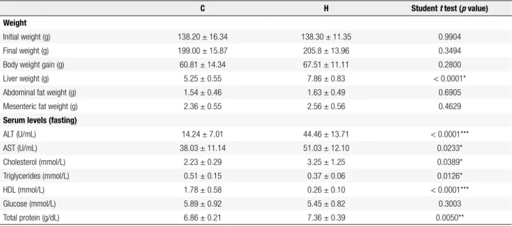
Table 2. Body, liver, and fat weights and serum levels after eight weeks on the experimental diets (mean ± SD)
C H Student t test (p value)
Weight
Initial weight (g) 138.20 ± 16.34 138.30 ± 11.35 0.9904
Final weight (g) 199.00 ± 15.87 205.8 ± 13.96 0.3494
Body weight gain (g) 60.81 ± 14.34 67.51 ± 11.11 0.2800
Liver weight (g) 5.25 ± 0.55 7.86 ± 0.83 < 0.0001*
Abdominal fat weight (g) 1.54 ± 0.46 1.63 ± 0.49 0.6905
Mesenteric fat weight (g) 2.36 ± 0.55 2.56 ± 0.56 0.4629
Serum levels (fasting)
ALT (U/mL) 14.24 ± 7.01 44.46 ± 13.71 < 0.0001***
AST (U/mL) 38.03 ± 11.14 51.03 ± 12.10 0.0233*
Cholesterol (mmol/L) 2.23 ± 0.29 3.25 ± 1.25 0.0389*
Triglycerides (mmol/L) 0.51 ± 0.15 0.37 ± 0.06 0.0126*
HDL (mmol/L) 1.78 ± 0.58 0.26 ± 0.10 < 0.0001***
Glucose (mmol/L) 5.89 ± 0.92 5.45 ± 0.82 0.3003
Total protein (g/dL) 6.86 ± 0.21 7.36 ± 0.39 0.0050**
Data are expressed as mean ± standard deviation. (n = 8). C: group that received the standard diet; H: group received the hypercholesterolemic diet. Statistical difference between means with * p ≤ 0.05; ** p ≤ 0.005; *** p ≤ 0.0001.
Table 3. Effects of the experimental diets on hepatic histology at eight weeks
Microvesicular steatosis
n (%)
Macrovesicular steatosis
n (%)
Inlammation n (%)
Fibrosis n (%)
C (n = 8) 1 (12.5) 0 (00) 3 (37.5) 0 (00)
H (n = 8) 8 (100) 8 (100) 8 (100) 0 (00)
C: group that received the standard diet; H: group that received the hypercholesterolemic diet.
The experimental model is a good model to study simple steatosis
According to table 3 we can notice that all animals in the H group had microvesicular and macrovesicular steatosis, demonstrating that a hypercholesterolemic diet during eight weeks induces steatosis, but does not inluence either inlammatory iniltrate or ibrosis.
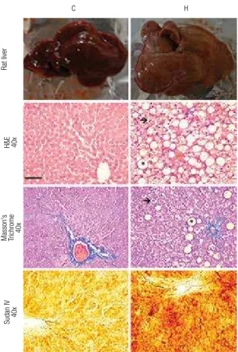
The liver sections showed the following histologi-cal features: the C group presented normal liver his-tology, and the H group presented steatosis with insigniicant inlammatory iniltrate and absence of i-brosis, characterizing simple steatosis (Figure 2).
The mRNA expression of NADPH oxidase is altered in livers with simple steatosis
Quantitative PCR analysis showed increased expression of NADPH oxidase subunits p22PHOX and p47PHOX in hypercholesterolemic rats, compared with the C group (Figure 3).
Figure 1. Metabolites and serum proteins (fast) after eight weeks of treatment with the experimental diets. C: group that received the standard diet; H: group received the hypercholesterolemic diet. For the statistical analysis, we used Student t test. Data are expressed as means ± standard deviations. (n = 8). Statistical differences between means with * p ≤ 0.05, ** p ≤ 0.005, *** p ≤ 0.0001.
80
60
40
20
0
C H
***
AL
T (U/mL)
5 4 3 2 1 0
C H
*
Cholesterol (mmol/L) Triglyceride (mnol/L)
2.5 2.0 1.5 1.0 0.5 0
C H
***
HDL (mmol/L)
Glucose (mmol/L)
8
6
4
2
0
C H
Total protein (g/dL)
10
** 8
6 4 2 0
C H
0.8
0.6
0.4
0.2
0
C H
* 80
60
40
20
0
C H
*
Cop
yright
© ABE&M t
odos os dir
eit
os r
eser
vados
.
6
4
2
C H
PHOX GP 91
C H
PHOX P 22
C H
PHOX P 47
C H
PHOX P 40
C H
PHOX P 67
Relative mRNA expression
0
*
*
Figure 3. Liver mRNA expression of NADPH oxidase subunits in livers after eight weeks of treatment with the experimental diets. C: group that received the standard diet; H: group that received the hypercholesterolemic diet. For the statistical analysis, we used Student t test. Data are expressed as mean ± standard deviation. (n = 8). Statistical difference between means with * P ≤ 0.05.
80
60
40
20
0
C H
Catalase (U/mg)
20000
15000
10000
5000
0
C H
Total glutatlhione (mnoles/mL) TBARS (U/mL/mg ptrotein) 5
4
*** 2
3
1
0
C H
SOD (U/mL)
1.5 *
**
1.0
0.5
0
C H
Figure 4. Antioxidant activity of enzymes and biomarkers of lipid peroxidation after eight weeks of treatment with the experimental diets. C:
group that received standard diet; H: group that received the hypercholesterolemic diet. For the statistical analysis, we used Student t
test for SOD and total glutathione. Data are expressed as means ± standard deviations. For the catalase and TBARS, we used the Mann-Whitney test. Data are expressed as medians (n = 8). Statistical difference between means with * P ≤ 0.05, ** p ≤ 0.005, *** p ≤ 0.0001.
Figure 5. Liver mRNA expression of antioxidant enzymes after eight weeks of treatment with the experimental diets. C: group that received standard diet; H: group that received the hypercholesterolemic diet. For the statistical analysis, we used Student t test. Data are expressed as means ± standard deviations (n = 8). Statistical difference between means with * P ≤ 0.05.
1.5
1.0
0.5
C H C H C H C H
Catalase Cu/Zn-SOD GPx γGCS
Relative mRNA expression
0.0
*
* *
Figure 2. Photomicrographs representative of rat livers and sections of livers stained with H & E an Masson’s trichrome and obtained after eight weeks in each of the experimental diets (C), group that received standard diet; (H), group that received the hypercholesterolemic diet. The arrows (→) indicate hepatocytes with microvesicular steatosis and asterisks (*) hepatocytes with macrovesicular steatosis. Bar = 50 μm.
The activity and expression of antioxidant enzymes and TBARS are also altered by lipid accumulation in the liver
Catalase activity did not show signiicant differences between groups, but mRNA expression was decreased on H group. On the contrary, SOD presented reduced activity and mRNA expression was unchanged. The to-tal hepatic glutathione content was 1.19-fold in the H group, and the mRNA expression decreased, compared with the control. TBARS levels are widely used as bio-markers of lipid peroxidation. Compared with control animals, the rats of the H group showed a 1.3-fold in-crease in TBARS (Figures 4 and 5).
C
Rat liver
Masson's Trichrome 40x
Sudan IV 40x H&E 40x
Cop
yright
© ABE&M t
odos os dir
eit
os r
eser
vados
.
DISCUSSION
Studies in animals have demonstrated strong associa-tions between diet composition and balance and the development of fatty liver. Because of this, it has been postulated that dietary habits may promote NAFLD in humans. The mechanisms by which diet may play a role include modulation of hepatic triglyceride accumula-tion and regulaaccumula-tion of the antioxidant activity, as well as changes in insulin sensitivity and postprandial triglyce-ride metabolism (1,24,25). Both excessive carbohydra-te intake (26) and excessive fat intake could play a role in increasing blood glucose, free fat acids, and insulin concentrations, independently or together (1,6).
The diet containing 25% soybean oil and 1% choles-terol promoted hypercholescholes-terolemia in rats and simple steatosis.
There is no single biochemical marker that can con-irm a NAFLD diagnosis or distinguish between ste-atosis, NASH, and cirrhosis, but liver function tests abnormalities are common in patients with NAFLD, with elevations in ALT and AST usually no greater than four times the upper limit of normal (3,27,28). Liver biopsy is the gold standard for the diagnosis of NAFLD, considering that it is the only method that can distinguish between simple steatosis, NASH, and the degree of ibrosis (29,30). Histologically, NASH is similar to alcohol-induced hepatitis, with the presence of macrovesicular steatosis, mixed inlammatory cell iniltration in the lobules, hepatocyte ballooning and necrosis, Mallory bodies, and perisinusoidal ibrosis or cirrhosis (5,31).
As expected, our results indicated that the hyper-cholesterolemic diet caused liver damage, and increased oxidative stress and cholesterol levels in female rats. Liver injury was characterized by hepatomegaly and increased activities of AST and ALT enzymes. Hepatic histology indicates that animals fed the hypercholes-terolemic diet presented lipid accumulation as shown by H&E and Sudan IV staining. This can be viewed as macrovesicular steatosis, represented by large white vesicles and marked by the intensity of red color, indi-cating the presence of lipids, respectively. Masson’s tri-chrome revealed no difference between the groups re-garding the presence of ibrosis. Thus, it is evident that animals fed the hypercholesterolemic diet only showed steatosis, without progression to NASH and cirrhosis, proving that the hypercholesterolemic diet consisting of 25% soybean oil and 1% cholesterol administered for
eight weeks was effective in inducing hepatic steatosis in Fischer rats.
During the development of NAFLD, there is an in-creased production of ROS, often leading to greater hepatic lipid peroxidation (32,33). Our results showed that consumption of the hypercholesterolemic diet increased liver TBARS, indicating increased oxidative stress. It is known that oxidative stress can occur by increasing of pro-oxidant systems and/or by lowering antioxidant enzymes. Increased NADPH oxidase activ-ity has been reported in animal models of NASH, in which dietary antioxidants or NADPH oxidase inhibi-tors ameliorated the progression of the disease (34-36). Thus, we decided investigate if there would be changes in gene expression of the subunits of this enzyme com-plex in this model.
The subunit p47PHOX of NADPH oxidase is the main responsible for transporting the cytosolic com-plex from the cytosol to the membrane during oxi-dase activation. Before the cytosolic oxioxi-dase compo-nents can be transferred to the membrane, however, p47PHOX must be extensively phosphorylated (37). When p47PHOX is phosphorylated, it binds to p22PHOX, an interaction that is probably responsible for activat-ing the oxidase (38).
The p22PHOX functions as an integral subunit of the inal electron transporter from NADPH to heme to molecular oxygen in generating superoxide anion (39). In this study, we found increased expression of p47PHOX and p22PHOX subunits of NADPH oxidase in the H group, thus indicating that this increase could be contributing to the activation of this enzyme complex, favoring the generation of ROS.
Cop
yright
© ABE&M t
odos os dir
eit
os r
eser
vados
.
Together, our data show a change in pro-oxidant/ antioxidant systems that resulted in increased TBARS. TBARS are by-products of oxidative stress that indi-cate lipid peroxidation. The production of 4-hydroxy-2-nonenal (HNE) and malondialdehyde (MDA), types of TBARS, up-regulate liver ibrosis via activation of stel-late cells and result in increased production of transform-ing growth factor-beta (3). Horoz and cols. (40) showed that patients with steatosis alone, and with NASH have higher levels of TBARS, corroborating our results.
The hypercholesterolemic diet consisting of 25% soy-bean oil and 1% cholesterol and administered for eight weeks was effective in inducing hepatic steatosis in rats, constituting a good model for the study of steatosis.
Deinitively, hepatic steatosis should not be con-sidered benign. In our experimental indings, we can prove that steatosis led to changes in mRNA expression and, consequently, an changed in the redox balance.
Understanding that steatosis alone may bring signif-icant changes is an important step to deine new strate-gies for the treatment of NAFLD in the future.
Acknowledgements: The authors declare that there is no poten-tial conlict of interest. This study was supported by the Fundação de Amparo à Pesquisa de Minas Gerais (Fapemig, Minas Gerais, Brazil) and Conselho Nacional de Desenvolvimento Cientíico e Tecnológico (CNPq, Brazil). The authors would like to thank Nara Nunes Laje and Poliane Silva Maciel for their early contri-butions to the study, Jair Pastor Mota and Clodoaldo Pereira dos Santos for maintaining the animal facilities.
REFERENCES
1. Lazo M, Clark JM. The epidemiology of nonalcoholic fatty liver disease: a global perspective. Semin Liver Dis. 2008;28:339-50. 2. Ferré P, Foufelle F. Hepatic steatosis: a role for de novo
lipogene-sis and the transcription factor SREBP-1c. Diabetes Obes Metab. 2010;12:83-92.
3. Lewis JR, Mohanty SR. Nonalcoholic fatty liver disease: a review and update. Dig Dis Sci. 2010;55:560-78.
4. Takahashi Y, Inui A, Fujisawa T, Takikawa H, Fukusato T. Histopatho-logical characteristics of non-alcoholic fatty liver disease in chil-dren: comparison with adult cases. Hepatol Res. 2011;41:1066-74. 5. Matteoni CA, Younossi ZM, Gramlich T, Boparai N, Liu YC, Mc-Cullough AJ. Nonalcoholic fatty liver disease: a spectrum of clini-cal and pathologiclini-cal severity. Gastroenterology. 1999;116:1413-9. 6. Finelli C, Tarantino G. Is there any consensus as to what diet or
lifestyle approach is the right one for NAFLD patients? J Gastroin-testin Liver Dis. 2012;21:293-302.
7. Ibrahim MA, Kelleni M, Geddawy A. Nonalcoholic fatty liver dise-ase: current and potential therapies. Life Sciences. 2013;92:114-8. 8. Paik Y-H, Brenner DA. NADPH oxidase mediated oxidative stress
in hepatic ibrogenesis. Korean J Hepatol. 2011;17:251-7. 9. Jiang JX, Venugopal S, Serizawa N, Chen X, Scott F, Li Y, et al.
Reduced nicotinamide adenine dinucleotide phosphate oxidase 2
plays a key role in stellate cell activation and liver ibrogenesis in vivo. Gastroenterology. 2010;139:1375-84.
10. de Mochel NS, Seronello S, Wang SH, Ito C, Zheng JX, Liang TJ, et al. Hepatocyte NAD(P)H oxidases as an endogenous source of reactive oxygen species during hepatitis C virus infection. Hepa-tology. 2010;52:47-59.
11. Evans JL, Goldine ID, Maddux BA, Grodsky GM. Oxidative stress and stress-activated signaling pathways: a unifying hypothesis of type 2 diabetes. Endocr Rev. 2002;23:599-622.
12. Zhu W, Jia Q, Wang Y, Zhang Y, Xia M. The anthocyanin cyanidin-3-O-β-glucoside, a lavonoid, increases hepatic glutathione syn-thesis and protects hepatocytes against reactive oxygen species during hyperglycemia: Involvement of a cAMP-PKA-dependent signaling pathway. Free Radic Biol Med. 2012;52:314-27.
13. Levin I, Petrasek J, Szabo G. The presence of p47phox in liver pa-renchymal cells is a key mediator in the pathogenesis of alcoholic liver steatosis. Alcohol Clin Exp Res. 2012;36:1397-406.
14. dela Pena A, Leclercq IA, Williams J, Farrell GC. NADPH oxidase is not an essential mediator of oxidative stress or liver injury in mu-rine MCD diet-induced steatohepatitis. J Hepatol. 2007;46:304-13. 15. Hardwick RN, Fisher CD, Canet MJ, Lake AD, Cherrington NJ. Di-versity in antioxidant response enzymes in progressive stages of human nonalcoholic fatty liver disease. Drug Metab Dispos. 2010;38:2293-301.
16. Savard C, Tartaglione EV, Kuver R, Haigh WG, Farrell GC, Subra-manian S, et al. Synergistic interaction of dietary cholesterol and dietary fat in inducing experimental steatohepatitis. Hepatology. 2013;57:81-92.
17. Ioannou GN, Morrow OB, Connole ML, Lee SP. Association be-tween dietary nutrient composition and the incidence of cirrho-sis or liver cancer in the United States population. Hepatology. 2009;50:175-84.
18. Xiong Q1, Xie P, Li H, Hao L, Li G, Qiu T, et al. Acute effects of mi-crocystins exposure on the transcription of antioxidant enzyme genes in three organs (liver, kidney, and testis) of male Wistar rats. J Biochem Mol Toxicol. 2010;24:361-7.
19. Guerra JF, Magalhães CL, Costa DC, Silva ME, Pedrosa ML. Die-tary açai modulates ROS production by neutrophils and gene expression of liver antioxidant enzymes in rats. J Clin Biochem Nutr. 2011;49:188-94.
20. Rossoni Júnior JV, Araújo GR, Pádua Bda C, Magalhães CL, Chaves MM, Pedrosa ML, et al. Annatto extract and β-carotene enhances antioxidant status and regulate gene expression in neutrophils of diabetic rats. Free Radic Res. 2012;46:329-38. 21. Aebi H. Catalase in vitro. Methods Enzymol. 1984;105:121-6. 22. Buege JA, Aust SD. Microsomal lipid peroxidation. Methods
En-zymol. 1978;52:302-10.
23. Lowry OH, Rosebrough NJ, Farr AL, Randall RJ. Protein measure-ment with the folin-phenol reagent. J Biol Chem. 1951;193:265-75. 24. Jenkins DJ, Josse AR, Labelle R, Marchie A, Augustin LS, Kendall
CW. Nonalcoholic fatty liver, nonalcoholic steatohepatitis, ectopic fat, and the glycemic index. Am J Clin Nutr. 2006;84(1):3-4. 25. Cave M, Deaciuc I, Mendez C, Song Z, Joshi-Barve S, Barve S, et
al. Nonalcoholic fatty liver disease: predisposing factors and the role of nutrition. J Nutr Biochem. 2007;18(3):184-95.
26. Solga S, Alkhuraishe AR, Clark JM, Torbenson M, Greenwald A, Diehl AM, et al. Dietary composition and nonalcoholic fatty liver disease. Dig Dis Sci. 2004;49(10):1578-83.
27. Pratt DS, Marshall M, Kaplan MM. Evaluation of abnormal liver-enzyme results in asymptomatic patients. N Engl J Med. 2000;342:1266-271.
Cop
yright
© ABE&M t
odos os dir
eit
os r
eser
vados
.
29. Saadeh S, Younossi ZM, Remer EM, Gramlich T, Ong JP, Hurley M, et al. The utility of radiological imaging in nonalcoholic fatty liver disease. Gastroenterology. 2002;123:745-50.
30. Adams LA, Angulo P. Treatment of non-alcoholic fatty liver disea-se. Postgrad Med J. 2006;82:315-22.
31. Lee RG. Nonalcoholic steatohepatitis: tightening the morpholo-gical screws on a hepatic rambler. Hepatology. 1995;21:1742-3. 32. Ludwig J, McGill DB, Lindor KD. Review: nonalcoholic
steatohe-patitis. J Gastroenterol Hepatol. 1997;12:398-403.
33. Morán-Ramos S, Avila-Nava A, Tovar AR, Pedraza-Chaverri J, López-Romero P, Torres N. Opuntia icus indica (nopal) attenuates hepatic steatosis and oxidative stress in obese Zucker (fa/fa) rats. J Nutr. 2012;142(11):1956-63.
34. Carmiel-Haggai M, Cederbaum AI, Nieto N. A high-fat diet leads to the progression of non-alcoholic fatty liver disease in obese rats. FASEB J. 2005;19:136-8.
35. Lu LS, Wu CC, Hung LM, Chiang MT, Lin CT, Lin CW, et al. Apocy-nin alleviated hepatic oxidative burden and reduced liver injury in hypercholesterolaemia. Liver Int. 2007;27:529-37.
36. Sancho P, Martín-sanz P, Fabregat I. Reciprocal regulation of NA-DPH oxidases and the cyclooxygenase-2 pathway. Free Radical Bio Med. 2011;5:1789-98.
37. Babior BM. NADPH oxidase: an update. Blood. 1999;93:1464-76. 38. Babior BM. NADPH oxidase. Curr Opin Immunol. 2004;16:42-7. 39. Fukui T, Ishizaka N, Rajagopalan S, Laursen JB, Capers Q 4th,
Taylor WR, et al. p22phox mRNA expression and NADPH oxidase activity are increased in aortas from hypertensive rats. Circ Res. 1997;80:45-51.