Fructose Mediated Non-Alcoholic Fatty Liver
Is Attenuated by HO-1-SIRT1 Module in
Murine Hepatocytes and Mice Fed a High
Fructose Diet
Komal Sodhi1*, Nitin Puri2, Gaia Favero3, Sarah Stevens1, Charles Meadows1, Nader G. Abraham4, Rita Rezzani3, Hayden Ansinelli1, Edward Lebovics4, Joseph I. Shapiro1
1Departments of Medicine and Surgery, Joan C. Edwards School of Medicine, Marshall University, Huntington, West Virginia, United States of America,2Department of Physiology & Pharmacology, University of Toledo College of Medicine, Toledo, Ohio, United States of America,3Department of Clinical and Experimental Sciences, Division of Anatomy and Physiopathology, University of Brescia, Brescia, Italy,
4Departments of Medicine and Gastroenterology, New York Medical College, Valhalla, New York, United States of America
Abstract
Background
Oxidative stress underlies the etiopathogenesis of nonalcoholic fatty liver disease (NAFLD), obesity and cardiovascular disease (CVD). Heme Oxygenase-1 (HO-1) is a potent endoge-nous antioxidant gene that plays a key role in decreasing oxidative stress. Sirtuin1 (SIRT1) belongs to the family of NAD-dependent de-acyetylases and is modulated by cellular redox.
Hypothesis
We hypothesize that fructose-induced obesity creates an inflammatory and oxidative envi-ronment conducive to the development of NAFLD and metabolic syndrome. The aim of this study is to determine whether HO-1 acts through SIRT1 to form a functional module within hepatocytes to attenuate steatohepatitis, hepatic fibrosis and cardiovascular dysfunction.
Methods and Results
We examined the effect of fructose, on hepatocyte lipid accumulation and fibrosis in murine hepatocytes and in mice fed a high fructose diet in the presence and absence of CoPP, an inducer of HO-1, and SnMP, an inhibitor of HO activity. Fructose increased oxidative stress markers and decreased HO-1 and SIRT1 levels in hepatocytes (p<0.05). Further fructose supplementation increased FAS, PPARα, pAMPK and triglycerides levels; CoPP negated this increase. Concurrent treatment with CoPP and SIRT1 siRNA in hepatocytes increased FAS, PPARα, pAMPK and triglycerides levels suggesting that HO-1 is upstream of SIRT1 and suppression of SIRT1 attenuates the beneficial effects of HO-1. A high fructose diet increased insulin resistance, blood pressure, markers of oxidative stress and lipogenesis OPEN ACCESS
Citation:Sodhi K, Puri N, Favero G, Stevens S, Meadows C, Abraham NG, et al. (2015) Fructose Mediated Non-Alcoholic Fatty Liver Is Attenuated by HO-1-SIRT1 Module in Murine Hepatocytes and Mice Fed a High Fructose Diet. PLoS ONE 10(6): e0128648. doi:10.1371/journal.pone.0128648
Editor:Hervé Guillou, INRA, FRANCE
Received:January 7, 2015
Accepted:April 29, 2015
Published:June 22, 2015
Copyright:© 2015 Sodhi et al. This is an open access article distributed under the terms of the
Creative Commons Attribution License, which permits unrestricted use, distribution, and reproduction in any medium, provided the original author and source are credited.
Data Availability Statement:All data underlying the findings are fully available upon request without restriction. All relevant data are within the paper and its supporting information files.
Funding:Dr. Shapiro provided the funds to accomplish this study. Funds were taken from National Institutes of Health grant NIH-1RO1HL109015-01. The funding institution was Marshall University. Author who received funding is JS.
along with fibrotic markers in mice (p<0.05). Increased levels of HO-1 increased SIRT1 lev-els and ameliorated fructose-mediated lipid accumulation and fibrosis in liver along with decreasing vascular dysfunction (p<0.05 vs. fructose). These beneficial effects of CoPP were reversed by SnMP.
Conclusion
Taken together, our study demonstrates, for the first time, that HO-1 induction attenuates fructose-induced hepatic lipid deposition, prevents the development of hepatic fibrosis and abates NAFLD-associated vascular dysfunction; effects that are mediated by activation of SIRT1 gene expression.
Introduction
Non-alcoholic fatty liver disease (NAFLD) is deposition of excess fat in hepatocytes that is not associated with alcoholism. Prevalence of NAFLD, and associated steatohepatitis, is steadily increasing in developed countries. In the United States, prevalence of NAFLD has been reported to be between 10 and 46% with NASH (biopsy-based) occurring in 3–5% of the popu-lation [1,2]. The impact of NAFLD diagnostic criteria on reported prevalence is evident in the National Health and Nutrition Examination Surveys (NHANES). NHANES 1988–1994 reported prevalence of NAFLD at 5.5%, NHANES 1999–2004 9.8%, and NHANES2005-2008 11%. These were based on elevated serum aminotransferases. A reevaluation of NHANES 1988–1994 based on hepatic steatosis on ultrasound and the absence of excessive alcohol con-sumption found the prevalence to actually be 19% [3].
SIRT1 is a class III protein deacetylase, a crucial cellular survival protein in combating meta-bolic imbalance [29]. It regulates glucose and lipid metabolism through its deacetylase activity and via its direct and indirect involvement in insulin signaling. Activation of SIRT1 decreases fatty liver by a reducing expression of lipogenic enzymes [30]. Hepatocyte specific loss of SIRT1 caused peroxisome proliferator-activated receptorα(PPARα) signal failure and decrease in fatty acidβ-oxidation [31]. Liver-specific SIRT1 deficiency caused hepatic glucose overproduction, chronic hyperglycemia and increased ROS production. Importantly, SIRT1 is amenable to redox manipulations and is suppressed by ROS [32]. Other investigators, includ-ing our lab, have shown SIRT1 rescue by antioxidants in a variety of tissues under oxidative stress. In this regard, the heme-heme oxygenase system (HO) is one of the key cellular-antioxi-dant defenses that lowers ROS by the breakdown of heme (pro-oxicellular-antioxi-dant) to carbon monoxide (CO) and biliverdin (BV). BV is rapidly reduced to the antioxidant, bilirubin [33,34]. There are two HO isoforms: the constitutive, Heme-Oxygenase isoenzyme 2 (HO-2); and, the induc-ible, HO-1. HO-1 is up-regulated during alterations in cellular redox and plays a role in myriad of pathological conditions, including metabolic syndrome [35]. Increasing HO activity results in the reversal of oxidative stress and a decrease in liver damage (reviewed in [36]). Increased HO-1 levels increase the phosphorylation of AMP-activated protein kinase (AMPK), and decrease fatty acid synthase (FAS) resulting in an increase in insulin sensitivity and the lower-ing of fatty acid levels [37,38]. Also, we have recently shown that induction of HO-1 attenuated the development of fatty liver and decreased lipid droplet size in obese mice [39], thus substan-tiating a significant role of HO-1 against heme-mediated adiposity and fatty liver. For this study we have hypothesized that HO-1, a critical anti-oxidant, forms a cytoprotective module with SIRT1 and together, they counteract diet-activated pathways in the liver that lead to NAFLD and NASH.
For this study we have hypothesized that HO-1, a critical anti-oxidant, forms a cytoprotec-tive module with SIRT1 and together, they counteract diet-activated pathways in the liver that lead to NAFLD. The aim of the study is to demonstrate that HO-1 induction in the liver reduces diet-induced metabolic imbalance, ROS, insulin resistance, and hepatic lipid deposi-tion and also prevent the development of hepatic fibrosis; effects that are mediated by activa-tion of SIRT1 gene expression. We believe that understanding the interacactiva-tions between HO-1 and SIRT1 in NAFLD and related hepatic fibrosis will lead to the development of new bio-markers and therapeutic strategies to fight hepatic dysfunction. This will result in improved quality of life and life expectancy in the obese, insulin resistant patient.
Material and Methods
Experimental design for
in vitro
experiment
Frozen mouse hepatocytes (AML12, CRL-2254) were purchased from ATTC. For experiments they were cultured in DMEM and Ham's F12 medium with supplements. Cells were plated in 12-well dishes and 75-cm2flasks at a density of 1-2X10⁴cells and were treated every alternate day for 5 days with and without fructose (500μM) in the absence and presence of CoPP
(5μM), small interfering RNA (siRNA) for SIRT1 (or non-specific siRNA), and SnMP (5μM)
Experimental design for
in vivo
experiment
All animal studies were approved by the Marshall University Animal Care and Use Committee in accordance with the National Institutes of Health Guidelines for Care and Use of Laboratory Animals. Forty, eight week–old C57Bl6 male mice were used in the studies. Mice were fed a HFr diet for 8 weeks, a time frame in which the manifestations of fatty liver are present, and were divided into four groups: 1) control diet; 2) HFr diet; 3) HFr diet treated for the last 4 weeks with Cobalt Protoporphyrin (CoPP) (5 mg/kg, twice a week); and 4) HFr diet treated for the last 4 weeks with CoPP (5 mg/kg, twice a week) and Tin mesoporphyrin (SnMP) (20mg/kg, twice a week). Control chow (Harlan, Teklad Lab Animal Diets, US) contained kcal from protein 30%, carbohydrate 57% and fat 13%. HFr diet (Harlan, Teklad Lab Animal Diets, US) contained kcal from protein 20.2%, carbohydrate 66.8% and fat 12.9%. Fat content of the two diets is similar (both derived from porcine). The control diet fat composition includes cholesterol ppm 209, Linoleic acid 1.05%, Linolenic acid 0.09%, Arachidonic acid 0.02%, Omega-3-Fatty Acid 0.3%, Total Saturated Fatty Acids 1.48% and Total Unsaturated Fatty Acids 1.62%. The HFr diet fat composition includes cholesterol ppm 950, Linoleic acid 0.59%, Linolenic acid 0.04%, Arachidonic acid 0.01%, Omega-3-Fatty Acid 0.05%, Total Saturated Fatty Acids 1.91% and Total Unsaturated Fatty Acids 1.75%. HFr diet is in accordance with published reports using similar diets to induce hepatic steatosis and steatohepatitis. Mice were weighed every week and blood pressure determined by the tail cuff method every 4 weeks dur-ing the course of the experiment. Prior to the experiment, mice were all acclimated to the tail cuff method. Mice were placed in a heat-controlled box (36–38°C) for approximately 10 mins before applying the tail cuff. The mean of a minimum of 5 measurements was obtained from each mouse. All measurements were determined at the same time of day for all mice. At the end of the 8-week period, mice were placed on an 8-hour fast, anesthetized with sodium pento-barbital (65 mg/kg, i.p.) and blood was obtained from the tail vein for measurement of glucose using a glucometer (Lifescan Inc., Miligitas, CA) and measurement of insulin using ELISA assay kit (Abcam, Cambridge, MA). Blood samples were collected in K3EDTA tubes at sacrifice and the plasma was separated. Alanine Aminotransferase (ALT) was measured in mice plasma to study liver function test. Liver and aorta tissues were flash frozen in liquid nitrogen and maintained at -80°C until assayed.
A second experiment was done in which mice were placed on a HFr diet for 20 weeks in order to study the prolonged effect of a HFr diet on the progression of hepatic fibrosis. The choice of time was predicated upon previous studies in mice that have shown hepatic fibrosis is clearly evident after 16–20 weeks of a HFr diet. Mice were divided into four groups: 1) control diet; 2) HFr diet; 3) HFr diet treated for the last 4 weeks with CoPP (5 mg/kg, twice a week); and 4) HFr diet treated for the last 4 weeks with CoPP (5 mg/kg, twice a week) and SnMP (20mg/kg, twice a week). After 20 weeks, liver tissue was flash frozen in liquid nitrogen and maintained at -80°C until assayed.
Measurement of Isoprostane, Heme, and Cytokine Levels
Measurement of Superoxide Levels for
in vitro
experiment
Hepatocytes were cultured on 96-well plates until they achieved approximately 70% conflu-ence. After treatment with or without fructose (500μM) in the absence and presence of CoPP
(5μM) and SnMP (5μM) for 2 days, the cells were incubated with 10μM dihydroethidium
(DHE) for 30 min at 37°C. Fluorescence intensity was measured using a Perkin-Elmer Lumi-nescence Spectrometer at excitation/emission filters of 530/620 nm.
Measurement of Triglyceride Levels for
in vitro
experiment
Hepatocytes were cultured in 75-cm2 flasks until they achieved approximately 70% confluence. After treatment for 5 days with or without fructose (500μM) in the absence and presence of
CoPP (5μM) and SnMP (5μM), the cells were collected and washed in ice-cold
phosphate-buff-ered saline (PBS). Triglyceride levels were determined in hepatocytes using a commercially available kit (Abcam, Cambridge, MA).
Determination of homeostasis model assessment of insulin resistance
The homeostasis model assessment of insulin resistance (HOMA-IR) was calculated from mice blood using glucose and insulin concentrations obtained after 8 h of food withdrawal, using the following formula: HOMA-IR = [fasting insulin (μU/ mL) × fasting glucose (mmol/L)] / 22.5.Determination of Triglyceride, Cholesterol content in hepatic tissue
Liver samples were homogenized in ice-cold PBS. Tissue lipids were extracted with methanol/ chloroform (1:2), dried, and resuspended in 5% fat-free bovine serum albumin. Triglyceride and Cholesterol levels were determined using a commercially available kit according to the manufacture’s protocol (Abcam, Cambridge, MA).Determination of Free Fatty Acids levels in hepatic tissue
Liver tissue (10mg) was homogenized in 1% (w/v) Triton X-100 in chloroform solution. After centrifugation of samples, the lower organic phase was collected and dried to remove chloro-form. The dried lipids were dissolved in Fatty acid assay buffer and FFA levels were determined using a commercially available kit according to the manufacture’s protocol (Sigma-Aldrich, St. Louis, MO).
Liver Oil Red-O staining
Frozen liver tissue sections (6μm thick) were stained with NovaUltra Oil Red O Stain Kit (IHC
World, LLC, Woodstock, MD, USA) according to the protocol provided by manufacturer. Liver tissue microphotographs were taken on a Nikon Eclipse 80i microscope equipped with a Nikon camera head DS-Fi1 (Nikon, Japan). For quantitative analysis, two or three random field/slide was taken from three liver samples from each group at magnification of 40x. The total area of red pixels on the Oil-Red-O stained tissue section was measured by using the Image J software provided by NIH. The data were expressed as mean±SEM of percentage of the Oil-Red-O stained areas with respect to total area.
Immunohistochemistry for in vivo experiments
conventional light microscopy to examine fibrosis and collagen accumulation in hepatic tis-sues. The stained sections were examined by microscope (Olympus, Japan).
RNA extraction and real-time PCR for
in vitro
and
in vivo
experiments
Total RNA was extracted from murine hepatocytes and mice liver tissue using RNeasy Protect Mini kit (QIAGEN, Maryland, USA) according to manufacturer’s instructions. Total RNA (1μg) was transcribed into cDNA using GeneAmp kit (Applied Biosystems, Branchburg, NJ,USA) reverse transcription reagents. Total RNA was analyzed by a quantitative real time poly-merase chain reaction (qRT-PCR). Real-time PCR was performed using SYBR Green PCR Master Mix (Applied Biosystems) on a 7500 HT Fast Real-Time PCR System (Applied Biosys-tems). Specific primers used were HO-1, PPARα, FAS, SIRT1, ACC, Srebp-1c, Elvol6, SCD-1 and actin. Each reaction was performed in triplicate. The comparative threshold cycle (Ct) method was used to calculate the fold amplification as specified by the manufacturer. Appropi-ate positive and negative controls for siRNAs were used for the experiments. All experimental samples were normalized using actin as an internal control and normalization was performed in separate reactions.
Western blot analysis
Murine hepatocytes pellets, liver and aortic tissue were pulverized under liquid nitrogen and placed in a homogenization buffer comprising (mmol/l): 10 phosphate buffer, 250 sucrose, 1 EDTA, 0.1 PMSF and 0.1% v/v tergitol, pH 7.5. Homogenates were centrifuged at 27,000xg for 10 minutes at 4°C. The supernatant was isolated and protein levels were assayed (Bradford Method) and immunoblotting was performed as described previously [29,42,43]. The super-natant was used for the determination of HO-1, SIRT1, Insulin receptor-β, IR Tyr 1146, pAKT (ser473), AKT, G6Pase, FAS, aP2, TGFβ, MMP2, gp phox91, peNOS, iNOS, pAMPK and AMPK.βActin was used to ensure adequate sample loading for all Western blots.
Statistical analyses
Statistical significance was determined using one-way analysis of variance followed by Tukey-Kramer post hoc test. P<0.05 was considered to be significant. Data are expressed as
means ± S.E.M.
Results
High-fructose treatment increases oxidative stress markers and
decreases expression of HO-1 and SIRT1 in cultured murine
hepatocytes; induction of HO-1 reverses these effects
In accordance with our hypothesis our results showed that cultured murine hepatocytes treated with HFr increased isoprostane levels obtained from conditioned media as compared to the control. CoPP decreased isoprostane levels (p<0.05) (Fig 1A) and concurrent treatment with
SnMP reversed the beneficial effects of CoPP. Similarly heme and superoxide levels were increased in murine hepatocytes treated with HFr as compared to the control. CoPP decreased heme and superoxide levels as compared to HFr treatment (Fig 1B and 1Crespectively, p<0.05) and concurrent treatment with SnMP reversed the beneficial effects of CoPP,
indicat-ing that HO activity is required for the reduction in these oxidative markers.
Hepatocytes treated with HFr displayed a marked decrease in HO-1 levels as compared to the control (Fig 1D, p<0.05). CoPP increased HO-1 levels and SnMP also increased HO-1
increase in HO-1 expression, is a potent inhibitor of HO activity, as shown previously [35,42,
43]. SIRT1 expression was decreased in hepatocytes treated with fructose while induction of HO-1, via CoPP, rescued SIRT1 and increased the expression of SIRT1 significantly as com-pared to cells treated with fructose. Furthermore, SnMP reversed the beneficial effect of CoPP and decreased the expression of SIRT1 (Fig 1E, p<0.01).
Potent antioxidants Biliverdin and tempol, rescues SIRT1 expression
As per our central hypothesis, HO-1 rescues cellular SIRT1 expression primarily via its antioxi-dant effects. To establish the“proof of concept”for this central hypothesis, we treated the hepa-tocytes with other antioxidants (antioxidant product of the HO system, BV and SOD-mimetic, tempol) in presence of fructose and studied the expression of SIRT1 gene. As expected our results showed that hepatocytes treated with fructose decreased SIRT1 expression (Fig 1F). Importantly, BV, (10μM concentration) and tempol (100μM), potent antioxidants, rescuedSIRT1 from fructose induced oxidative stress (Fig 1F; p<0.01). These results support our
Fig 1. Effect of high-fructose (HFr) supplementation (500μM) on markers of oxidative stress and HO-1 and SIRT1 gene expressions in hepatocytes treated with and without CoPP (5μM) and SnMP (5μM).(A) Isoprostanes level in condition media of hepatocytes (B) heme content measurement in hepatocytes (C) superoxide measurement in hepatocytes. (D) HO-1 mRNA levels (E) SIRT-1 protein levels. (F) Effect of Biliverdin and Tempol on SIRT1 expression in fructose (Fr)-treated hepatocytes. Hepatocytes were treated with biliverdin (10μM) and with Tempol (100μM) in presence of fructose (500μM) for 24 hours. Results are mean±SE, n = 6/group.*p<0.05 vs CTR; #p<0.05 vs HFr, +p<0.05 vs HFr+CoPP.
notion that hepatocyte HO-1-induction will restore cellular redox balance, which is impaired in NAFLD and will increase cellular SIRT1 expression.
HO-1 induction increases pAMPK and PPAR
α
and decreases FAS
levels in cultured murine hepatocytes; SIRT-1 knockdown decreases
pAMPK and PPAR
α
and increases FAS levels
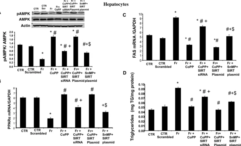
To assess whether HO-1 requires the participation of SIRT1 to mediate and/or amplify its actions, we studied the effect of SIRT1 siRNA and SIRT plasmid in hepatocytes treated with fructose. Our results showed that fructose decreased pAMPK and PPARαlevels and increased the expression of FAS (Fig 2A, 2B and 2Crespectively); this effect of fructose treatment was negated by treatment with CoPP. Interestingly, concurrent treatment with CoPP and SIRT1 siRNA decreased pAMPK and PPARαand increased FAS levels suggesting that HO-1 is upstream of SIRT1 and that suppression of SIRT1 attenuates the beneficial effects of increased levels of HO-1. We also utilized plasmid SIRT1 to assess if increased expression of SIRT1 (in the absence of HO-1 up-regulation) is sufficient to prevent the detrimental effects of HFr on lipid accumulation and metabolic imbalance. Treatment of hepatocytes with fructose, SnMP and SIRT plasmid decreased pAMPK and PPARαand increased FAS levels as compared to
Fig 2. Effect of CoPP with and without SIRT1-siRNA, and with and without SIRT plasmid on pAMPK, PPARα, FAS expression and triglyceride levels in fructose (Fr)-treated hepatocytes.(A) pAMPK/AMPK expression by western blot analysis. (B) PPARαmRNA levels. (C) FAS mRNA levels measured by RT-PCR in hepatocytes. Results are mean±SE, n = 4/group.*p<0.05 vs CTR; #p<0.05 vs HFr, +p<0.05 vs HFr+CoPP, $ vs Fr+CoPP+SIRT Plasmid. (D) Triglyceride levels measured by RT-PCR in hepatocytes. Results are mean±SE, n = 4/group.*p<0.05 vs CTR; #p<0.05 vs HFr, +p<0.05 vs HFr+CoPP, $ vs Fr+CoPP+SIRT Plasmid.
hepatocytes treated with fructose, CoPP and SIRT plasmid (Fig 2A, 2B and 2Crespectively; p<0.05). In agreement with our hypothesis, our results further showed that hepatocytes treated
with fructose, CoPP and SIRT plasmid did not significantly decrease pAMPK, PPARαand FAS levels as compared to cells treated with fructose and CoPP alone indicating a HO-1 depen-dent activation of SIRT1 expression.
HO-1 induction decreases triglycerides levels in cultured murine
hepatocytes; SIRT-1 knockdown attenuates the inhibitory action and
increases triglycerides levels
As seen inFig 2D, fructose increased triglycerides content in hepatocytes; this increase was negated by treatment with CoPP. Concurrent treatment with CoPP and SIRT1 siRNA increased triglycerides levels further suggesting that HO-1 is upstream of SIRT1. Treatment of hepatocytes with fructose, SnMP and SIRT plasmid increased triglycerides levels as compared to hepatocytes treated with fructose, CoPP and SIRT plasmid (Fig 2D; p<0.05). Our results
further showed that hepatocytes treated with fructose, CoPP and SIRT plasmid did not signifi-cantly decrease triglycerides levels as compared to cells treated with fructose and CoPP alone indicating a HO-1 dependent activation of SIRT1 expression.
Effect of HO-1 induction on metabolic profile and liver function in mice
fed a high-fructose diet
A HFr diet increased blood pressure in mice compared to their control group, (p<0.05) (Fig
3A), an effect reversed via CoPP. Similarly our results showed that fasting blood glucose levels were increased in mice fed a HFr diet as compared to the control (Fig 3B; p<0.05). CoPP
decreased blood glucose levels and concurrent treatment with SnMP reversed the beneficial effects of CoPP. Correspondingly, HOMA-IR was increased in mice fed a HFr diet as com-pared to the control mice (Fig 3C; p<0.05). CoPP significantly decreased HOMA-IR as
com-pared to mice fed a HFr diet. Further ALT levels were significantly increased in mice fed HFr diet (Fig 3D) as compared to the control group and this increase was negated by treatment with CoPP. Furthermore, SnMP reversed the beneficial effect of CoPP and decreased ALT lev-els in plasma (p<0.01).
Effect of HO-1 induction on hepatic lipid content in mice fed a
high-fructose diet
To examine whether HO-1 induction can suppress the formation of hepatic steatosis, the levels of triglycerides and cholesterol in hepatic tissue were measured. Our results showed that tri-glycerides and cholesterol content (Fig 3E and 3Frespectively; p<0.05) was significantly
increased in mice fed a HFr diet as compared to control mice. As expected, CoPP decreased tri-glycerides and cholesterol content as compared to mice fed a HFr diet and concurrent treat-ment with SnMP reversed the beneficial effects of CoPP.
Effect of HO-1 induction on hepatic lipogenesis and FFA levels in mice
fed a high-fructose diet
As shown inFig 4A, mice fed a HFr diet have significantly (p<0.05) more lipid accumulation
compared to the control mice. CoPP decreased FFA levels in hepatic tissue as compared to mice fed a fructose diet (Fig 4B; p<0.05). Expression of genes involved in hepatic fatty acid
synthesis; Elvol6 and Srebp-1c were induced in mice fed with a high-fructose diet compared to control group. Administration of CoPP significantly reduced the increased mRNA expressions to near control levels (Fig 4C). Similarly, ACC and SCD-1 mRNA expressions were signifi-cantly increased in mice fed a HFr diet as compared to the control mice and this increase was negated by treatment with CoPP (Fig 4D). Furthermore, SnMP reversed the beneficial effect of CoPP and decreased ACC and SCD-1 levels in hepatic tissue (p<0.01).
Fig 3. Effect of induction of HO-1 (CoPP) and inhibition of HO (SnMP) on metabolic profile and hepatic lipid content in mice fed a high fructose diet for 8 weeks.(A) Blood pressure. (B) Fasting blood glucose levels. (C) HOMA-IR (D) Plasma ALT levels. (E) Triglycerides levels in hepatic tissue. (F) Cholesterol levels in hepatic tissue. Results are mean±SE, n = 6/group.*p<0.05 vs CTR; #p<0.05 vs HFr, +p<0.05 vs HFr+CoPP.
doi:10.1371/journal.pone.0128648.g003
Fig 4. Effect of induction of HO-1 (CoPP) and inhibition of HO (SnMP) in mice fed a high fructose diet for 8 weeks on hepatic lipogenesis and FFA levels.(A) Oil Red O staining of lipids in liver and quantitative analysis of different groups, magnifications: 40X (n = 4). A representative section for each group is shown; (B) Hepatic FFA levels. (C) Elvol6 and Srebp-1c mRNA levels measured by RT-PCR and (D) ACC and SCD-1 mRNA expressions measured by RT-PCR. Results are mean±SE, n = 6/group.*p<0.05 vs CTR; #p<0.05 vs HFr, +p<0.05 vs HFr+CoPP.
Effect of HO-1 induction on hepatic SIRT1 expression and markers of
oxidative stress in mice fed a high-fructose diet
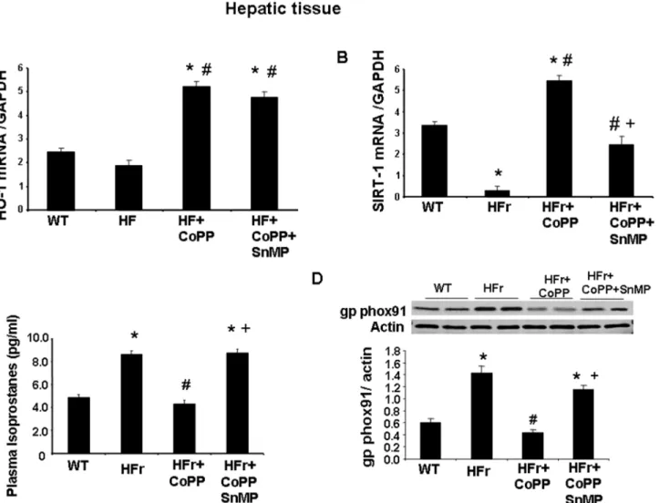
Mice fed a HFr diet and concurrently treated with CoPP exhibited increased hepatic HO-1 expression as compared to the control (Fig 5A). SnMP also increased HO-1 expression. How-ever, these findings are not surprising as SnMP, which induced a significant increase in HO-1 expression, remains a potent inhibitor of HO activity, as shown previously [35,42,43]. Mice fed a HFr diet exhibited decreased hepatic SIRT1 expression as compared to the control (Fig 5B). Furthermore, SnMP reversed the beneficial effect of CoPP and decreased the expression of SIRT1 (p<0.05). Mice fed a HFr diet had increased plasma isoprostane levels and an increased
expression of the hepatic NADPH-oxidase-subcomponent, gp/phox91 (Fig 5C and 5D respec-tively; p<0.05), a potent marker of oxidative stress, compared to the control mice. CoPP
reduced isoprostane and gpphox 91 levels as compared to mice fed a fructose diet (p<0.05).
SnMP reversed the effect of CoPP and increased the markers of oxidative stress.
Fig 5. Effect of induction of HO-1 (CoPP) and inhibition of HO (SnMP) in mice fed a high fructose diet for 8 weeks on HO-1 mRNA, SIRT1 mRNA, plasma isoprostane and gp phox91 protein expression.(A) HO-1 mRNA levels measured by RT-PCR. (B) SIRT1 mRNA levels measured by RT-PCR. (C) Plasma isoprostane levels and (D) gp phox91 protein expression. Results are mean±SE, n = 6/group.*p<0.05 vs CTR; #p<0.05 vs HFr, +p<0.05 vs HFr+CoPP.
Effect of HO-1 induction on hepatic insulin receptors, pAKT, G6Pase
levels and lipogenic markers in mice fed a high-fructose diet
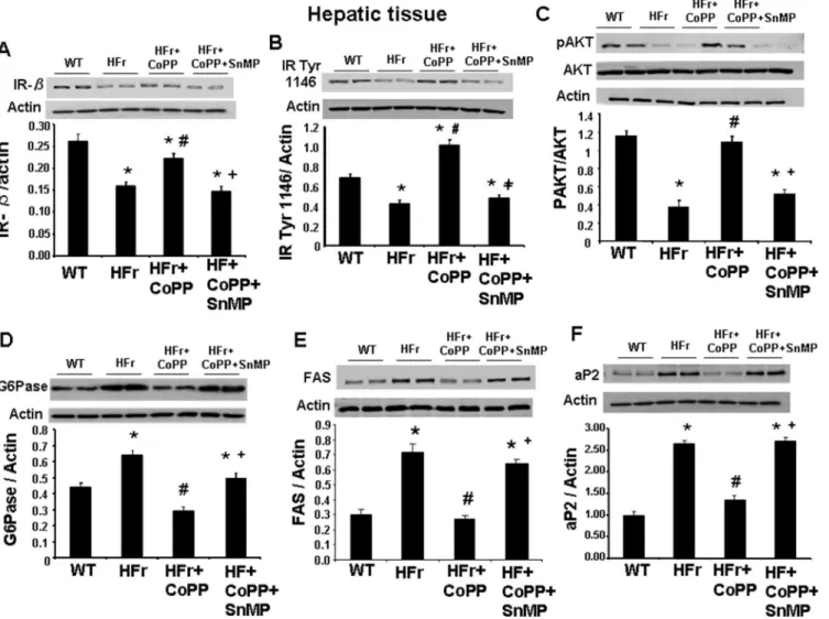
Western blots analyses of generic insulin receptor-beta (IR-β) (Fig 6A) and insulin receptor phosphorylated at tyrosine 1466 (Fig 6B) showed a significant decreased expression in mice fed a HFr diet compared with their controls. This decrease was blocked by the administration of CoPP while the co-administration of CoPP and SnMP reversed the effect of CoPP. Similarly, mice fed a HFr diet showed reduced phosphorylation of AKT in liver when compared to con-trol mice (Fig 6C). CoPP restored the phosphorylation of AKT to levels comparable to control mice while SnMP reversed the beneficial effects of CoPP on AKT phosphorylation (p<0.05).
Further our results showed that mice fed a HFr diet had higher mRNA expression of G6Pase, an important marker of gluconeogenesis, in hepatic tissue as compared to the control mice and this increase was negated by treatment with CoPP (Fig 6D; p<0.05). Also our results
showed that a HFr diet increased expression of lipogenic markers, FAS, (p<0.05) (Fig 6E) and
aP2 (Fig 6F), in hepatic tissue compared to their control group. Further our results indicate
Fig 6. Effect of induction of HO-1 (CoPP) and inhibition of HO (SnMP) in mice fed a high fructose diet for 8 weeks on western blot and densitometry analysis.(A) insulin receptor-β. (B) Insulin receptor phosphorylated at tyrosine 1146. (C) pAKT/AKT levels. (D) G6Pase. (E) FAS and (F) aP2 expression. Data are shown as mean band density normalized toβ-actin. Results are mean±SE, n = 4/group.*p<0.05 vs CTR; #p<0.05 vs HFr, +p<0.05 vs HFr+CoPP.
that mice treated with CoPP had decreased FAS and aP2 levels in hepatic tissue as compared to mice fed a HFr diet alone (Fig 6E and 6Frespectively; p<0.05). Furthermore, mice treated with
SnMP along with CoPP had increased FAS (p<0.05) and aP2 expression demonstrating the
beneficial effect of the HO-1-SIRT axis.
HO-1 induction attenuates high-fructose diet-induced inflammatory and
fibrotic markers in mice fed a high-fructose diet for 20 weeks
Immunohistochemistry was done on liver samples obtained from mice treated for 20 weeks with a HFr diet. No fibrosis was observed in the control mice (Fig 7Aa). The mice fed a HFr diet showed 10% fibrosis (Fig 7Ab). Further our results showed that administration of SnMP to CoPP treated mice fed a HFr diet reversed the beneficial effect of CoPP and had 9% fibrosis (Fig 7Ad). Mice fed a HFr diet had a significant increase in TNFα, a potent inducer of collagen synthesis (Fig 7B) compared to control mice. CoPP reduced TNFαlevels as compared to mice
Fig 7. Effect of induction of HO-1 (CoPP) and inhibition of HO (SnMP) on hepatic fibrosis, markers of hepatic fibrosis in mice fed high-fructose diet for 20 weeks.(A) Masson’s trichrome staining in liver and quantitative analysis of WT, high fructose, high fructose treated with CoPP, and high fructose treated with CoPP and SnMP, magnifications: 40× (n = 4) (*Indicates fibrosis). A representative section for each group is shown. (B) Plasma TNFαlevels. (C) MMP2 protein expression and (D) TGFβprotein expression on western blot analysis. Data are shown as mean band density normalized toβ-actin. Results are mean±SE, n = 4/group.*p<0.05 vs CTR; #p<0.05 vs HFr, +p<0.05 vs HFr+CoPP.
fed a fructose diet (p<0.05). SnMP abolished the CoPP effect suggesting the HO activity is
required for the beneficial effects of CoPP. Moreover, mice fed a HFr diet showed a significant increase in MMP-2 and TGF1βexpression (Fig 7C and 7Drespectively; p<0.05), compared to
the control mice. Treatment with CoPP reduced MMP-2 and TGF1βexpression as compared to mice on a HFr diet (Fig 7C and 7Drespectively). Administration of SnMP to CoPP treated mice fed a HFr diet reversed the beneficial effect of CoPP and increased markers of hepatic fibrosis.
HO-1 induction attenuates high-fructose diet-induced redox markers in
the aorta
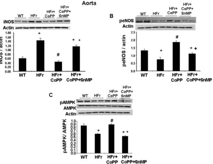
Our results showed activation of inflammatory pathways (iNOS) in the aorta of mice fed a HFr diet (Fig 8A). CoPP increased the aortic expression of peNOS (Fig 8B; p<0.05); the concurrent
administration of SnMP with CoPP decreased the expression of peNOS. Therefore CoPP increased NO bioavailability, restoring the balance of aortic eNOS and iNOS isoforms and the redox state. Further mice fed a HFr diet had a decreased expression of pAMPK (Fig 8C;
Fig 8. Effect of induction of HO-1 (CoPP) and inhibition of HO (SnMP) in mice fed a high fructose diet for 8 weeks on western blot and densitometry analysis of aortic tissue.(A) iNOS protein expression. (B) peNOS protein expression and (C) pAMPK protein expression. Data are shown as mean band density normalized toβ-actin. Results are mean±SE, n = 4/group.*p<0.05 vs CTR; #p<0.05 vs HFr, +p<0.05 vs HFr+CoPP.
p<0.05) as compared to the control group. CoPP increased pAMPK levels (p<0.05) as
com-pared to mice fed a fructose diet. SnMP abolished the beneficial effects of CoPP.
The results of the control group treatments are included in tables A to H in supporting information (S1 File).
Discussion
This study establishes the protective role of the heme-HO system in counteracting pathologies brought about by a HFr diet, specifically; reduction in hepatic lipid accumulation, improve-ment in insulin sensitivity and metabolic balance, and attenuation of hepatic fibrosis. Impor-tantly, our results show that this protection, at least in part, is mediated by the
HO-1-dependent rescue of hepatic SIRT1. We demonstrate that HO-1 acts through SIRT1 to form a functional module within hepatocytes to attenuate steatohepatitis, hepatic fibrosis and meta-bolic imbalance. Thus our results allude to the presence of a hepatic HO-1-SIRT1 axis that attenuates hepatic steatotic pathways and has systemic effects including, improvement of vas-cular function and restoration of insulin sensitivity.
The first key finding of the study is the redox-dependent attenuation of hepatocyte SIRT1 that is rescued by HO-1. High-sugar diets alter redox state of hepatocytes and eventually cause increased lipid accumulation in these cells [44,45]. This is confirmed in our study by elevated oxidative stress in hepatocytes cultured with high-fructose supplementation. This is accompa-nied by suppression of hepatocyte SIRT1 levels. ROS-mediated suppression of SIRT1 has been reported by us and by other investigators [32]; also, the antioxidant properties of the heme-HO system are well documented [36,46–49]. Thus, protective effects of HO-1-induction on hepa-tocyte SIRT1 are novel but not surprising. This is the first report showing hepatic SIRT1 rescue by the up-regulation of the heme-HO system in hepatocytes stressed by high-fructose supple-ment. Antioxidant properties of SIRT1 are primarily ascribed to BV, which has the ability to quench variety of free radicals. Tempol, on the other hand, is a SOD-mimetic and primarily reduces superoxide levels. SIRT1 rescue by both, biliverdin and tempol, lead us to propose that increased superoxide levels could bring about high-fructose mediated SIRT1 suppression. However, it is important to point out that the precise molecular mechanisms of this antioxi-dant-rescue of SIRT1 are not fully understood.
component of the effects of HO-1. But overall, ourin vitroresults indicate that at least part of the protective actions the HO system on the metabolic pathways in hepatocytes is via SIRT1-rescue. Importantly, SIRT1 up-regulates these metabolic regulators in multiple settings [30,31,54]. Pos-sible downstream targets for the HO1-SIRT1 module could include AMPK and PPARα. With regards to these proteins, p-AMPK/AMPK and PPARαare known to modulate the hepatic meta-bolic pathways [37,54] and their activation leads to reduction in hepatic glucose output, suppres-sion of pro-lipogenic pathways and improvement in liver function.
A murine model of diet-induced hepatic steatosis and fibrosis confirms ourin vitrofindings. Third key finding of the study highlights the hepato-protective effect of HO-1 in mice fed a HFr diet. CoPP treated mice showed significant improvement in hepatic steatosis, fibrosis and metabolic balance. Hepatic FAS levels were down regulated while insulin signaling improved, in mice with increased levels of HO-1 induction. Although HFr mediated NAFLD is well estab-lished, the precise molecular mechanisms remain incompletely understood. Up-regulation of HO-1 attenuate adiposity in mice fed high-fat diet by reprogramming adipocyte phenotype to functional health adipocyte [55]. HO-1 induction reversed fructose-mediated increase in oxi-dants, isoprostane production and adipocyte dysfunction [56]. HO-1 gene targeting either adi-pocytes or vascular system attenuates adiposity, ROS and vascular dysfunction in mice fed a high-fat diet [55,57]. Our results, showing high redox potential in hepatic tissues of mice fed HFr, are in line with these reports and lead us to believe that ROS-dependent pathways are cen-tral to the pathophysiology of NAFLD [58]. ROS-induced SIRT1 suppression is one of these candidate pathways. By interfering with this NAD-dependent deacetylase, high oxidative stress alters cellular metabolic balance and HO-1 system is the first line of defense against such inju-ries. We demonstrate in this study that induction of HO-1 leads to a reduction in lipid accumu-lation and FFA, a decrease in blood glucose levels and a decrease in ROS and inflammation in hepatocytes, a major cause of insulin resistance. It is important to note that our findings are in contrast with the recent work by Jaiset al. The authors showed that liver-specific KO of HO-1 decreases hepatic lipid accumulation and that overexpression of HO-1 in hepatocytes results in insulin resistance. At this time we are not fully able to explain the dissimilarities in our results; however, certain differences in the experimental design do stand out. First, Jaiset alused a model of high-fat diet to induce hepatic steatosis whereas HFr was used in ours. It could be that insulin resistance and hepatic steatosis brought on by these diets engage distinct cellular defense mechanisms and adaptive responses. Additionally, activation of compensatory responses during HO-1 KO, such as HO-2, may contribute to the observed differences in our findings. Secondly, Jaiset alused adenoviral constructs to show that acute overexpression of HO-1 (7 days) in hepatocytes results in insulin resistance. We have used a model of chronic up regulation of HO-1 and temporal changes in the role of this system may occur during meta-bolic homeostasis; further studies are needed to fully resolve this issue.
Hepatic steatosis also increases the risk for CVD [13,15] leading to endothelial dysfunction, atherosclerosis and hypertension [14,61,62]. Non-alcoholic fatty liver might contribute in the pathogenesis of CVD through the systemic release of inflammatory and oxidative-stress media-tors or through the contribution of hepatic steatosis to insulin resistance and atherogenic dysli-pidemia. Our results showed that HO-1-SIRT1 axis salvaged endothelial dysfunction by modulating signaling and survival pathways to improve NAFLD-induced CVDs. Increased lev-els of HO-1 are associated with an increase in peNOS, and NO bioavailability [63,64]. Also an increase in AMPK signaling is considered an important metabolic response key to the attenua-tion of ROS-mediated endothelial dysfuncattenua-tion, since pAMPK utilizes eNOS as a substrate and enhances the levels of peNOS [64,65]. In agreement with these reports, our results demon-strate that the HO-1-SIRT1 axis increases the level of peNOS and pAMPK to ameliorate vascu-lar dysfunction. Taken together, these observations solidify our notion that a HFr diet affects inflammatory and redox pathways in both, liver and vascular tissues and that activation of the HO-1-SIRT1 module has the ability to counteract these changes. This insight may prove to have profound therapeutic implications for treatment of nonobese type 2 diabetics who have already attempted lifestyle modification with diet and exercise since directly targeting NAFLD could be a particularly high yield intervention for them, decreasing glycemia and risk of adverse cardiovascular events.
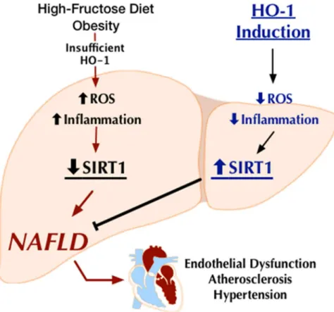
In conclusion (Fig 9), this study demonstrates that HO-1 induction in the liver reduced fructose-induced hepatic lipid deposition, prevented the development of hepatic fibrosis and abated NAFLD-associated metabolic and vascular imbalance; effects that are mediated by
Fig 9. Schematic demonstrating HO-1 induction attenuates fructose-induced hepatic lipid deposition, prevent the development of hepatic fibrosis and abate NAFLD-associated vascular dysfunction; effects that are mediated by activation of the SIRT1 gene expression.
activation of the SIRT1 gene expression. It is evident that the SIRT family of genes is key play-ers in redox biology. Further studies are needed, however, to fully elucidate the HO-1-SIRT1 interactions in intact animals. Hepatocyte-specific SIRT1 KO mice will further our understand-ing of these interactions. We believe that elucidatunderstand-ing these interactions will lead to the develop-ment of new biomarkers and therapeutic strategies to fight hepatic dysfunction associated with NAFLD.
Supporting Information
S1 File. The results of the treatments done in control groups are provided as supporting information in supporting tables. Table A: Control groups fromFig 1.Table B: Control groups fromFig 2.Table C: Control groups fromFig 3.Table D: Control groups fromFig 4. Table E: Control groups fromFig 5.Table F: Control groups fromFig 6.Table G: Control groups fromFig 7.Table H: Control groups fromFig 8.
(DOCX)
Acknowledgments
The authors wish to thank Ms. Jennifer Brown and Kyle Maxwell for their assistance in the preparation of this manuscript.
Author Contributions
Conceived and designed the experiments: KS NP. Performed the experiments: KS NP GF SS HA. Analyzed the data: KS NP. Contributed reagents/materials/analysis tools: JIS NGA. Wrote the paper: KS NP. Helped in editing the manuscript: EL RR CM.
References
1. Vernon G, Baranova A, Younossi ZM. Systematic review: the epidemiology and natural history of non-alcoholic fatty liver disease and non-non-alcoholic steatohepatitis in adults. Aliment Pharmacol Ther 2011; 34:274–285. doi:10.1111/j.1365-2036.2011.04724.xPMID:21623852
2. Williams CD, Stengel J, Asike MI, Torres DM, Shaw J, Contreras M, et al. Prevalence of nonalcoholic fatty liver disease and nonalcoholic steatohepatitis among a largely middle-aged population utilizing ultrasound and liver biopsy: a prospective study. Gastroenterology 2011; 140:124–131. doi:10.1053/j. gastro.2010.09.038PMID:20858492
3. Lazo M, Hernaez R, Eberhardt MS, Bonekamp S, Kamel I, Guallar E, et al. Prevalence of nonalcoholic fatty liver disease in the United States: the Third National Health and Nutrition Examination Survey, 1988–1994. Am J Epidemiol 2013; 178:38–45. doi:10.1093/aje/kws448PMID:23703888
4. Qureshi K, Abrams GA. Metabolic liver disease of obesity and role of adipose tissue in the pathogene-sis of nonalcoholic fatty liver disease. World J Gastroenterol 2007; 13:3540–3553. PMID:17659704
5. Marchesini G, Bugianesi E, Forlani G, Cerrelli F, Lenzi M, Manini R, et al. Nonalcoholic fatty liver, stea-tohepatitis, and the metabolic syndrome. Hepatology 2003; 37:917–923. PMID:12668987
6. Adams LA, Angulo P. Recent concepts in non-alcoholic fatty liver disease. Diabet Med 2005; 22:1129– 1133. PMID:16108837
7. Delgado JS. Evolving trends in nonalcoholic fatty liver disease. Eur J Intern Med 2008; 19:75–82. doi: 10.1016/j.ejim.2007.02.034PMID:18249301
8. Bugianesi E, Moscatiello S, Ciaravella MF, Marchesini G. Insulin resistance in nonalcoholic fatty liver disease. Curr Pharm Des 2010; 16:1941–1951. PMID:20370677
9. Fon TK, Rozman D. Nonalcoholic Fatty liver disease: focus on lipoprotein and lipid deregulation. J Lip-ids 2011; 2011:783976. doi:10.1155/2011/783976PMID:21773052
11. Targher G, Byrne CD. Clinical Review: Nonalcoholic fatty liver disease: a novel cardiometabolic risk factor for type 2 diabetes and its complications. J Clin Endocrinol Metab 2013; 98:483–495. doi:10. 1210/jc.2012-3093PMID:23293330
12. Abel T, Feher J. [Non-alcoholic fatty liver disease and cardiovascular risk]. Orv Hetil 2008; 149:1299– 1305. doi:10.1556/OH.2008.28418PMID:18617457
13. Gaggini M, Morelli M, Buzzigoli E, DeFronzo RA, Bugianesi E, Gastaldelli A. Non-alcoholic fatty liver disease (NAFLD) and its connection with insulin resistance, dyslipidemia, atherosclerosis and coronary heart disease. Nutrients 2013; 5:1544–1560. doi:10.3390/nu5051544PMID:23666091
14. Targher G, Arcaro G. Non-alcoholic fatty liver disease and increased risk of cardiovascular disease. Atherosclerosis 2007; 191:235–240. PMID:16970951
15. Oni ET, Agatston AS, Blaha MJ, Fialkow J, Cury R, Sposito A, et al. A systematic review: burden and severity of subclinical cardiovascular disease among those with nonalcoholic fatty liver; should we care? Atherosclerosis 2013; 230:258–267. doi:10.1016/j.atherosclerosis.2013.07.052PMID: 24075754
16. Pettinelli P, Obregon AM, Videla LA. Molecular mechanisms of steatosis in nonalcoholic fatty liver dis-ease. Nutr Hosp 2011; 26:441–450. doi:10.1590/S0212-16112011000300003PMID:21892559
17. Abdelmalek MF, Lazo M, Horska A, Bonekamp S, Lipkin EW, Balasubramanyam A, et al. Higher dietary fructose is associated with impaired hepatic adenosine triphosphate homeostasis in obese individuals with type 2 diabetes. Hepatology 2012; 56:952–960. doi:10.1002/hep.25741PMID:22467259
18. Abdelmalek MF, Suzuki A, Guy C, Unalp-Arida A, Colvin R, Johnson RJ, et al. Increased fructose con-sumption is associated with fibrosis severity in patients with nonalcoholic fatty liver disease. Hepatology 2010; 51:1961–1971. doi:10.1002/hep.23535PMID:20301112
19. Basaranoglu M, Basaranoglu G, Sabuncu T, Senturk H. Fructose as a key player in the development of fatty liver disease. World J Gastroenterol 2013; 19:1166–1172. doi:10.3748/wjg.v19.i8.1166PMID: 23482247
20. Lim JS, Mietus-Snyder M, Valente A, Schwarz JM, Lustig RH. The role of fructose in the pathogenesis of NAFLD and the metabolic syndrome. Nat Rev Gastroenterol Hepatol 2010; 7:251–264. doi:10.1038/ nrgastro.2010.41PMID:20368739
21. Nomura K, Yamanouchi T. The role of fructose-enriched diets in mechanisms of nonalcoholic fatty liver disease. J Nutr Biochem 2012; 23:203–208. doi:10.1016/j.jnutbio.2011.09.006PMID:22129639
22. Basaranoglu M, Basaranoglu G, Sabuncu T, Senturk H. Fructose as a key player in the development of fatty liver disease. World J Gastroenterol 2013; 19:1166–1172. doi:10.3748/wjg.v19.i8.1166PMID: 23482247
23. Longato L. Non-alcoholic fatty liver disease (NAFLD): a tale of fat and sugar? Fibrogenesis Tissue Repair 2013; 6:14. doi:10.1186/1755-1536-6-14PMID:23866299
24. Rebollo A, Roglans N, Alegret M, Laguna JC. Way back for fructose and liver metabolism: bench side to molecular insights. World J Gastroenterol 2012; 18:6552–6559. doi:10.3748/wjg.v18.i45.6552 PMID:23236229
25. Johnson RJ, Perez-Pozo SE, Sautin YY, Manitius J, Sanchez-Lozada LG, Feig DI, et al. Hypothesis: could excessive fructose intake and uric acid cause type 2 diabetes? Endocr Rev 2009; 30:96–116. doi:10.1210/er.2008-0033PMID:19151107
26. Garcia-Galiano D, Sanchez-Garrido MA, Espejo I, Montero JL, Costan G, Marchal T, et al. IL-6 and IGF-1 are independent prognostic factors of liver steatosis and non-alcoholic steatohepatitis in morbidly obese patients. Obes Surg 2007; 17:493–503. PMID:17608262
27. Theret N, Lehti K, Musso O, Clement B. MMP2 activation by collagen I and concanavalin A in cultured human hepatic stellate cells. Hepatology 1999; 30:462–468. PMID:10421655
28. Kato J, Sato Y, Inui N, Nakano Y, Takimoto R, Takada K, et al. Ethanol induces transforming growth factor-alpha expression in hepatocytes, leading to stimulation of collagen synthesis by hepatic stellate cells. Alcohol Clin Exp Res 2003; 27:58S–63S. PMID:12960509
29. Puri N, Sodhi K, Haarstad M, Kim DH, Bohinc S, Foglio E, et al. Heme induced oxidative stress attenu-ates sirtuin1 and enhances adipogenesis in mesenchymal stem cells and mouse pre-adipocytes. J Cell Biochem 2012; 113:1926–1935. doi:10.1002/jcb.24061PMID:22234917
30. Yamazaki Y, Usui I, Kanatani Y, Matsuya Y, Tsuneyama K, Fujisaka S, et al. Treatment with SRT1720, a SIRT1 activator, ameliorates fatty liver with reduced expression of lipogenic enzymes in MSG mice. Am J Physiol Endocrinol Metab 2009; 297:E1179–E1186. doi:10.1152/ajpendo.90997.2008PMID: 19724016
32. Chung S, Yao H, Caito S, Hwang JW, Arunachalam G, Rahman I. Regulation of SIRT1 in cellular func-tions: role of polyphenols. Arch Biochem Biophys 2010; 501:79–90. doi:10.1016/j.abb.2010.05.003 PMID:20450879
33. Berberat PO, Katori M, Kaczmarek E, Anselmo D, Lassman C, Ke B, et al. Heavy chain ferritin acts as an antiapoptotic gene that protects livers from ischemia reperfusion injury. FASEB J 2003; 17:1724– 1726. PMID:12958189
34. Li M, Kim DH, Tsenovoy PL, Peterson SJ, Rezzani R, Rodella LF, et al. Treatment of obese diabetic mice with a heme oxygenase inducer reduces visceral and subcutaneous adiposity, increases adipo-nectin levels, and improves insulin sensitivity and glucose tolerance. Diabetes 2008; 57:1526–1535. doi:10.2337/db07-1764PMID:18375438
35. Cao J, Peterson SJ, Sodhi K, Vanella L, Barbagallo I, Rodella LF, et al. Heme oxygenase gene target-ing to adipocytes attenuates adiposity and vascular dysfunction in mice fed a high-fat diet. Hyperten-sion 2012; 60:467–475. doi:10.1161/HYPERTENSIONAHA.112.193805PMID:22753217
36. Abraham NG, Kappas A. Pharmacological and clinical aspects of heme oxygenase. Pharmacol Rev 2008; 60:79–127. doi:10.1124/pr.107.07104PMID:18323402
37. Chau MD, Gao J, Yang Q, Wu Z, Gromada J. Fibroblast growth factor 21 regulates energy metabolism by activating the AMPK-SIRT1-PGC-1alpha pathway. Proc Natl Acad Sci U S A 2010; 107:12553– 12558. doi:10.1073/pnas.1006962107PMID:20616029
38. Stienstra R, Mandard S, Patsouris D, Maass C, Kersten S, Muller M. Peroxisome proliferator-activated receptor alpha protects against obesity-induced hepatic inflammation. Endocrinology 2007; 148:2753– 2763. PMID:17347305
39. Hinds TD Jr, Sodhi K, Meadows C, Fedorova L, Puri N, Kim DH, et al. Increased HO-1 levels ameliorate fatty liver development through a reduction of heme and recruitment of FGF21. Obesity (Silver Spring) 2013.
40. Abraham NG, Camadro JM, Hoffstein ST, Levere RD. Effects of iron deficiency and chronic iron over-loading on mitochondrial heme biosynthetic enzymes in rat liver. Biochim Biophys Acta 1986; 870:339– 349. PMID:3955059
41. da Silva JL, Tiefenthaler M, Park E, Escalante B, Schwartzman ML, Levere RD, et al. Tin-mediated heme oxygenase gene activation and cytochrome P450 arachidonate hydroxylase inhibition in sponta-neously hypertensive rats. Am J Med Sci 1994; 307:173–181. PMID:8160707
42. Botros FT, Schwartzman ML, Stier CT Jr, Goodman AI, Abraham NG. Increase in heme oxygenase-1 levels ameliorates renovascular hypertension. Kidney Int 2005; 68:2745–2755. PMID:16316349
43. Sodhi K, Puri N, Inoue K, Falck JR, Schwartzman ML, Abraham NG. EET agonist prevents adiposity and vascular dysfunction in rats fed a high fat diet via a decrease in Bach 1 and an increase in HO-1 lev-els. Prost Other Lipid Mediat 2012; 98:133–142.
44. Estall JL, Ruas JL, Choi CS, Laznik D, Badman M, Maratos-Flier E, et al. PGC-1alpha negatively regu-lates hepatic FGF21 expression by modulating the heme/Rev-Erb(alpha) axis. Proc Natl Acad Sci U S A 2009; 106:22510–22515. doi:10.1073/pnas.0912533106PMID:20018698
45. Krahenbuhl L, Lang C, Ludes S, Seiler C, Schafer M, Zimmermann A, et al. Reduced hepatic glycogen stores in patients with liver cirrhosis. Liver Int 2003; 23:101–109. PMID:12654132
46. Burgess A, Li M, Vanella L, Kim DH, Rezzani R, Rodella L, et al. Adipocyte heme oxygenase-1 induc-tion attenuates metabolic syndrome in both male and female obese mice. Hypertension 2010; 56:1124–1130. doi:10.1161/HYPERTENSIONAHA.110.151423PMID:21041703
47. Nicolai A, Li M, Kim DH, Peterson SJ, Vanella L, Positano V, et al. Heme Oxygenase-1 Induction Remodels Adipose Tissue and Improves Insulin Sensitivity in Obesity-Induced Diabetic Rats. Hyper-tension 2009; 53:508–515. doi:10.1161/HYPERTENSIONAHA.108.124701PMID:19171794
48. Kim DH, Burgess AP, Li M, Tsenovoy PL, Addabbo F, McClung JA, et al. Heme oxygenase-mediated increases in adiponectin decrease fat content and inflammatory cytokines, tumor necrosis factor-alpha and interleukin-6 in Zucker rats and reduce adipogenesis in human mesenchymal stem cells. J Phar-macol Exp Ther 2008; 325:833–840. doi:10.1124/jpet.107.135285PMID:18334666
49. Yang H, Zhao LF, Zhao ZF, Wang Y, Zhao JJ, Zhang L. Heme oxygenase-1 prevents liver fibrosis in rats by regulating the expression of PPARgamma and NF-kappaB. World J Gastroenterol 2012; 18:1680–1688. doi:10.3748/wjg.v18.i14.1680PMID:22529699
50. Elliott SS, Keim NL, Stern JS, Teff K, Havel PJ. Fructose, weight gain, and the insulin resistance syn-drome. Am J Clin Nutr 2002; 76:911–922. PMID:12399260
51. Abraham N, Tsenovoy P, McClung J, Drummond G. Heme oxygenase: a target gene for anti-diabetic and obsesity. Curr Pharm Des 2008; 14:412–421. PMID:18289068
autophagy. Am J Physiol Heart Circ Physiol 2012; 302:H1394–H1409. doi:10.1152/ajpheart.00584. 2011PMID:22245770
53. Armutcu F, Coskun O, Gurel A, Kanter M, Can M, Ucar F, et al. Thymosin alpha 1 attenuates lipid per-oxidation and improves fructose-induced steatohepatitis in rats. Clin Biochem 2005; 38:540–547. PMID:15885234
54. Kim E, Choi Y, Jang J, Park T. Carvacrol Protects against Hepatic Steatosis in Mice Fed a High-Fat Diet by Enhancing SIRT1-AMPK Signaling. Evid Based Complement Alternat Med 2013; 2013:290104.
55. Abraham NG, Sodhi K, Silvis AM, Vanella L, Favero G, Rezzani R, et al. CYP2J2 Targeting to Endothe-lial Cells Attenuates Adiposity and Vascular Dysfunction in Mice Fed a High-Fat Diet by Reprogram-ming Adipocyte Phenotype. Hypertension 2014.
56. Khitan Z, Harsh M, Sodhi K, Shapiro JI, Abraham NG. HO-1 Upregulation Attenuates Adipocyte Dys-function, Obesity, and Isoprostane Levels in Mice Fed High Fructose Diets. J Nutr Metab 2014; 2014:980547. doi:10.1155/2014/980547PMID:25295182
57. Cao J, Sodhi K, Inoue K, Quilley J, Rezzani R, Rodella L, et al. Lentiviral-human heme oxygenase tar-geting endothelium improved vascular function in angiotensin II animal model of hypertension. Hum Gene Ther 2011; 22:271–282. doi:10.1089/hum.2010.059PMID:20836698
58. McCullough AJ. Pathophysiology of nonalcoholic steatohepatitis. J Clin Gastroenterol 2006; 40 Suppl 1:S17–S29. PMID:16540762
59. Jiang JX, Chen X, Fukada H, Serizawa N, Devaraj S, Torok NJ. Advanced glycation endproducts induce fibrogenic activity in nonalcoholic steatohepatitis by modulating TNF-alpha-converting enzyme activity in mice. Hepatology 2013; 58:1339–1348. doi:10.1002/hep.26491PMID:23703665
60. Vasko R, Xavier S, Chen J, Lin CH, Ratliff B, Rabadi M, et al. Endothelial Sirtuin 1 Deficiency Perpe-trates Nephrosclerosis through Downregulation of Matrix Metalloproteinase-14: Relevance to Fibrosis of Vascular Senescence. J Am Soc Nephrol 2013.
61. Targher G. Non-alcoholic fatty liver disease, the metabolic syndrome and the risk of cardiovascular dis-ease: the plot thickens. Diabet Med 2007; 24:1–6.
62. Xirouchakis E, Sigalas A, Manousou P, Calvaruso V, Pleguezuelo M, Corbani A, et al. Models for non-alcoholic fatty liver disease: a link with vascular risk. Curr Pharm Des 2008; 14:378–384. PMID: 18289064
63. Cao J, Sodhi K, Puri N, Monu SR, Rezzani R, Abraham NG. High fat diet enhances cardiac abnormali-ties in SHR rats: Protective role of heme oxygenase-adiponectin axis. Diabetol Metab Syndr 2011; 3:37. doi:10.1186/1758-5996-3-37PMID:22196253
64. Li Q, Guo Y, Ou Q, Cui C, Wu WJ, Tan W, et al. Gene transfer of inducible nitric oxide synthase affords cardioprotection by upregulating heme oxygenase-1 via a nuclear factor-{kappa}B-dependent pathway. Circulation 2009; 120:1222–1230. doi:10.1161/CIRCULATIONAHA.108.778688PMID:19752329