P R A C T I C A L A D V I C E F O R P H Y S I C I A N S
UDC: 616.21-06:617.7 DOI: 10.2298/VSP141118141J
Silent sinus syndrome – one more reason for an ophthalmologist to
have a rhinologist as a good friend
Sindrom tihog sinusa – još jedan razlog za oftalmologa da ima rinologa kao
dobrog prijatelja
Ljiljana Jovančević*†, Vladimir Čanadanović*‡, Slobodan Savović*†,
Biljana Zvezdin*§, Zoran Komazec*†
*Faculty of Medicine, University of Novi Sad, Novi Sad, Serbia; †Otorhinolaryngology Clinic, ‡Ophthalmology Clinic, Clinical Centre of Vojvodina, Novi Sad, Serbia;
§
Institute for Pulmonary Diseases of Vojvodina, Sremska Kamenica, Serbia
Key words:
maxillary sinus; syndrome; enophthalmos; diagnosis; ophthalmologic surgical procedures; otorhinolaryngologic surgical procedures; treatment outcome.
Ključne reči:
maksilarni sinus; sindrom; enoftalmus; dijagnoza; hirurgija, oftalmološka, procedure; hirurgija, otorinolaringološka, procedure; lečenje, ishod.
Introduction
Silent sinus syndrome (SSS) is a rare condition invol-ving the maxillary sinus, characterized by unilateral collapse of the maxillary sinus and orbital floor, associated with nega-tive antral pressure in the absence of sinus symptoms 1, 2. It is also known as imploding antrum syndrome and typical radio-logical findings are ipsilateral depression of the orbital floor and opacification of a collapsed maxillary sinus 1, 2. There has been some more than 105 cases of SSS published in En-glish literature so far (1). The largest case series with 22 pa-tients was published by Kass et al. 3 in 1997.
SSS is characterized by spontaneous and progressive enophthalmos (“sunken” eye-eye recession into globe) and hypoglobus (globe displaced downward; a drop in the pupillary level), so it is common that these patients first pre-sent to ophthalmologist 1, 2, 4. Its development is gradual and progressive, so after a few months up to a few years may be-come symptomatic 1. Since patients present with ophthalmo-logical complains, without any nasal or sinus symptoms, with painless course and slow development, the term “silent sinus” was introduced 5.
The first report of this entity was in 1964 in a paper written by Montgomery 6. His report was about patients who had diplopia and enophthalmos associated with collapse of the maxillary sinus. Wilkins and Kulwin 7, in their paper pu-blished in 1981, emphasized that there was no orbital trauma in patients as a cause of the clinical simptoms and signs, alt-hough up till then it was known it happens only as a
consequence of orbital trauma. Soparkar et al. 5, in their pa-per published in 1994, introduced the term silent sinus syndrome. They described a large group of 14 patients with spontaneous, unilateral enophthalmos and hypoglobus asso-ciated with “asymptomatic, bone thinning, maxillary sinus disease” [seen on computed tomography (CT) scans].
Imploding antrum syndrome can be primary or secondary. Primary or spontaneous (SSS) is idiopathic, whereas secondary may arise from mid-face trauma (including surgery), rhinosurgery, chronic rhinosinusitis and has also been reported in less than 1% of patients after orbital decompression in Graves ophthalmopathy ( thyroid eye disease) 1, 5, 8, 9.
SSS most commonly presents unilaterally, although there are reports on it being bilateral 4, 10. It occurs exclusively in maxillary sinus (there is one report about it in the frontal si-nus) 1, 11. SSS presents in the third to forth decades of life and seems to affect both genders equally 9. The incidence of SSS is similar in the left and right maxillary sinuses (there might be a slight predominance for presenting on the right side – (57%) 4, 9. The average duration of the progressive, characteristic orbi-topathies until presentation is 3 months (range 10 days – 2 years) 5, 9. Average enophthalmos at presentation is 2,96 mm (± 0.16 mm), average hypoglobus at presentation is 2.78 (± 0.25 mm) 5, 9, 12. Although mostly observed in adults, there have been reports on SSS in children 9, 13, 14.
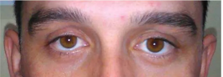
Fig. 1 – Typical ocular (facial) asymmetry in silent sinus syndrome (SSS) – hypoglobus and enophthalmos (3 mm) on the left side; upper-lid retraction, deepened upper-lid sulcus.
are abnormal eyelid signs (retraction, ptosis, absent crease), lid lag, and dry eyes from lagophthalmos 1, 2, 8, 15–17.
Pathophysiology
SSS has two main theorized mechanisms: maxillary sinus atelectasis (MSA) which could be idiopathic, posttraumatic, or post-surgery, and maxillary sinus hypoplasia (MSH) 4.
The exact pathophysiology of SSS is unknown, and so far, there are three main theories 1, 3, 5, 9, 15, 17.
The first theory is associated with prolonged negative pres-sure (continued negative prespres-sure within the sinus) 3, 5, 15, 17–19. A complete obstruction of the MS ostium results in hypoventilation and accumulation of secretion. In this enclosed cavity subsequent gas resorption leads to subatmospheric pressu-re that cpressu-reates vacuum, thus cpressu-reating a suction effect of negative pressure within the maxillary sinus, vacuum may induce osteo-penia, bone remodeling and sinus walls retraction (like eustachi-an tube dysfunction causing retraction in the middle ear) 15. The chronic negative pressure in the sinus slowly retracts the orbital floor, altering orbital anatomy and affecting the function of orbi-tal contents. Continued negative pressure within the sinus activa-tes the osteoclasts; in turn, these make the sinus walls thinner. The enophtalmos and hypoglobus are induced because the thin-ned orbital floor cannot support the pressure from the overlying orbital content, which gradually expands into the sinus 3, 5, 15, 17–19.
There is a hypothesis suggesting that lateralized middle turbinate may act as one-way pneumatic valve, leading to progressive reduction of air in the antrum and subsequent collapse of the maxillary sinus 1, 16.
However, given the rarity of SSS and the very high pre-valence of maxillary sinus ostiumobstruction, Hourany et al. 20 has placed this first theory explanation under question. The prolonged negative pressure theory also fails to offer an explanation for the exclusive involvement of the maxillary sinus, so he supposed that some other compounding factors such as trauma or anatomic predisposition play a role 20.
The second theory of SSS pathogenesis is inflammatory ero-sion 15. Chronic inflammation could induce the erosion of the
orbi-hypoplastic maxillary sinus, but one third of the patients have no history of sinus disease in childhood and SSS occurs in normal and well-developed maxillary sinus.
Diagnosis
The diagnosis of SSS is made by typical clinical features – gradual onset of enophthalmos and/or hypoglobus, in the absen-ce of orbital trauma (including surgery) or sinus disease, nasal endoscopy and CT scans of the nose and paranasal sinuses 1, 9.
Nasal endoscopy will show either a completely normal finding, or one of the two typical pictures: firstly, widened middle meatus on the affected side with inward retraction of the uncinate process 15 and secondly, middle turbinate latera-lized (middle meatus obscured due to lateral displacement of the middle turbinate toward the uncinate process) 9, 15, 21.
ret-Fig. 2 – Coronal computed tomography (CT) scan: late-ralized left uncinate process and medial maxillary sinus
wall, enlarged left middle meatus, completely opacified left maxillary sinus, and its volume decreased.
Fig. 3 – Axial computed tomography (CT) scan: inward retracted walls of the left maxillary sinus, left maxillary sinus completely opacified, and its volume decreased.
raction of anterior, posterior and medial wall into sinus lu-men. There is also patchy loss of mineralisation. Typically, all 4 walls of the sinus are retracted, though one of the medi-al, anterior, or posterolateral walls may be spared 20.
The orbital floor is always retracted, commonly thin-ned, while the other walls may be thinthin-ned, normal, or slightly thickened 1, 4, 20, 25. Orbital floor thickening is also a possibility. Considering the meaning of the thinned or thic-kened sinus walls in SSS, Hourany et al. 20 discused the issue in their paper, illustrating it with the case that supports the notion that SSS is an aquired condition, since thickening of the sinus walls is probably related to chronic inflammation and not to underlying developmental hypoplasia 20.The opa-cification of the MS can be complete or near complete (total or near total) 16, 18. Sanchez et al. 25 described an image of a “pseudo-pneumo-orbit” that can also be seen due to air trap-ped under the upper eyelid.
The cases of lateralized uncinate process and increased orbital volumes observed on CT scans, but lacking clinical enophthalmos and hypoglobus exist, and are a matter of dis-cussion if it is, or it is not the SSS. Wise et al. 26 in their pa-per published in 2007 conclude that it potentially represents early SSS, before the development of clinical orbital fin-dings, so such cases should be considered and treated as an early stage of SSS.
Differential diagnosis
The changed architecture of maxillary sinus seen in
SSS should be differentiated from the MSH and chronic
maxillary atelectasis (CMA) 27.
Maxillary sinus hypoplasia or failure of development
(arrested pneumatisation), is an infrequent congenital
anomaly, and can occur in the absence of disease or surgery 27. This is often accompanied by hypoplasia of the uncinate process. The prevalence of 10.4% has been
descri-bed 4, with a proposed classification of the degree of
hypoplasia based on CT appearances in three types 4, 27.
The distinction between the imaging appearance of hypoplastic maxillary sinus and silent sinus syndrome is not well understood. Some authors believe that a congenital un-derdevelopment of the maxillary sinus is responsible for the development of SSS, but the acquired nature of this conditi-on is now more readily apparent 20.
Chronic maxillary atelectasis is the term that describes a persistent decrease in the sinus volume from inwardly bowing antral walls 4, 12. CMA was also differentiated in 3 stages based on the observed anatomical changes on CT scans. Stage 3 CMA (clinical deformity) is diagnosed when enophthalmos, hypoglobus, and/or midfacial deformity is noted. The presence of sinus-related symptoms distinguishes CMA stage III from SSS. Brandt and Wright 12 are some of the authors that support the concept that SSS and CMA are the same clinical entity, be-cause SSS fits within the staging classification of CMA. They suggested abandoning the term SSS and recommend universal adoption of the CMA staging system, which uses nomenclature that more accurately portrays the pathophysiology and natural history of this condition 12.
A clinician treating a patient presenting with enophthalmos and hypoglobus, must also consider a wide range of diseases 9. The differential diagnosis for SSS includes trauma to the orbit (especially blow out fracture), prior orbital decompression for Graves orbithopathy, chronic rhinosinusitis, osteomyelitis, Wegener granulomatosis, orbital metastasis, human immunodeficiency virus (HIV) lipodystrophy, and prior orbital radiation therapy 9, 17, 18, 28. There are also some really rare condi-tions to consider like orbital fat atrophy, Recklinghausen disease (the absence of the sphenoid wing), linear scleroderma, Parry-Romberg syndrome (progressive hemifacial atrophy) and pseu-doenophthalmos 9, 17, 18, 20, 28.
Treatment
of injury . Then, a wide meatalantrostomy must be made, which provides aeration to the maxillary sinus. Antrostomy typically results in the release of negative sinus pressure and re-expansion of the collapsed cavity leading to the reduction of enopthalmos. A wide antrostomy prevents future reobstructions, and good reaeration of the sinus helps to avoid recurrent enop-hthalmos 4, 16, 29. So, a rhinosurgeon should perform a complete uncinectomy, anterior ethmoidectomy (adds exposure of the hia-tus semilunaris and medial orbital wall), trimming of the inferior third of the middle nasal turbinate with gentle medial displace-ment (if it is lateralized)to prevent reocclusion of the natural maxillary ostium and wide middle meatal antrostomy 1, 16, 29, 30. In some cases an inferior meatal antrostomy with even endosco-pic medial maxillectomy are done 9. Inside the maxillary sinus with SSS, the mucus secretion (thick glue like) is often found, and removed 15, 30.
Besides the described traditional sinus treatment with FESS in SSS, there is a report on succesfull ball treatment with balloon sinuplasty 31.
The second stage in the treatment of SSS is a surgical procedure done to restore orbital volume and symmetry 1.
natural resolution of orbital findings and subjective compla-ins happen. The recommendation today is to do the two-stage approach to orbital repair, as described, and do the second operation, if necessary, after at least 6 months 1, 9. If a clinically significant and symptomatic enophthalmos or hypoglobus persist at 6 months after sinus surgery, the orbi-tal floor repair is absolutely indicated 1, 9.
Conclusion
Silent sinus syndrome is a rare entity of spontaneous progressive asymptomatic collapse of the maxillary sinus. The diagnosis is based on the gradual onset of enophthalmos and/or hypoglobus, in the absence of orbital trauma (inclu-ding surgery) or prior symptoms of sinus disease. Treatment is surgical, meaning functional endoscopic sinus surgery as the first and necessary step, and orbital floor repair perfor-med in some cases, as the second step.
Silent sinus syndrome describes a constellation of ocu-lar and sinonasal findings, so both otorhinoocu-laryngologists and ophtalmologists should be familiar with it.
R E F E R E N C E S
1. Babar-Craig H, Kayhanian H, De Silva DJ, Rose GE, Lund VJ. Spon-taneous silent sinus syndrome (imploding antrumsyndrome): Case series of 16 patients. Rhinology 2011; 49(3): 315−7.
2. Rose GE, Sandy C, Hallberg L, Moseley I. Clinical and radiologic characteristics of the imploding antrum, or "silent sinus", syn-drome. Ophthalmology 2003; 110(4): 811−8.
3. Kass ES, Salman S, Rubin PA, Weber AL, Montgomery WW. Chronic maxillary atelectasis. Ann Otol Rhinol Laryngol 1997; 106(2): 109−16.
4. Guillen DE, Pinargote PM, Guarderas JC. The silent sinus syn-drome: Protean manifestations of a rare upper respiratory dis-order revisited. Clin Mol Allergy 2013; 11(1): 5.
5. Soparkar CN, Patrinely JR, Cuaycong MJ, Dailey RA, Kersten RC, Rubin PA, et al. The silent sinus syndrome. A cause of sponta-neous enophthalmos. Ophthalmology 1994; 101(4): 772−8. 6. Montgomery WW. Mucocele of the maxillary sinus causing
enophthalmos. Eye Ear Nose Throat 1964; 43: 41−4. 7. Wilkins RB, Kulwin DR. Spontaneous enophthalmos associated with
chronic maxillary sinusitis. Ophthalmology 1981; 88(9): 981−5. 8. Rose GE, Lund VJ. Clinical features and treatment of late
enophthalmos after orbital decompression. A condition
sug-gesting cause for idiopathic imploding antrum (silent sinus) syndrome. Ophthalmology 2003; 110(4): 819−26.
9. Georgalas C, Fokkens W. Rhinology and scull base surgery: From the lab to the operating room: an evidence -based ap-proach. Thieme 2013; 952: 700.
10.Suh JD, Ramakrishnan V, Lee JY, Chiu AG. Bilateral silent sinus syndrome. Ear Nose Throat J. 2012; 91(12): 19−21.
11.Naik RM, Khemani S, Saleh HA. Frontal silent sinus syndrome. Otolaryngol Head Neck Surg 2013; 148(2): 354−5.
12.Brandt MG, Wright ED. The silent sinus syndrome is a form of chronic maxillary atelectasis: A systematic review of all re-ported cases. Am J Rhinol 2008; 22(1): 68−73.
13.Yip CC, McCulley TJ, Kersten RC, Tami TA, Kulwin DR. Silent sinus syndrome as a cause of diplopia in a child. J Pediat-rOphthalmol Strabismus 2003; 40(5): 309−11.
14.Chang DT, Truong MT. A child with silent sinus syndrome and spontaneous improvement after sinus surgery. Int J Pediatr Otorhinolaryngol 2014; 78(11): 1993−5.
16.Bossolesi P, Autelitano L, Brusati R, Castelnuovo P. The silent sinus syndrome: Diagnosis and surgical treatment. Rhinology 2008; 46(4): 308−16.
17.Numa WA, Desai U, Gold DR, Heher KL, Annino DJ. Silent si-nus syndrome: A case presentation and comprehensive review of all 84 reported cases. Ann Otol Rhinol Laryngol 2005; 114(9): 688−94.
18.Annino DJ, Goguen LA. Silent sinus syndrome. Curr Opin Oto-laryngol Head Neck Surg 2008; 16(1): 22−5.
19.Davidson JK, Soparkar CN, Williams JB, Patrinely JR. Negative si-nus pressure and normal predisease imaging in silent sisi-nus syndrome. Arch Ophthalmol 1999; 117(12): 1653−4.
20.Hourany R, Aygun N, Della Santina CC, Zinreich SJ. Silent sinus syndrome: An acquired condition. AJNR Am J Neuroradiol 2005; 26(9): 2390−2.
21.De Sousa A, Sandrea M, Salas A, Medina FB. Maxillary silent si-nus syndrome: A retrospective review of 18 cases. Internet J Otorhinolaryngol 2009; 10(2): 1−12.
22.Illner A, Davidson HC, Harnsberger HR, Hoffman J. The silent si-nus syndrome: Clinical and radiographic findings. AJR Am J Roentgenol 2002; 178(2): 503−6.
23.Bas A, Tutar O, Samanci C, Kepek F. Silent sinus syndrome: CT and MRI findings. BMJ Case Rep 2012; 2012. pii: bcr2012007492.
24.Gaudino S, Di Lella GM, Piludu F, Martucci M, Schiarelli C, Africa E, et al. CT and MRI diagnosis of silent sinus syndrome. Ra-diol Med 2013; 118(2): 265−75.
25.Sánchez-Dalmau BF, Pascual L, Lao X, Maiz J. Sinus syndrome, an uncommon cause of enophthalmos. Arch Soc Esp Oftal-mol 2008; 83(2): 125−8.
26.Wise SK, Wojno TH, Delgaudio JM. Silent sinus syndrome: Lack of orbital findings in early presentation. Am J Rhinol 2007; 21(4): 489−94.
27.Lund VJ, Stammberger H, Fokkens WJ, Beale T, Bernal-Sprekelsen M, Eloy P, et al. European position paper on the anatomical terminology of the internal nose and paranasal sinuses. Rhinol Suppl 2014; (24): 1−34.
28.Hamedani M, Pournaras JA, Goldblum D. Diagnosis and man-agement of enophthalmos. Surv Ophthalmol 2007; 52(5): 457−73.
29.Sciarretta V, Pasquini E, Tesei F, Modugno GC, Farneti G. Endo-scopic Sinus Surgery for the Treatment of Maxillary Sinus Ate-lectasis and Silent Sinus Syndrome. J Otolaryngol 2006; 35(01): 60−4.
30.Jovančević LJ, Savović S, Sotirović-Seničar S, Buljčik-Čupić M. Silent sinus syndrome - one more indication for functional endo-scopic sinus surgery. Med Pregl 2014; 67(1): 65−8.
31.Kilty SJ. Maxillary sinus atelectasis (silent sinus syndrome): Treatment with balloon sinuplasty. J Laryngol Otol 2014; 128(2): 189−91.
32.Cardesín A, Escamilla Y, Romera M, Molina JA. Single surgical step for endoscopic surgery and orbital reconstruction of a si-lent sinus syndrome. Acta Otorrinolaringol Esp 2013; 64(4): 297−9.
Received on November 18, 2014. Accepted on October 16, 2015.