372
Carvalho BV et al. Paranasal sinuses mucocele at computed tomography
Radiol Bras. 2013 Nov/Dez;46(6):372–375
Typical and atypical presentations of paranasal sinus mucocele
at computed tomography
*
Apresentações típicas e atípicas de mucocele dos seios paranasais na tomografia computadorizada
Bruna Vilaça de Carvalho1, Izabella de Campos Carvalho Lopes1, James de Brito Corrêa1, Laura Filgueiras Mourão Ramos2, Emília Guerra Pinto Coelho Motta2, Renata Lopes Furletti Caldeira Diniz2
Mucoceles are cystic masses that generally affect the sinuses. It occurs as a result from obstruction of the ostium of a sinus and consequential accumulation of mucus. Frontal and ethmoid sinuses are mostly affected. Usually, the clinical symptoms are insidious, varying with the extent of the affected region. The treatment is surgical and endoscopic surgery is the method of choice in most cases. The present study is aimed at describing the main characteristics of paranasal sinuses mucoceles, demonstrating and illustrating a series of atypical presentations with emphasis on imaging findings.
Keywords: Paranasal sinuses; Mucocele; Presentation; Atypical.
As mucoceles são formações císticas que podem afetar os seios paranasais. Ocorrem quando há obstrução na drena-gem de um seio, com resultante acúmulo de secreção. Os seios frontal e etmoidal são os mais acometidos. A apresen-tação clínica normalmente tem sintomas insidiosos que variam com a extensão da região acometida. O tratamento é cirúrgico, sendo a cirurgia endoscópica o método de escolha na maioria dos casos. O objetivo do nosso estudo é descrever as principais características das mucoceles dos seios paranasais, bem como demonstrar e ilustrar uma série de apresentações atípicas, com ênfase nos achados de imagem.
Unitermos: Seios paranasais; Mucocele; Apresentação; Atípica.
Abstract
Resumo
* Study developed at Instituto de Pesquisa e Pós-Gradua-ção da Faculdade de Ciências Médicas de Minas Gerais / Hospi-tal Mater Dei, Belo Horizonte, MG, Brazil
1. MDs, Residents, Department of Radiology and Imaging Diagnosis, Hospital Mater Dei, Belo Horizonte, MG, Brazil.
2. MDs, Radiologists, Hospital Mater Dei, Belo Horizonte, MG, Brazil.
Mailing Address: Dra. Bruna Vilaça de Carvalho. Rua Per-nambuco, 753, Savassi. Belo Horizonte, MG, Brazil, 30130-151. E-mail: [email protected].
Received November 23, 2012. Accepted after revision April 5, 2013.
Carvalho BV, Lopes ICC, Corrêa JB, Ramos LFM, Motta EGPC, Diniz RLFC. Typical and atypical presentations of paranasal sinus muco-cele at computed tomography. Radiol Bras. 2013 Nov/Dez;46(6):372–375.
0100-3984 © Colégio Brasileiro de Radiologia e Diagnóstico por Imagem ICONOGRAPHIC ESSAY
PATHOGENESIS
The exact etiology and pathogenesis of mucoceles still remain uncertain. Muco-celes may develop as the communication between the sinuses and the nasal cavity is obstructed(3,5).
Usually, mucoceles are post-inflamma-tory complications (allergy, chronic infec-tion, chronic inflammatory condiinfec-tion, mucociliary dysfunction) but, less usually, may be secondary to neoplastic obstruction (osteoma, juvenile nasoangiofibroma, car-cinoma), inflammatory processes (Cald-well-Luc procedure) and post-traumatic processes (iatrogenic accidents), generally with long-term progression(2,4).
CLINICAL CONDITION
The clinical presentation is variable, ac-cording to the affected region, normally with insidious symptoms and slow course, possibly causing facial pain, headache, fa-cial pressure, nasal obstruction, dental pain, ophthalmological alterations and dacryo-diagnosis is of paramount relevance(1). Its
incidence is higher between the third and fourth decades of life, but mucoceles may occur at any age range, with no predomi-nance of sex. Such condition is rarely ob-served in the pediatric population, except for patients with predisponent factors for obstruction such as trauma, surgery, expansile lesion, chronic sinusitis, allergy or cystic fibrosis(1–3).
Mucoceles primarily occur in the fron-tal sinuses (60% to 65%)(2), but may also
be found in ethmoid sinuses (20% to 25%), maxillas (10%) and sphenoid sinuses (1% to 2%)(1,3,4). Many times, the primary site of
mucoceles cannot be determined because of the progressive osteolysis of the adjacent bone walls, with significant destruction of anatomical parameters, besides veiling of other sinuses caused by obstruction of their drainage ostia(3).
The present pictorial essay describes the main characteristics of paranasal sinuses mucoceles, as well as demonstrates and il-lustrates a series of typical presentations with emphasis on imaging findings.
INTRODUCTION
Paranasal sinuses mucoceles are benign, expansile cystic masses covered by respi-ratory epithelium, resulting from accumu-lation and retention of mucus secretion in cases where the sinus drainage is ob-structed(1,2). Mucoceles rarely occur in the
paranasal sinuses, but such pathology is the most common expansile lesion of these si-nuses(2).
In spite of being benign, the expansile nature of mucocele may cause paranasal si-nuses bone erosion(3) by compression and
373
Carvalho BV et al. Paranasal sinuses mucocele at computed tomography
Radiol Bras. 2013 Nov/Dez;46(6):372–375
cystitis. As mucocele becomes infected, it is called mucopyocele and may course with sinusitis, orbital cellulitis, erythema, fever and pain. Rarely, intracranial extension may be found in association with meningitis, subdural or cerebral abscess, pneumo-cephalus or liquoric fistula(2).
IMAGING FINDINGS
Mucoceles can be diagnosed by means of computed tomography (CT) and mag-netic resonance imaging (MRI), and sug-gested by facial sinuses radiography.
CT offers detailed information on the bone structure, and is considered to be the complementary method of choice in the investigation of mucoceles(2,4). Usually,
mucoceles is seen as an isodense/mildly hyperdense sinus opacity in relation to the cerebral tissue (Figure 1), but in cases of acute infection, it may appear as a more dense and peripherally enhanced image(3).
Thus, considering that mucocele is not en-hanced by contrast agents, it is possible to make the differential diagnosis with neo-plasms(1). The neighboring bone structure
is remodeled with areas of thickening and erosion(3). Additionally, in the areas of
greater fragility, one may observe hernia-tion into adjacent structures(4).
Figure 1. CT with coronal reconstruction showing a slightly hyperdense content as related to the ce-rebral tissue within the left frontal sinus, with her-niation into the orbital cavity.
Figure 2. Coronal MRI showing expansile lesion occupying the left frontal sinus with extension to-wards the orbital cavity.
Figure 3. CT with coronal reconstruction identify-ing the presence of a mass in the medial corner at left, with erosion of the ipsilateral orbital wall. Also, partial obstruction of the inferior nasal fossa by soft tissue which might represent an inferior extension of the mucocele.
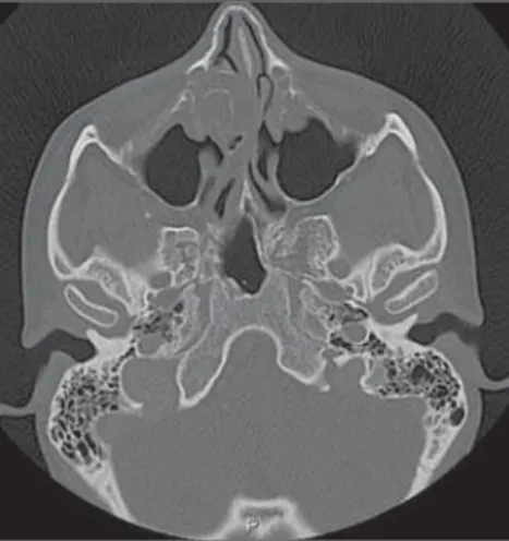
Figure 4. CT with axial section demonstrating the presence of an expansile lesion in the left nasolac-rimal duct.
association with osteoma and hematoma, among others.
The involvement of the nasolacrimal duct is uncommon and is found principally in the pediatric population, causing nasal obstruction and respiratory discomfort. Na-solacrimal duct mucocele is caused by ob-struction of such duct, interrupting the nor-mal flow of tears and resulting in the de-velopment of a mass in the medial corner of the orbit (Figures 3 to 6). Typically, pa-tients with nasolacrimal mucoceles present the following triad of imaging findings a) mass in the medial corner of the orbit, rep-resenting nasolacrimal duct enlargement; b) nasolacrimal bone duct enlargement sec-ondary to soft tissues which enlarge the
mended in following situations: CT image showing bone erosion, mucocele extending out from the sinus, and partial loss of vi-sion(1).
The radiological appearance of muco-celes at MRI varies with the time of evolu-tion of the disease. Initially, the contents will be predominantly aqueous, so the cor-responding image will be hypointense on T1-weighted, and hyperintense on T2-weighted sequences. Over time, the protein contents may increase, resulting in hyperintense images both on T1- and T2-weighted sequences.
The differential diagnosis for mucocele is made with innumerable inflammatory, congenital, cystic and neoplastic condi-tions, such as Rathke’s cleft cyst, dermoid cyst, cysticercosis, hypophyseal adenoma, craniopharyngioma, optic glioma, besides neoplastic lesions of skull base, facial si-nuses and nasopharynx(1). It is important to
observe the differentiation between mucoceles and retention cysts: mucoceles develop from sinus ostium obstruction, and the cavity becomes completely filled with mucus involved by the sinusal mucosa; on the other hand, retention cysts do not fill completely the cavity and are formed by the ductal epithelium and by the gland capsule.
ATYPICAL PRESENTATIONS
Paranasal sinuses mucoceles may be found in atypical locations, such as naso-lacrimal duct, intersinus cell, turbinate, in MRI is useful to identify the relation
be-tween mucocele, brain tissue, orbit and soft tissues (Figure 2)(5). In spite of not offering
recom-374
Carvalho BV et al. Paranasal sinuses mucocele at computed tomography
Radiol Bras. 2013 Nov/Dez;46(6):372–375
nasolacrimal duct; c) intranasal mass rep-resenting inferior extension of the muco-cele(6).
Association between mucocele and os-teoma is a rarity; only 21 cases with intrac-ranial extension are reported in the litera-ture. In such cases, osteoma is considered as the primary lesion and mucocele, sec-ondary. In most cases, the diagnosis is based on the clinical symptoms associated with intraorbital or intracranial extension of the disease (Figures 7 and 8)(7).
Mucoceles may also occur after trauma, and their differentiation is important in cases of coexistence with hematoma. In such cases, the higher density of the he-matoma can be observed as compared with the density of the mucocele (Figure 9).
Intersinus cell is defined as pneumati-zation at the median line or paramedian bone lamella (septum) between the frontal sinuses. A mucocele may develop within
Figure 5. CT with sagittal reconstruction, demon-strating the presence of an expansile lesion in the left nasolacrimal duct.
Figure 6. CT with coronal reconstruction demon-strating no significant enhancement of a lesion in the left nasolacrimal duct.
Figure 8. CT with coronal reconstruction for soft tissues. Lesion within the left frontal sinus with extension into the orbital cavity.
Figure 7. CT with coronal reconstruction for bone structures, demonstrating the presence of an os-teoma in the left frontal sinus as well as disconti-nuity of the superolateral wall of the orbit.
Figure 11. CT with coronal reconstruction for bone structures. Volumetric increase of the right middle turbinate secondary to the presence of mucocele in the concha pneumatization.
Figure 10. CT with coronal reconstruction. Pres-ence of soft tissue inside the intersinus cell.
Figure 9. CT with axial section. Higher density is observed on the posterior hematoma as compared with the anterior intraorbital mucocele at left.
such structure and be either sufficiently large to block unilaterally the frontal sinus ostium, or increase to form a giant muco-cele involving both frontal sinuses. Gener-ally, such mucocele erodes the anterior and/ or posterior sinuses tables (Figure 10)(8).
The pneumatization of the middle tur-binate occurs as an extension of the ethmoi-dal cells, which extends through the supe-rior meatus or through the frontal recess. The interior of the concha bullosa is lined by the respiratory epithelium and generally drains into the middle meatus. In case of obstruction of such drainage, infection may develop progressing to mucocele (Figures 11 to 14)(9).
MANAGEMENT
The management of mucoceles is surgi-cal, aiming at restoring the sinus drainage, and the approach may be either by means of conventional methods (transcranial and transeptal) or by nasal endoscopy(1,2).
375
Carvalho BV et al. Paranasal sinuses mucocele at computed tomography
Radiol Bras. 2013 Nov/Dez;46(6):372–375
the treatment of choice for paranasal si-nuses mucoceles(2). The main objective of such method is the complete removal of both the lesion and the sinusal mucosa, thus preventing disease recurrence. CT or MRI may be necessary in the suspicion of recur-rence(1).
CONCLUSION
It is important to know the common findings as well as the atypical presenta-tions of mucoceles for a correct diagnosis and treatment of sinusal and extrasinusal
lesions, thus avoiding possible pre- and postoperative clinical complications.
REFERENCES
1. Tinoco P, Pereira JC, Lourenço Filho RC, et al. Tra-tamento nasoendoscópico da mucocele de seio es-fenoidal. Arq Int Otorrinolaringol. 2009;13:336–9. 2. Marambaia O, Gomes AM, Marambaia PP, et al. Tratamento endoscópico das mucoceles frontoet-moidais. Rev Bras Otorrinoloaringol. 2008; 74(Supl).
3. Price HI, Batnitzky S, Karlin CA, et al. Computed tomography of benign disease of the paranasal si-nuses. Radiographics. 1983;3:107–40. 4. Towbin R, Dunbar JS. The paranasal sinuses in
childhood. Radiographics. 1982;2:253–79.
5. Cavazza S, Bocciolini C, Laudadio P, et al. Two anomalous localizations of mucocele: clinical pre-sentation and retrospective review. Acta Otorhino-laryngol Ital. 2007;27:208–11.
6. Koch BL. Case 73: Nasolacrimal duct mucocele. Radiology. 2004;232:370–2.
7. Sakamoto H, Tanaka T, Kato N, et al. Frontal si-nus mucocele with intracranial extension associ-ated with osteoma in the anterior cranial fossa. Neurol Med Chir. 2011;51:600–3.
8. Som PM, Lawson W. Interfrontal sinus septal cell: a cause of obstructing inflammation and muco-celes. AJNR Am J Neuroradiol. 2008;29:1369–71. 9. Sala CR, Fuster MA, Molina JV, et al. Mucocele de concha bullosa con afectación orbitaria. Acta Otorrinolaringol Esp. 2002;53:46–9.
Figura 14. TC com reconstrução coronal para par-tes moles. Aumento volumétrico da concha nasal média direita secundário à presença de mucocele no interior da pneumatização da concha. Figura 13. TC com corte axial. Aumento
volumé-trico da concha nasal média direita secundário à presença de mucocele no interior da pneumatiza-ção da concha.