Submitted29 April 2016
Accepted 26 May 2016
Published30 June 2016
Corresponding author
Naguib Salleh,
Academic editor
Esta Sterneck
Additional Information and Declarations can be found on page 14
DOI10.7717/peerj.2145
Copyright
2016 Loh et al.
Distributed under
Creative Commons CC-BY 4.0
OPEN ACCESS
Sub-chronic testosterone treatment
increases the levels of epithelial sodium
channel (ENaC)-
α
,
β
and
γ
in the
kidney of orchidectomized adult male
Sprague–Dawley rats
Su Yi Loh, Nelli Giribabu and Naguib Salleh
Department of Physiology, University of Malaya, Kuala Lumpur, Malaysia
ABSTRACT
Testosterone has been reported to cause blood pressure to increase. However mech-anisms that underlie the effect of this hormone on this physiological parameter are currently not well understood. The aims of this study were to investigate effects of testosterone on expression ofα,β andγ-epithelial sodium channel (ENaC) proteins and messenger RNAs (mRNAs) in kidneys, the channel known to be involved in Na+ reabsorption, which subsequently can affect the blood pressure.
Methods.Adult male Sprague–Dawley (SD) rats were orchidectomized fourteen days prior to receiving seven days treatment with testosterone propionate (125µg/kg/day or
250µg/kg/day) with or without flutamide (androgen receptor blocker) or finasteride
(5α-reductase inhibitor). Following sacrifice, the kidneys were removed and were subjected forα,β andγ-ENaC protein and mRNA expression analyses by Western blotting and Real-time PCR (qPCR) respectively. The distribution ofα,βandγ-ENaC proteins in kidneys were observed by immunofluorescence.
Results.Theα,β andγ-ENaC proteins and mRNA levels in kidneys were enhanced in rats which received testosterone-only treatment. In these rats, α,β and γ-ENaC proteins were distributed in the distal tubules and collecting ducts of the nephrons. Co-treatment with flutamide or finasteride resulted in the levels ofα,βandγ-ENaC proteins and mRNAs in kidneys to decrease. In conclusions, increases inα,βandγ -ENaC protein and mRNA levels in kidneys mainly in the distal tubules and collecting ducts under testosterone influence might lead to enhance Na+ reabsorption which subsequently might cause an increase in blood pressure.
SubjectsMolecular Biology, Nephrology, Pharmacology
Keywords Testosterone, Alpha ENaC, Beta ENaC, Gamma ENaC, Kidneys
INTRODUCTION
Epithelial sodium channel (ENaC), which consists of three homologous subunits (α,β andγ) plays an important role in Na+
Kamynina & Staub,2002). Mutation of ENaC gene could lead to hypotension, while its prolonged activation could lead to severe hypertension (Bubien,2010). Expression of ENaC in kidneys could also be affected by sex hormones i.e., estrogen and progesterone (Gambling et al.,2004). There were evidences which suggest the involvement of testosterone in regulating kidney ENaC expression. Quan and colleagues (2004) reported that dihydrotestosterone (DHT) injection to adult male Sprague–Dawley rats could increase Na+
reabsorption in kidney proximal tubules, suggesting that this could be mediated via ENaC. Meanwhile, administration of testosterone in spontaneous hypertensive (SHR) rats was found to decrease the pressure-induced natriuresis, again pointing towards the involvement of ENaC (Reckelhoff, Zhang & Granger,1998).
To date, the information with regard to effect of testosterone on ENaC expression in kidneys were far from complete. Quinkler et al. (2005) reported that sub-chronic (14 days) treatment of adult male Wistar rats with testosterone resulted in elevatedα-Enac
mRNA levels in kidney homogenates. Meanwhile, administration of dihydrotestosterone (DHT) causing the same effects, but was lesser than testosterone. Additionally, their study also showed that incubation of human kidney proximal tubule cell line with testosterone but not DHT caused increase inα-ENaC mRNA level with testosterone effect was being antagonized by flutamide. However, no expression forβandγ-ENaC was detected in these cells. In the meantime, testosterone and DHT were found to decrease expression ofα,β andγ-ENaC subunits in ovariectomised female Wistar rat kidney (Kienitz et al.,2009). Based on these findings, we hypothesized that testosterone affects expression of all ENaC subunits i.e.,α,β andγ in the kidneys, in which their co-existence will lead to a fully functioning ENaC channel, thus would augment Na+
reabsorption which might result in the rise in blood pressure under testosterone influence (Hanukoglu & Hanukoglu,2016). Therefore, the aims of this study were to investigate testosterone effect onα,βandγ ENaC protein and mRNA expression levels in kidneys. In addition, the possible involvement of androgen receptor and DHT in mediating testosterone effects were also investigated.
MATERIALS AND METHODS
Animal preparation and hormonal treatment
Eight weeks old male Sprague–Dawley (SD) rats were housed in a clean and well ventilated environment under light-dark cycle (12/12 h). The animals were fed with standard rat diet (Harlan, Germany) and tap waterad libitum. All procedures were approved by Institutional Animal Care and Use Committee (IACUC), University of Malaya (ethics number: 2014-05-07/physio/R/NS). Sham operation and orchidectomies were performed under ketamine/xylazine anesthesia. Two (2) weeks following recovery, hormonal treatments were initiated, which coincide with the age of the rats at 10 weeks old. Rats were divided into eight (8) treatment groups with eight (8) rats per group and were given the following treatment for seven (7) consecutive days.
Group 1: Intact, sham-operated receiving peanut oil only—S
Group 2: Orchiectomized, receiving peanut oil only—O
Group 4: Orchiectomized, receiving 250µg/kg/day testosterone—T250
Group 5: Orchiectomized, receiving 125 µg/kg/day testosterone plus flutamide
(8 mg/kg/day)—T125+FU
Group 6: Orchiectomized, receiving 250 µg/kg/day testosterone plus flutamide
(8 mg/kg/day)—T250+FU
Group 7: Orchiectomized, receiving 125 µg/kg/day testosterone plus finasteride
(5 mg/kg/day)—T125+FN
Group 8: Orchiectomized, receiving 250µg/kg/day testosterone plus finasteride
(5 mg/kg/day)—T250+FN
At the end of the treatment, rats were sacrificed by cervical dislocation and kidneys were removed for molecular biological and histological analyses.
Quantification of EnacmRNA levels in kidneys by high throughput
qPCR (Fluidigm)
Immediately following removal, the kidneys from n=4 rats per group were stored in RNAlater solution (Ambion, Foster City, CA, USA) in order to preserve the RNA integrity. Tissues were then weighted and disrupted in a tissue lysis buffer by using a rotor-stator homogenizer (Heidolph DIAX 600). RNA extraction was performed by using a Macherey-Nagel Nucleo Spin RNA kit (Düren, Germany) according to the manufacturer’s guidelines. cDNA synthesis was then carried out by using a Bio-Rad iScript Reverse Transcription Supermix for RT-qPCR (Biorad, Hercules, CA, USA). A high throughput qPCR-based microfluid dynamic array technology (Fluidigm) was performed to evaluate the changes in gene expression. The converted cDNA was pre-amplified by using Applied Biosystems PreAmp Master Mix, first, one cycle 95 ◦
C for 10 min, then 10 cycles, 95 ◦
C for 15 s and finally one cycle, 60 ◦
C for 4 min. The pre-amplified cDNA was diluted accordingly and was then used to perform the qPCR in Fluidigm dynamic array chip following the manufacturer’s protocol with the aid of applicant specialists. The chip was run in the BioMark Instrument for qPCR at 95 ◦C for 10 min, followed by 40 cycles at 95 ◦C for 15 sec and lastly at 60 ◦
C for 1 min.Gapdhwas selected as a house-keeping gene as its expression were the most stable in kidneys. Taqman primers and probes forGapdh, Scnn1a(Enac-α),Scnn1b(Enac-β) andScnn1g (Enac-γ) were purchased from Applied Biosystems, CA, USA. All experiments were carried out in triplicates and the data were analyzed by using a Fluidigm Real-Time PCR analysis software.
(PVDF) membrane (Bio-Rad, USA), then blocked in 5% bovine serum albumin (BSA) (Sigma-Aldrich, USA) for 60 min at room temperature. The membranes were then probed with primary antibodies againstα-ENaC,β-ENaC,γ-ENaC (sc-22239, sc-25354, sc-21014, Santa Cruz Biotechnology, CA, USA respectively) for 90 min at room temperature. This was followed by incubation in horseradish peroxidase (HRP) conjugated secondary antibody (Santa Cruz Biotechnology, Santa Cruz, CA, USA) for 60 min at room temperature. The blots were then developed by using a Thermo Scientific Super Signal West Pico Chemiluminescent Substrate (Rockford, USA) according to the manufacturer’s manual. Chemiluminescent signals were captured by using a highly sensitive CCD camera-based imager (BioSpectrum Imaging System). The band intensity of each target was analyzed by using Image J software. GAPDH (Santa Cruz Biotechnology) was used as endogenous control. The experiment was performed in triplicate and the average ratios of target protein/GAPDH band intensity were then determined.
Whole body perfusion fixation and detection of ENaC distribution in kidneys by immunofluorescence
Another set of animals (n=4 rats per group), which were duplicate of those used for Western blotting and qPCR analyses, were anaesthetized with ketamine/xylazine anesthesia and a transcardial perfusion was performed with injection of 4% paraformaldehyde (PFA) in phosphate buffer saline (PBS). The animals were then killed and the kidneys were removed and fixed in 30% sucrose for three days. The tissues were then processed and embedded in a paraffin wax. The kidneys were sectioned coronally into 5µm by using a
microtome. Sections were then mounted onto poly-L-lysine coated glass slides. Sections were then de-parafinized and antigens were retrieved by boiling the sections in 1mM EDTA (BioVision InC.), pH 8.0 containing 0.05% (v/v) Tween20 (Sigma Aldrich). Sections were then blocked with appropriate 10% (v/v) normal serum (Santa Cruz Biotechnology) in PBS for 1 h at room temperature and were then incubated with target primary antibodies (as above) at the ratio of 1:100 at 4 ◦C, overnight. After three times of washing with PBS, sections were then incubated in appropriate fluorophore conjugated secondary antibodies at a dilution of 1:200, for 1 h at room temperature and then sealed with UltraCruz mounting medium (Santa Crus Biotechnology). Secondary antibodies used in this experiment were goat anti-mouse IgG FITC (sc-2010, Santa Crus Biotechnology, CA, USA), donkey anti-goat lgG (H+L) conjugated with DyLight 550 and donkey anti-rabbit lgG (H+L) conjugated with DyLight 488 (SA5-10087, SA5-10038, Thermo Scientific, Rockford, IL, USA). The images were viewed and captured by using a confocal laser scanning microscope (Leica TCS SP5 II) at a fixed exposure time. All experiments were carried out in four (4) replicates and the representative images were selected. All images were taken at 63x magnification and the slides without tissue were used as white balancer.
Measurement of serum testosterone level by enzyme-linked immunoassay (ELISA)
Immediately following sacrifice and prior to transcardial perfusion, the blood from
by enzyme-linked immunosorbent assay (ELISA) kit (BioSource International, Inc., Camarillo, CA, USA), according to the manufacturer guideline.
Statistical analysis
All data were analyzed by SPSS software with mean±standard error of mean (S.E.M) obtained. Statistical significant between groups was evaluated by Studentt-test followed by one-way analysis of variance (ANOVA). Statistically significant difference were denoted asp<0.05. Tukey’s post-hoc test was used to examine adequacy of the samples and all values were >0.08 which indicate adequate sample size.
RESULTS
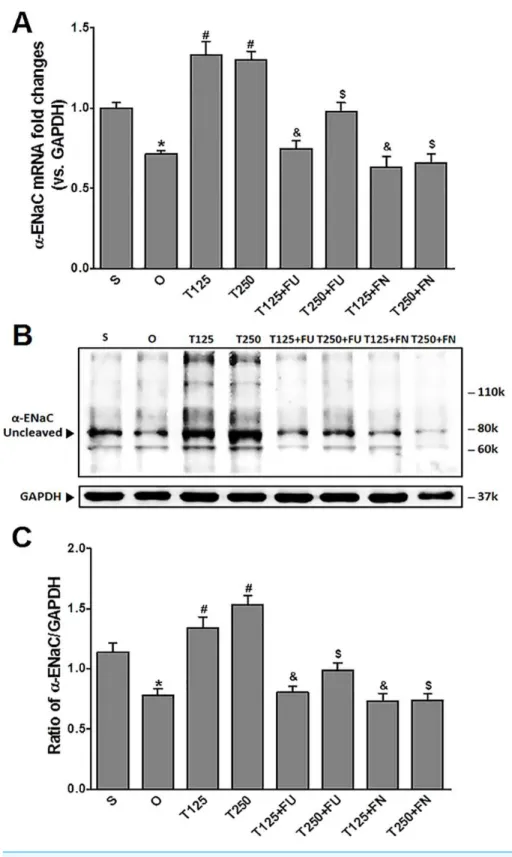
Testosterone increasesα-ENaC protein and mRNA levels in kidneys
InFig. 1A, the levels ofα-EnacmRNA were highest following testosterone-only treatment. Co-treatment with flutamide or finasteride caused α-Enac mRNA levels to decrease (p<0.05). The effect of flutamide was greater in rats which received 125µg/kg/day
testosterone when compared to 250 µg/kg/day testosterone. However, the effect of
finasteride in rats treated with 125µg/kg/day testosterone- was not significantly different
when compared to its effect in rats treated with 250µg/kg/day testosterone.
In Figs. 1Band1C, expression levels ofα-ENaC protein was highest in rats which received 250 µg/kg/day testosterone. In these rats, co-administration of flutamide or
finasteride caused α-ENaC protein expression level to decrease (p<0.05). Flutamide effect was greater in rats treated with 125 µg/kg/day testosterone than in rat treated
with 250µg/kg/day testosterone. However, following co-administration of finasteride, no
significant difference inα-ENaC protein expression level was observed between rats which received 125 mg/kg/day testosterone and rats which received 250µg/kg/day testosterone.
Testosterone increasesβ-ENaC protein and mRNA levels in kidneys
InFig. 2A, the levels ofβ-EnacmRNA were highest in rats which received testosterone-only treatment. In these rats, co-administration of flutamide or finasteride resulted inβ-Enac
mRNA levels to decrease (p<0.05). The effect of flutamide was greater in rats which received 125µg/kg/day testosterone when compared to rats which received 250µg/kg/day
testosterone. However, co-administration of finasteride did not causeβ-EnacmRNA levels in rats which received 125µg/kg/day testosterone to be difference from rats which received
250µg/kg/day testosterone.
In Figs. 2Band2C, the levels of expression ofβ-ENaC protein were highest in rats which received 250µg/kg/day testosterone. In these rats, co-administration of flutamide or
finasteride resulted inβ-ENaC protein expression level to decrease (p<0.05). The effects of flutamide were greater in rats which received 125µg/kg/day testosterone when compared to
rats which received 250µg/kg/day testosterone. However, co-administration of finasteride
did not cause β-ENaC protein expression level in rats which received 125µg/kg/day
Figure 1 (A)α-EnacmRNA level (B) whole membrane image ofα-ENaC protein band and (C) ratio of α-ENaC/GAPDH protein band intensity in kidneys. Values represent mean±S.E.M of four rats.∗
p< 0.05 compared to sham-operated rats,#p<0.05 compared to orcidectomized control rats,&p<0.05
compared to T125.$p<0.05 compared to T250. S, sham operated; O, orchiectomized non-treated; T125,
Figure 2 (A)β-EnacmRNA level (B) whole membrane image ofβ-ENaC protein band and (C) ratio of β-ENaC/GAPDH protein band intensity in kidneys. Values represent mean±S.E.M of four rats.∗
p< 0.05 compared to sham-operated rats,#p<0.05 compared to orcidectomized control rats,&p<0.05
compared to T125.$p<0.05 compared to T250. S, sham operated; O, orchiectomized non-treated, T125,
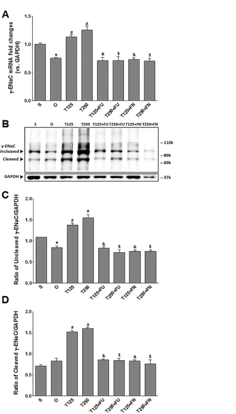
Testosterone increasesγ-ENaC protein and mRNA levels in kidneys InFig. 3A, the levels ofγ-EnacmRNA were highest in rats which received 250µg/kg/day
testosterone. In these rats, co-administration of flutamide or finasteride resulted inγ
-Enac mRNA levels to decrease (p<0.05). The effect of flutamide was not significantly different between rats which received 125µg/kg/day testosterone and rats which received
250µg/kg/day testosterone. Similarly, the effect of finasteride in rats which received
125µg/kg/day testosterone was not significantly different from its effect in rats which
received 250 mg/kg/day testosterone.
In Figs. 3Band3C, the levels ofγ-ENaC protein was highest in rats which received 250µg/kg/day testosterone. In these rats, co-administration of flutamide or finasteride
resulted inγ-ENaC protein expression level to decrease (p<0.05). Co-administration of flutamide with testosterone did not causeγ-ENaC protein expression level in rats which received 125µg/kg/day testosterone to be different from rats which received 250µg/kg/day
testosterone. In contrast, co-administration of finasteride with testosterone caused greater decrease in the level of this protein in rats which received 250µg/kg/day testosterone when
compared to rats which received 125µg/kg/day testosterone.
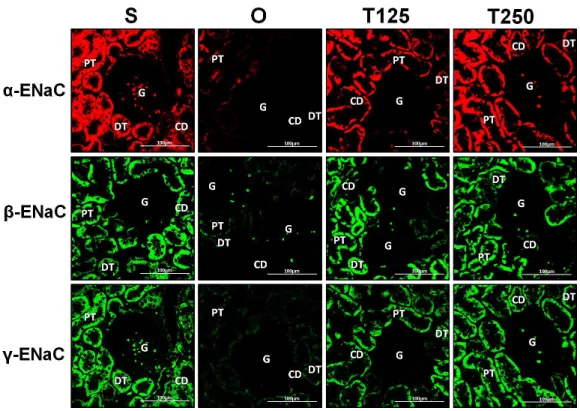
Distribution ofα-ENaC protein in nephrons
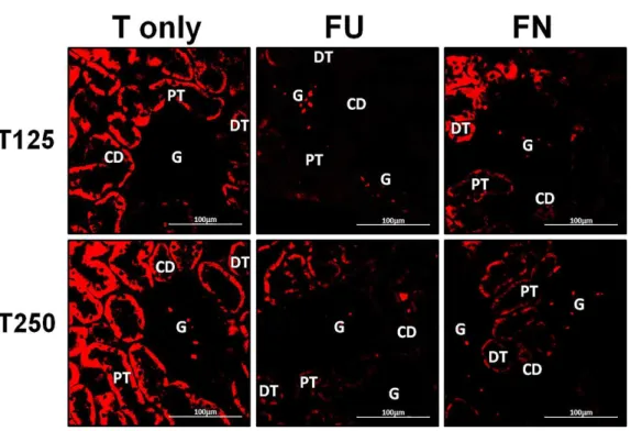
InFig. 4,α-ENaC protein was seen to be highly distributed in distal tubules and collecting ducts of rats which received 125 and 250 µg/kg/day testosterone. In these rats,
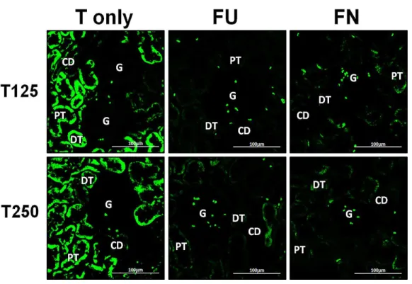
co-administration of flutamide or finasteride resulted inα-ENaC protein distribution level to be relatively lower (Fig. 5).
Distribution ofβ-ENaC protein in nephrons
InFig. 4,β-ENaC protein was highly distributed in distal tubules and collecting ducts in rats which received 250µg/kg/day testosterone. However, co-administration of flutamide
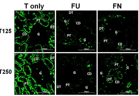
or finasteride with testosterone resulted in relatively lowerβ-ENaC protein distribution level when compared to rats which received testosterone-only treatment (Fig. 6).
Distribution ofγ-ENaC protein in nephrons
InFig. 4,γ-ENaC protein was highly distributed in distal tubule and collecting ducts in rats which received 125 and 250µg/kg/day testosterone. However, co-administration of
flutamide with testosterone resulted in relatively lowerγ-ENaC protein distribution when compared to testosterone-only treatment (Fig. 7). Similar effects could be seen in rats following concomitant co-administration of finasteride with testosterone.
Serum testosterone level
In Table 1, administration of 125µg/kg/day testosterone resulted in serum level of this
hormone to be 13.5 fold greater than its level in non-testosterone-treated orchiectomised rats. Treatment with 250µg/kg/day testosterone resulted in serum testosterone level to be
16.5 fold greater than in non-testosterone treated orchiectomized rats.
DISCUSSION
Figure 3 (A)γ-EnacmRNA level (B) whole membrane image ofγ-ENaC protein band and (C) ratio ofγ-ENaC/GAPDH protein band intensity in kidneys. γ-ENaC possesses a cleaved and an un-cleaved forms. Values represent mean±S.E.M of four rats.∗
p<0.05 compared to sham-operated rats,#p<
0.05 compared to orcidectomized control rats,&p<0.05 compared to T125.$p< 0.05 compared to
T250. S, sham operated; O, orchiectomized non-treated; T125, 125µg/kg/day testosterone-treated; T250, 250µg/kg/day testosterone-treated rats; FU, flutamide, FN, finasteride. Molecular weight ofγ-ENaC=
Figure 4 Immunofluorescenceimages showing distribution ofα,βandγ-ENaC proteins in nephrons.
Red or green fluorescence signals indicate the sites where ENaC subunit proteins were expressed. G, glomeruli; PT, prximal tubule; DT, distal tubule, CD, collecting duct. S, sham operated; O, orchiectomized non-treated; T125, 125µg/kg/day testosterone-treated; T250, 250 mg/kg/day testosterone-treated rats. Scale bar=100µM.
Table 1 Serum level of testosterone in different experimental groups.Values were expressed as mean±S.E.M of eight (8) different observations.
Groups Serum testosterone levels (ng/ml)
Sham 2.89±0.38
ORX 0.25a±0.20
T125 3.37b±0.40
T250 4.12b±0.55
Notes.
ap<0.05 compared to shamoperated rats. bp<0.05 compared to orchidectomized control rats.
Sham, Sham-operated; ORX, Orchidectomized control; T125, 125µg/kg/day testosterone; T250, 250µg/kg/day testos-terone.
kidneys of orchidectomised adult male ratsin-vivo. The dose-dependent increase in serum testosterone levels was observed when testosterone was subcutaneously injected to these rats at increasing doses which suggested that this hormone most likely was not metabolized as it bypassed the first-pass effect. It was found that equal amount ofα-ENaC protein was expressed in the orchidectomised male rats’ kidneys following administration of 125 and 250µg/kg/day testosterone, however higher expression ofβ andγ ENaC proteins
Figure 5 Effects of flutamide and finasteride on expression ofα-ENaC in nephrons.Red fluorescence signals indicate sites where ENaC subunit proteins were expressed. G, glomeruli; PT, prximal tubule; DT, distal tubule; CD, collecting duct. S, sham operated; O, orchiectomized non-treated; T125, 125µg/kg/day testosterone-treated; T250, 250µg/kg/day testosterone-treated rats. Scale bar=100µM.
to 125µg/kg/day testosterone. The latter findings were consistent with the findings by Kienitz et al.(2006) who reported that administration of testosterone at 500 mg/kg/ in male rats resulted in high level ofα-ENaC protein in kidneys. Another study has revealed that the levels ofα-ENaCmRNA in human kidney cell line was enhanced by testosterone, however this hormone was found to have no significant effect onβ andγ-ENaC mRNA levels (Quinkler et al., 2005). In contrast, we found that the levels of β andγ- ENaC proteins and mRNAs were highly up-regulated in the orchidectomised male rats’ kidneys by testosterone. We postulated that co-expression of all ENaC subunits (α,βandγ) would result in a fully operating channel as their co-existence was required for the maximal ENaC channel function (Hamm, Feng & Hering-Smith,2010).
In this study, we have found that the level of expression ofα,β andγ-ENaC proteins and mRNAs in the kidney was significantly decreased following co-administration of flutamide with testosterone. The effect of flutamide onαandβ-ENaC but notγ-ENaC was found greater in rats which received 125µg/kg/day when compared to 250µg/kg/day
testosterone. The reason behind this effect was unknown; however, we postulated that following high dose testosterone treatment i.e., at 250µg/kg/day, some of this hormone
Figure 6 Effects of flutamide and finasteride on expression ofβ-ENaC in nephrons.Green fluorescence signals indicate sites where ENaC subunit proteins were expressed. G, glomeruli; PT, prximal tubule; DT, distal tubule; CD, collecting duct. S, sham operated; O, orchiectomized non-treated; T125, 125µg/kg/day testosterone-treated; T250, 250µg/kg/day testosterone-treated rats. Scale bar=
100µM.
seen following high dose testosterone treatment. In the meantime, at high dose, there was a possibility that testosterone could exert a non-genomic effect which was not inhibited by flutamide (Mokhtar et al.,2014). Finasteride was found to inhibit testosterone effect in causing increase in ENaC subunits expression in the kidneys, which suggested that DHT was involved. In the absence of DHT, levels of ENaC subunits in kidneys would be markedly decreased. Besides DHT, other hormone which was also known to cause expression level ofα,βandγ-ENaC subunits in the kidneys to increase is aldosterone, which was reported to induce redistribution of all ENaC subunits to the apical membrane of kidneys’ distal tubule and collecting ducts (Stachowiak, Nussdorfer & Malendowicz,1991;Masilamani et al.,1999). Therefore, redistribution of all ENaC subunits to the apical membrane of distal tubule and collecting duct under testosterone influence might produce similar effect to that reported under aldosterone, in which this would result in fully functioning channels (Masilamani et al.,1999). The resultant effect would lead to increase in Na+
reabsorption. We have found that ENaC subunits’ protein were also found to be expressed in the cytoplasm under testosterone influence, in which these distributions could represent the channels at various stages of processing.
The effect of testosterone on ENaC subunit expression as observed in our study could help to explain the mechanisms underlying this hormone effects on kidneys’ Na+
Figure 7 Effectsof flutamide and finasteride on expression ofγ-ENaC in nephrons.Green fluorescence signals indicate sites where ENaC subunit proteins were expressed. G, glomeruli; PT, prximal tubule; DT, distal tubule; CD, collecting duct. S, sham operated; O, orchiectomized non-treated; T125, 125µg/kg/day testosterone-treated; T250, 250µg/kg/day testosterone-treated rats. Scale bar=100µM.
has been known, we have provided additional information in which in kidneys of male rat model, testosterone was also found to enhanceβ andγ-ENaC subunit expression in addition to the already known increase inα-ENaC expression (Quinkler et al.,2005). These testosterone effects would likely result in a fully functioning channel. However, more works need to be done in order to confirm the optimal ENaC channel function at the apical membrane of the distal tubule and collecting duct epithelia for example by using a patch clamp in which kidney epithelial cells obtained from animals treated with testosterone will be exposed to a specific ENaC inhibitor such as amiloride (Edinger et al.,
2012). If in the case where ENaC channels are functioning, inhibition by amiloride will result in reduced Na+
conductance. Additionally, the involvement of DHT can also be confirmed by administering this compound directly to the rats and the involvement of genomic pathway in mediating testosterone effect on ENaC expression can be confirmed by using androgen receptor knock-out animal model.
to high serum testosterone levels and this was further confirmed from the observations that ovariectomized female SHR rats that was given testosterone had increase in blood pressure. Furthermore, studies have shown that natriuresis were markedly reduced in intact male and testosterone-treated ovariectomised female SHR rats which indicated that higher amount of Na+
was reabsorbed in the kidney under the influence of this hormone (Reckelhoff, Zhang & Granger,1998). Similar findings were reported in humans whereby serum testosterone levels correlate with the blood pressure (Khaw & Barrett-Connor,1988).
In conclusions, our findings have provided evidences which might support the role of testosterone in causing a relatively higher blood pressure in males than females. Increased in kidney Na+ reabsorption which might occur secondary to testosterone-induced up-regulation of ENaC might predispose the males to hypertension and could perhaps be the reason why males have higher incidence of this disease compared to age-matched females before menopause. Finally, the differential effect of testosterone on α,β andγ-EnaC subunits expression in the kidney might contribute towards gender differences in blood pressure regulation (Reckelhoff,2001;Reckelhoff et al.,1999).
ADDITIONAL INFORMATION AND DECLARATIONS
Funding
This work was funded by the University of Malaya UMRG research fund (RPO11/13HTM) and a PPP grant (PG002-2014B). The funders had no role in study design, data collection and analysis, decision to publish, or preparation of the manuscript.
Grant Disclosures
The following grant information was disclosed by the authors: University of Malaya UMRG research fund: RPO11/13HTM. PPP grant: PG002-2014B.
Competing Interests
The authors declare there are no competing interests.
Author Contributions
• Su Yi Loh performed the experiments, analyzed the data, prepared figures and/or tables.
• Nelli Giribabu performed the experiments, prepared figures and/or tables.
• Naguib Salleh conceived and designed the experiments, analyzed the data, contributed
reagents/materials/analysis tools, wrote the paper, reviewed drafts of the paper.
Animal Ethics
The following information was supplied relating to ethical approvals (i.e., approving body and any reference numbers):
Data Availability
The following information was supplied regarding data availability: The raw data has been supplied asSupplemental Datasets.
Supplemental Information
Supplemental information for this article can be found online athttp://dx.doi.org/10.7717/ peerj.2145#supplemental-information.
REFERENCES
Bubien JK. 2010.Epithelial Na(+) channel (ENaC), hormones and hypertension.Journal
Biological Chemistry 285(31):23527–23531DOI 10.1074/jbc.R109.025049.
Edinger RS, Bertrand CA, Rondandino C, Apodaca GA, Johnson JP, Butterworth MB. 2012.The epithelial sodium channel (ENaC) establishes a trafficking vesicle pool responsible for its regulation.PLoS ONE7(9):e46593
DOI 10.1371/journal.pone.0046593.
Gambling L, Dunfor S, Wilson CA, McArdle HJ, Baines DL. 2004.Estrogen and progesterone regulateα,β, andγENaC subunit mRNA levels in female rat kidney.
Kidney International65(5):1774–1781DOI 10.1111/j.1523-1755.2004.00593.x.
Garty H. 2000.Regulation of the epithelial Na+channel by aldosterone: open questions and emerging answers.Kidney International57(4):1270–1276 DOI 10.1046/j.1523-1755.2000.00961.x.
Hamm LL, Feng Z, Hering-Smith KS. 2010.Regulation of sodium transport by ENaC in the kidney.Current Opinion in Nephrology and Hypertension19(1):98–105 DOI 10.1097/MNH.0b013e328332bda4.
Hanukoglu I, Hanukoglu A. 2016.Epithelial sodium channel (ENaC) family: phylogeny, structure-function, tissue distribution, and associated inherited diseases.Gene
579(2):95–132DOI 10.1016/j.gene.2015.12.061.
Kamynina E, Staub O. 2002.Concerted action of ENaC, Nedd4–2, and Sgk1 in transepithelial Na+transport.American Journal of Physiology-Renal Physiology
283(3):F377–F387DOI 10.1152/ajprenal.00143.2002.
Khaw KT, Barrett-Connor E. 1988.Blood pressure and endogenous testosterone in men: an inverse relationship.Journal of Hypertension6(4):328–332.
Kienitz T, Allolio B, Strasburger CJ, Quinkler M. 2006.Renal expression of alpha-ENaC is increased in testosterone treated rats [Abstract].Endocrine Abstracts11:P302.
Kienitz T, Allolio B, Strasburger CJ, Quinkler M. 2009.Sex-specific regulation of ENaC and androgen receptor in female rat kidney.Hormone and Metabolic Research = Hormon-und Stoffwechselforschung = Hormones et Métabolisme41(5):356–362 DOI 10.1055/s-0029-1192033.
Masilamani S, Kim GH, Mitchell C, Wade JB, Knepper MA. 1999.
Mokhtar HM, Giribabu N, Kassim N, Muniandy S, Salleh N. 2014.Testosterone decreases fluid and chloride secretions in the uterus of adult female rats via down-regulating cystic fibrosis transmembrane regulator (CFTR) expression and functional activity.The Journal of Steroid Biochemistry and Molecular Biology144:361–372 DOI 10.1016/j.jsbmb.2014.08.007.
Quan A, Chakravarty S, Chen JK, Chen JC, Loleh S, Saini N, Harris RC, Capdevila J, Quigley R. 2004.Androgens augment proximal tubule transport.American Journal of Physiology-Renal Physiology287(3):F452–F459DOI 10.1152/ajprenal.00188.2003.
Quinkler M, Bujalska IJ, Kaur K, Onyimba CU, Buhner S, Allolio B, Hughes SV, Hewi-son M, Stewart PM. 2005.Androgen receptor-mediated regulation of theα-subunit of the epithelial sodium channel in human kidney.Hypertension46(4):787–798 DOI 10.1161/01.HYP.0000184362.61744.c1.
Reckelhoff JF. 2001.Gender differences in the regulation of blood pressure.Hypertension
37(5):1199–1208.
Reckelhoff JF, Zhang H, Granger JP. 1998.Testosterone exacerbates hypertension and reduces pressure-natriuresis in male spontaneously hypertensive rats.Hypertension
31(1):435–439DOI 10.1161/01.HYP.31.1.435.
Reckelhoff JF, Zhang H, Srivastava K, Granger JP. 1999.Gender differences in hyper-tension in spontaneously hypertensive rats role of androgens and androgen receptor.
Hypertension34(4):920–923DOI 10.1161/01.HYP.34.4.920.
Schild L. 2010.The epithelial sodium channel and the control of sodium balance.
Biochimica Biophysica Acta1802(12):1159–1165DOI 10.1016/j.bbadis.2010.06.014.
Schulster M, Bernie AM, Ramasamy R. 2016.The role of estradiol in male reproductive function.Asian Journal of Andrology18(3):435–440DOI 10.4103/1008-682X.173932.
Stachowiak A, Nussdorfer GG, Malendowicz LK. 1991.Ovariectomy-induced changes in the adrenal cortex of spontaneously hypertensive rats.Histology Histopathology
6(2):257–259.