Binding of the Antagonist Caffeine to the
Human Adenosine Receptor hA
2A
R in Nearly
Physiological Conditions
Ruyin Cao1,2, Giulia Rossetti1,2,3,4*, Andreas Bauer5, Paolo CarIoni1,2,3
1German Research School for Simulation Sciences (joint venture of RWTH Aachen University and Forschungszentrum Jülich GmbH), D-52425, Jülich, Germany,2Computational Biomedicine, Institute for Advanced Simulation (IAS-5), Forschungszentrum Jülich GmbH, D-52425, Jülich, Germany,3Institute of Neuroscience and Medicine (INM-9), Forschungszentrum Jülich GmbH, D-52425, Jülich, Germany,4Jülich Supercomputing Centre (JSC), Forschungszentrum Jülich GmbH, D-52425, Jülich, Germany,5Institute of Neuroscience and Medicine (INM-2), Forschungszentrum Jülich GmbH, D-52425, Jülich, Germany
Abstract
Lipid composition may significantly affect membrane proteins function, yet its impact on the protein structural determinants is not well understood. Here we present a comparative mo-lecular dynamics (MD) study of the human adenosine receptor type 2A (hA2AR) in complex
with caffeine—a system of high neuro-pharmacological relevance—within different mem-brane types. These are POPC, mixed POPC/POPE and cholesterol-rich memmem-branes. 0.8-μs MD simulations unambiguously show that the helical folding of the amphipathic helix 8 depends on membrane contents. Most importantly, the distinct cholesterol binding into the cleft between helix 1 and 2 stabilizes a specific caffeine-binding pose against others visited during the simulation. Hence, cholesterol presence (~33%-50% in synaptic membrane in central nervous system), often neglected in X-ray determination of membrane proteins, af-fects the population of the ligand binding poses. We conclude that including a correct de-scription of neuronal membranes may be very important for computer-aided design of ligands targeting hA2AR and possibly other GPCRs.
Introduction
Increasingly emerging experiments point to an important role of membrane lipid composition in structure/function relationships of G-protein coupled receptors (GPCRs)—the largest mem-brane protein family in mammals [1–4]. For instance, in rhodopsin, adding (1-palmitoyl-2-oleoylphosphatidylethanolamine) POPE lipids into POPC (1-palmitoyl-2-oleoylphosphati-dylcholine) lipid bilayers affect the equilibrium between two sub-states of the rhodopsin func-tional cycle [5,6]. Another example is given by the class A GPCR serotonin1A receptor: its binding to the agonist 8-OH-DPAT decreases with cholesterol concentration [7]. This issue is crucial for pharmacological applications, as more than one quarter of FDA-approved drugs tar-get GPCRs [8].
OPEN ACCESS
Citation:Cao R, Rossetti G, Bauer A, CarIoni P (2015) Binding of the Antagonist Caffeine to the Human Adenosine Receptor hA2AR in Nearly
Physiological Conditions. PLoS ONE 10(5): e0126833. doi:10.1371/journal.pone.0126833
Academic Editor:Yang Zhang, University of Michigan, UNITED STATES
Received:January 13, 2015
Accepted:April 8, 2015
Published:May 20, 2015
Copyright:© 2015 Cao et al. This is an open access article distributed under the terms of theCreative Commons Attribution License, which permits unrestricted use, distribution, and reproduction in any medium, provided the original author and source are credited.
Data Availability Statement:All relevant data are within the paper and its Supporting Information files.
Funding:Jülich Supercomputing Centre (JSC) provided support in the form of a salary for author GR, but did not have any additional role in the study design, data collection and analysis, decision to publish, or preparation of the manuscript. The specific roles of all authors are articulated in the‘author contributions’section.
Molecular Dynamics (MD) simulations are being instrumental to assess the effect of native membrane environment on conformational properties of GPCRs. Indeed, MD studies of class A GPCR, beta-2 adrenergic receptor, using four different membrane types with or without cho-lesterol, have suggested that the stability of the receptor“ionic lock”[9] varies with different lipid compositions [4]. In addition, 1.6μs MD simulations of rhodopsin embedded in
(1-stear-oyl-2-phosphatidylcholine) SDPC/SDPE (1-stearoyl-2 docosahexaenoyl-phosphatidylethanolamine) lipid bilayer, in the presence of cholesterol, have lead to the con-clusion that specific cholesterol-rhodopsin binding modulates the TM1-TM2-TM7 helices/ helix 8 interactions, essential for the receptor’s activation [10]. Finally, MD simulations indi-cates that the stabilization of the amphipathic helix 8 (H8) of the class C GPCR mGluR2 recep-tor increases with cholesterol concentration and that such stabilization depends also on membrane thickness [3].
Here we use MD simulations to elucidate the effect of membrane composition on an antag-onist-bound neuronal GPCR. This is the class A GPCR human adenosine receptor type 2A (hA2AR) [11]—highly localized in the so-called“striatum”of the brain [12]—in complex with
the antagonist caffeine (CFF, chemical formula inS1 Fig). CFF binding to hA2AR may lead to
neuroprotection [13–17]. It prevents apoptotic cell death in a Parkinson’s' model [18]. The ef-fect on passing from artificial lipid bilayers to conditions near to real neuronal membranes, where cholesterol content varies from 33% to 50% [19], is investigated by performing 0.8μ
s-long MD simulations on three systems, named in sequence asI,IIandIII(seeTable 1).Iis composed by pure POPC lipid bilayer (seeS1 Fig), which is commonly used for MD studies on adenosine receptor [20–25].IIis a bilayer of equally mixed POPC and POPE lipids (S1 Fig). IIIresembles the synaptic membrane (42% POPC, 34% POPE and 25% of cholesterol mole-cules,S1 Fig), where hA2AR is expressed [26] and the binding to CFF exerts its beneficial
neu-roprotection effects [16]. Notably,IIImarkedly differs from the artificial membrane mimics (detergent n-nonyl-β-D-glucopyranoside), where the CFF/hA2AR complex is embedded for
X-ray structure determination [27].
Results and Discussion
Convergence Analysis
The degree of convergence of the 800 ns long MD simulations of systemsI,IIandIIIis here investigated by using the so-called‘all-to-all RMSD analysis’[28]. This assembles pairs of Cα
atoms' RMSDs in matrices along an MD simulation. In the last 400 ns, the matrices of the three systems (S2 Fig) show a "leave-and-return pattern", a converged-alike feature according to [28], see supporting information. On longer time-scales, this feature is not observed. This sug-gests that the protein conformations of the three systems might have reached a fair conver-gence in the last 400 ns timescale here. Consistently with this fact, the CαRMSDs oscillate
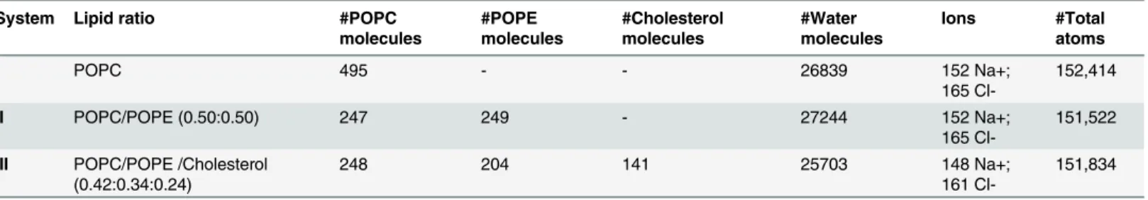
Table 1. Composition of the three systems simulated here.
System Lipid ratio #POPC
molecules
#POPE molecules
#Cholesterol molecules
#Water molecules
Ions #Total
atoms
I POPC 495 - - 26839 152 Na+;
165
Cl-152,414
II POPC/POPE (0.50:0.50) 247 249 - 27244 152 Na+;
165
Cl-151,522
III POPC/POPE /Cholesterol (0.42:0.34:0.24)
248 204 141 25703 148 Na+;
161
Cl-151,834
doi:10.1371/journal.pone.0126833.t001
around average values in such time scale (Panel A inS3 Fig). The properties presented here are therefore calculated for the last 400 ns.
Overall Fold
The typical seven transmembrane helices are maintained across the three systems during the overall trajectory (Panel B inS3 Fig). However, the fold of H8, located at the membrane-cytoplasm interface, which was not solved in the X-ray structure [27], shows membrane-sensitive conformations. Indeed residues 292–317 of H8 preserve a helical conformation inII andIIIonly (Fig 1). Yet, the helical content of H8 decreases inI: residues 305–317 unfolded into flexible loop after 250 ns MD (Fig 1). This is associated with two features, which are pres-ent only inI: a very large increase of the CαRMSD (Panel A inS3 Fig), and the presence of two
non-overlapping blocks in the matrix calculated with the all-to-all RMSD analysis (S2 Fig). The latter is a signature of a significant, irreversible transition between two distinct conforma-tions [28]. The different stability of H8 across the three systems is likely to be related to an in-crease of membrane thickness on passing fromItoII, and, more, toIII, observed here (see Section‘Membrane Structure’below for details). As results, H8 inIIandIIIis only half-exposed to the solvent, being the other half immersed in the membrane. This stabilizes H8, be-cause this helix is amphipathic (Panel B inS4 Fig). Such stabilization is not present for system I, where H8 is more solvent-exposed because of the thinner thickness of the bilayer. H8 is a key structural element for hA2AR function, as it connects the transmembrane helices interacting
with ligands with the cytoplasmic C terminus coupling with alpha-actinin (type 2), dopamine receptors (types 2 and 3), glutamate mGlu5 receptors and other regulatory GPCRs [29,30]. Hence, our simulations point to the importance of using proper membrane environment to study this neurotransmitter receptor. Our findings share similarities with an NMR study of the structurally-related class A GPCR humanβ2adrenergic receptor, where H8 is helical in
DMSO and disordered in water [31]. Also for the class C GPCR mGluR2 receptor [3], mixed Fig 1. Membrane-sensitive folding of H8. A)The cartoon representations of receptor’s backbone of systemsI–IIIare shown in blue to red according to residues’increased flexibility, as emerging from the so-called PAD index values [32]; the inserted panel shows the location of H8 (in yellow cartoon) in each membrane; POPC, POPE and cholesterol molecules are shown in red, blue, green lines respectively. The phosphorous atoms are shown as violet Van de Waals spheres.B)Secondary structure content of H8-including C segment (res 292 to 329)
(292REFRQTFRKIIRSHVLRQQEPFKAAAAHHHHHHHHHHH329) is reported as a function of the simulated time.βsheet,αhelix, coil and bend, and turn are
shown in red, blue, white, green and yellow, respectively.
POPC/cholesterol membrane was shown to stabilizes the helical structure of the H8, whereas the pure POPC membrane induces a disruption of H8.
The flexibility of the three systems, here analyzed in terms of the so-called PAD index for backbone atoms [32], is similar for the three systems, with the exceptions of the N-term of helix H1 and the second extracellular loop ECL2 (S5 Fig), which are significantly more flexi-ble inI.
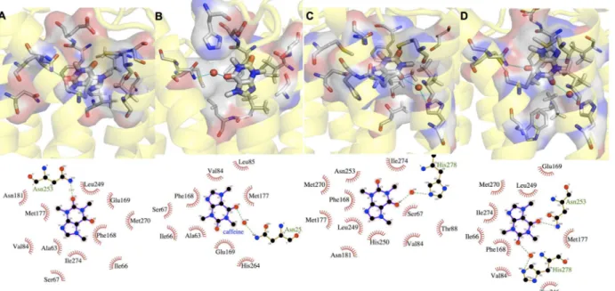
Binding Site
CFF exhibits multiple binding poses (A-D inFig 2) in receptor binding cavity across the three systems, comparable to what found for adenosine in this receptor [33]. Most of the identified binding poses are similar to those found in the 0.069-μs MD study of a H8-truncated CFF/
hA2AR complex [34]. Our poses yet differ from those in the 0.005-μs MD study [35], possibly
because of the large difference (more than two orders of magnitude) between our time scale and theirs. The population of the CFF poses depends on the type of membrane environment. A (38%), B (29%), and C (94%) are the most populated poses forI,II,III, respectively (seeFig 2
andTable 2). Notably, the pose in the X-ray structure (D) [27] (Fig 2), is not the most populat-ed one in any of the three systems (seeS1 Text). C is almost the only pose assumed by the Fig 2. CFF’s most populated binding poses in systems I-III (A-D).For each binding pose, the upper panel shows the protein backbone in yellow cartoon, CFF and residues interacting with CFF in thick and thin sticks, respectively. Water molecules forming H-bonds with CFF and residues are represented as red sphere; the lower panel shows the corresponding 2-d chart.
doi:10.1371/journal.pone.0126833.g002
Table 2. Populations of CFF binding poses (%) detected across systems I-III over the last 400 ns of MD simulated time.
BP Index I II III
A 38.1
-B - 29.1
-C 31.9 5.8 92.4
D - - 3.8
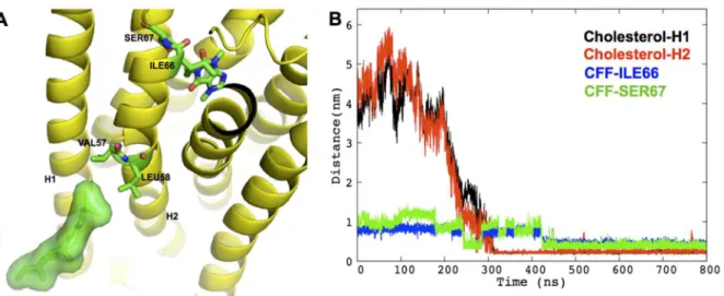
antagonist in the physiologically relevant systemIII. It is stabilized by hydrophobic contacts between the CFF/C5 methyl group and ILE66 and SER67 side chains on the extracellular side of H2 (panel A inFig 3). This stabilizing interaction may be triggered by the diffusion of a cho-lesterol molecule, already after 0.3μs, to the cleft between H1 and H2 (panel B inFig 3). Indeed,
this specific cholesterol binding induces conformational rearrangements of VAL57, LEU58, ILE66 and SER67 (S6 Fig), which in turn result in the enhanced stabilization of the hydropho-bic interaction between CFF and H2 residues. We conclude that the cholesterol very likely drives specific pose for CFF. The calculated lateral diffusion coefficient of cholesterol molecules around the receptor is not too dissimilar from that of cholesterol molecules in the proximity of the lipids (510−8cm2s-1, 810−8cm2s-1respectively,S2 TextandS7 Fig), suggesting that the observed cholesterol binding event is not strongly dependent on cholesterol’s starting location. Notably, in the absence of cholesterol molecules (systemsIandII), one POPC molecule replac-es cholreplac-esterol in the binding cleft. Hence, this receptor’s binding cleft seems to act like an anti-diffusion trap for lipids and, more, for cholesterol molecules, showing higher specificity for the latter. Interestingly, in theμs-MD simulations of apo hA2AR, Lyman et al. also detected the
spe-cific cholesterol presence between helices H1 and H2 [23].
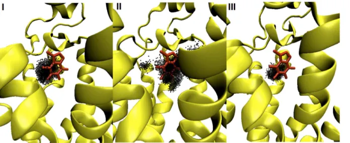
Next, we investigated the mobility of CFF, i.e. the different extent of roto-translation of the ligand inside the cavity across the three systems over the last 400 ns. This mobility is measured in terms of the orientational flipping angle (defined in the method section, seeS8 Fig) and the CFF center of mass (Fig 4), sampled along the simulation time. Not surprisingly,IIIfeatures the smallest fluctuations of both quantities, as the ligand is mostly in the C conformation.II ex-hibits the largest fluctuations whilstIfeatures intermediate values (seeFig 4andS8 Fig).
The MD-averaged number of water molecules in the binding cavity ofII(32 molecules) is much larger than those ofIand, more, ofIII(21 and 16 molecules, respectively, seeS9 Fig). Hence, this study corroborates the plausible hypothesis that enhanced hydration of binding cavity increases ligand dynamics inII[33].
Fig 3. Specific cholesterol binding to hA2AR A) Cartoon showing cholesterol-binding pose in H1/H2 cleft in system III.The receptor is shown in
yellow cartoon; the cholesterol molecule is shown as green sticks surrounded by its solvent accessible surface; CFF, cholesterol-interacting residues, VAL57, LEU58, as well as CFF-interacting residues ILE66, SER67 are shown as green sticks with oxygen and nitrogen atoms colored in red and blue, respectively.B) The diffusion of cholesterol into of the H1/H2 cleft enhances hydrophobic contacts between CFF and H2.The minimum distances between the specific cholesterol molecule and H1 (residues 5–34), between cholesterol and H2 (residues 41–67), between C5@CFF and heavy atoms of ILE66 and SER67 side chains, are shown in black, red, blue and green, respectively.
Membrane Structure
The primary amine group of POPE (inIIandIII) forms intra and intermolecular hydrogen bonding with the lipids' phosphate groups (S2 Table) [36]. Water- and receptor-lipid hydrogen bonds are comparable across the three systems (S2 Table).
The different membrane composition affects its thickness. The latter increases on passing fromItoII, and fromIItoIII. The latter observation is consistent with the experimental ob-servation that the presence of cholesterol causes an increase of thickness of lipid bilayers [37]. These features are shown by a plot of phosphate groups’density distributions (panel A inS4 Fig). The area per lipid decreases from 0.61 nm2(systemI) to 0.56 nm2(systemII) and 0.50 nm2(systemIII), seeS2 Table. This is consistent with the fact that the average area per lipid
of pure POPC is greater than that of pure POPE membrane [38]. Hence, the area per lipid is anti-correlated with the thickness of the membrane, consistently with what already found in ref. [39].
The POPC and headgroups’dipole moments turn out to be oriented differently on passing fromIto the other two systems. Let us define the PN vector (from the phosphorus atom to the nitrogen atom of one lipid headgroup, seeS10 Fig) and the angle (FPN) between the PN vector
and the axis perpendicular to the lipid bilayer surface (z-axis). The POPC headgroups’dipoles turn out to be perpendicular to z-axis in systemI, with a large standard deviation (FPN~ 90
(36) degrees, seeS11 Fig). Instead, in systemsIIandIII, these headgroups show a bivariate dis-tribution with two shallow peaks aroundFPN~ 65 degree andFPN~ 115. Interestingly, the
POPE headgroups are oriented perpendicular to the z-axis (FPN~ 90(28) degrees and 90(29)
for systemsIIandIII, respectively).
In systemIII, the POPC/POPE ratio is 1.2:1. This differs from that of systemII, which fea-tures a POPC/POPE ratio of 1:1. To test whether this difference in POPC/POPE ratio plays a role for protein structural, we performed an additional simulation where we replaced 24 POPE molecules of systemIIwith 24 POPC molecules. The results are rather similar to the ones of systemIIand are reported in SI (S12 Fig).
Fig 4. The distribution of CFF center of mass within the ligand-binding cavity of hA2AR across systems I-III.The receptor and CFF are shown as
yellow cartoon and red sticks, respectively. CFF center of mass at each collected frame over the last 400 ns of MD simulated time is depicted as one black dot.
Conclusion
Both hA2AR fold and CFF binding dynamics are sensitive to the lipid environment where
hA2AR is embedded. The lipid polar headgroups also exhibit varied dipole orientations in
dif-ferent membrane environments. Most importantly, the presence of cholesterol in the mem-brane is shown to drastically affect CFF binding pose population and mobility. X-ray studies commonly crystallize ligand/receptor complexes in detergent mimics [40], without the physio-logically high concentration of cholesterol. The artificial environment is found here to affect the population of ligand poses drastically: the pose found in the X-ray structure, at 3.6-Å of res-olution, is the most populated one in none of our 0.8μs-long MD simulations. This study
sug-gests that computer-aided studies of hA2AR in nearly physiological conditions may give key
contributions to the investigation of receptor's function as well as to the development of CFF derivatives that retain CFF's neuroprotective benefits with much higher affinity for the target than CFF [41,42].
Materials and Methods
Homology Modeling of hA
2AR
hA2AR is a class A GPCR, composed of 7 transmembrane helices (H1–H7) and a helix lying at
the membrane-cytoplasm interface (H8). The X-ray structure of the CFF/hA2AR complex has
been solved at a 3.6-Å resolution (PDBid: 3RFM) [27]. Amino acid sequence after residue 317 was deleted to remove the highly mobile cytoplasmic C terminus. The truncated sequence was joined by a polyhistidine tag (residues 318–329). Residues1MPIMGS6,150KEGKNHSQ157,
306HVLRQQEPFKAAAAHHHHHHHHHH329are not detected in the crystal structure.
More-over the crystalized receptor contains 8 mutations (A54L, T88A, R107A, K122A, L202A, L235A, V239A, S277A). The missing regions were complemented and the mutations were mu-tated back to wild type by multiple-template-based homology modeling (S1 Table).
12 X-ray structures of hA2AR are deposited in the Protein Data Bank [15,27,43–47]. Among
these we selected 3RFM and 4 other templates with resolution below 3.0 Å (3VG9, 3EML, 4EIY, 2YDV). Notice that the 2YDV template where hA2AR is bound with an agonist
N-ETHYL-5'-CARBOXAMIDO (NEC), is the only one used for modeling the residues 291–
325 in the C terminus without including any other residues, as it has the longest resolved helix H8 among all the available hA2AR X-ray structures. 100 models using the 5 templates were
gen-erated in Modeller 9.11 [48]. The best model, in terms of both DOPE score [49] and stereo-chemistry PROCHECK analysis [50] underwent to loop refinement procedure [48]. 500 models were generated. The best model was selected as the optimal initial model for MD simulations.
The first six residues in the N terminus are predicted as a helical segment. The missing 8 res-idues (150KEGKNHSQ157) in the loop connecting helix 4 and helix 5 are also so as to form a short helical segment. As for the missing residues at the C terminus, residues306
HVLRQQEPF-KAAAAHHHH323are modeled as the H8 [46]. The last six residues, all histidines, are modeled as a loop. The backbone Root Mean Square Deviation (RMSD) between the model and 3RFM is 0.9 Å. The backbone RMSD of the residues located in hA2AR ligand binding site (within 7.0
Å of CFF) against their counterparts in 3RFM is 3.3 Å.
Simulation Details
Membrane models in systemsI,IIandIIIwere generated using the MemBuilder tool [51]. The inflateGRO code [52] was used to pack lipids around the hA2AR constructed in the previous
this choice, the minimum distance between periodic images of the protein was larger than 1.5 nm in all systems. Water, sodium and chloride ions were added in order to solvate and neutral-ize the systems at an ionic strength of 0.15 M. The final systems comprised ~150,000 atoms (Table 1).
The AMBER99SB-ILDN force fields [53], the Slipids [54,55], the TIP3P [56] force fields were used for the protein and ions, the lipids, and the water molecules respectively. The Gener-al Amber force field (GAFF) parameters [57] were used for CFF, along with the RESP atomic charge using Gaussian 09 [58] with the HF-6-31Gbasis set [59,60]. MD simulations were per-formed using Gromacs v4.5.5 package [61] on JUROPA supercomputer. The Particle Mesh Ewald method [62] was used to treat the long-range electrostatic interaction with a real space cutoff of 1.2 nm. A 1.2 nm cutoff was used for the short-range non-bonded interaction. A time-step of 2 fs was set. The LINCS algorithm [63] was applied to constrain all bonds involving hy-drogen atoms. Constant temperature and pressure conditions were achieved via independently coupling protein, lipids, solvent and ions to Nosè-Hoover thermostat [64] at 310 K and Ander-sen-Parrinello-Rahman Barostat [65] at 1 atm. For each system, the receptor in the free state underwent minimization, 1-ns simulated annealing and 10-ns equilibration with positional re-straint using a force constant of 1000 kJ mol-1nm-2on the heavy atoms of the protein. 40-ns
equilibration was further carried out with positional restraint on the side chains of the residues within the binding cavity (residues within 7.0 Å of CFF on backbone alignment to 3RFM [27]). This allowed water molecules to diffuse into the ligand-binding cavity. Next, CFF was inserted so as to fit the conformation it has in the X-ray structure [27] using backbone alignment in Pymol [66]. Energy minimization, annealing, 20-ns equilibration with positional restraint on the side chains of the residues belonging to the binding cavity and the CFF were performed be-fore removing all the restraints. Then 0.8μs MD at 310 K and 1 atm was performed forI,II,
III, with one frame collected every 20 ps. The starting binding pose of CFF resembled fairly that in the X-ray counterpart (RMSD = 1.4 Å, 0.8 Å, 2.3 Å forI,II,III).
Trajectory Analysis
The RMSD, pairwise RMSD matrices [28] and secondary structure content are calculated over the entire trajectory with g_rms, do_dssp of the Gromacs v4.6.5 package [61]. The CFF orienta-tional flipping angle is defined as arccos(μτμι), whereμιis the vector in the plane of the bicyclic
core of CFF, chosen so that it faces toward the extracellular side in the initial frameιof the tra-jectory;μτis the vector at each frameτof the trajectory. Also this quantity is calculated over
the entire trajectory.
The following properties are calculated over the last 400 ns of the three MD simulations: (i) The PAD flexibility index, using a in house code [67,68]. (ii) The density profile of lipid phos-phate groups along the z axis, using g_density in Gromacs v4.6.5 package [61]. (iii) The CFF binding poses, identified using the Gromos cluster algorithm [69] with a 2-Å RMSD cutoff on alignment of protein backbone. The g_cluster module of Gromacs v4.6.5 [61] has been used. (iv) CFF center of mass is defined by the vector riof the coordinates of CFF center of mass at a
frame i, upon protein backbone alignment. (v) The average number of water molecules in the ligand binding cavity of hA2AR is calculated by following the similar procedure in [33,70].
atoms. (vii) The average area per lipid (APL) is calculated with the GridMat-MD program [71]. (viii) The PN vector of one lipid molecule is defined as the vector from the phosphorous atom to the nitrogen atom of its polar headgroup, as defined in [72]. TheFPNof one lipid molecule
is the angle between its PN vector and the z-axis. This is the axis perpendicular to the lipid bi-layer surface. For each system,FPNof each lipid molecule is sampled over the entire MD
simu-lation for the normalizedFPNdistributions of POPC and POPE lipids. (ix) the lateral diffusion
coefficients of cholesterol molecules inIIIare calculated using the Einstein relation [73] for a 60 ns simulation ofIIIin the NVT ensemble (seeS2 Textfor details). Cholesterol molecules are classified as molecules in close proximity of the protein if they have atoms within 0.35 nm of the receptor during the dynamics. The other molecules are classified as 'free'.
Molecular graphics are drawn using Pymol [66], VMD [74] and Ligplot+ [75].
Supporting Information
S1 Fig. Chemical structures of CFF, POPC, POPE and cholesterol molecules. (PDF)
S2 Fig. Pairwise RMSD matrix of Cαatoms. (PDF)
S3 Fig. Proteins' backbone. (PDF)
S4 Fig. Selected properties of systems I-III. (PDF)
S5 Fig. Flexibility of individual residues of hA2AR.
(PDF)
S6 Fig. Cholesterol-induced conformational transitions of H2 residues in system III. (PDF)
S7 Fig. Lateral Mean-square displacements (MSDs) of two groups of cholesterol molecules in system III.
(PDF)
S8 Fig. CFF orientational flipping angle. (PDF)
S9 Fig. The hydration of the ligand binding cavity of hA2AR.
(PDF)
S10 Fig. Orientation of POPC and POPE headgroups. (PDF)
S11 Fig. Distribution ofFPNof lipid headgroups for systems I-III.
(PDF)
S12 Fig. Changing the ratio between the lipids in system II. (PDF)
S1 Table. Available X-ray structures of hA2AR.
(PDF)
S1 Text. Description of most populated CFF binding poses A-D. (PDF)
S2 Text. Lateral diffusion coefficient of cholesterol molecules. (PDF)
Acknowledgments
We acknowledge the computing time granted by the supercomputer JUROPA at the Jülich Supercomputing Center.
Author Contributions
Conceived and designed the experiments: GR AB PC. Performed the experiments: RC. Ana-lyzed the data: RC GR PC. Contributed reagents/materials/analysis tools: RC GR PC. Wrote the paper: RC GR PC.
References
1. Niu S-L, Mitchell DC, Litman BJ (2002) Manipulation of cholesterol levels in rod disk membranes by methyl-β-cyclodextrin effects on receptor activation. Journal of Biological Chemistry 277: 20139–
20145. PMID:11889130
2. Stone W, Farnsworth C, Dratz E (1979) A reinvestigation of the fatty acid content of bovine, rat and frog retinal rod outer segments. Experimental eye research 28: 387–397. PMID:446567
3. Bruno A, Costantino G, de Fabritiis G, Pastor M, Selent J (2012) Membrane-sensitive conformational states of helix 8 in the metabotropic Glu2 receptor, a class C GPCR. PloS one 7: e42023. doi:10.1371/ journal.pone.0042023PMID:22870276
4. Mahmood MI, Liu X, Neya S, Hoshino T (2013) Influence of lipid composition on the structural stability of G-protein coupled receptor. Chemical and Pharmaceutical Bulletin 61: 426–437. PMID:23546002
5. Soubias O, Teague WE, Hines KG, Mitchell DC, Gawrisch K (2010) Contribution of membrane elastic energy to rhodopsin function. Biophysical Journal 99: 817–824. doi:10.1016/j.bpj.2010.04.068PMID: 20682259
6. Soubias O, Gawrisch K (2012) The role of the lipid matrix for structure and function of the GPCR rho-dopsin. Biochimica et Biophysica Acta (BBA)-Biomembranes 1818: 234–240. doi:10.1016/j.bbamem. 2011.08.034PMID:21924236
7. Pucadyil TJ, Chattopadhyay A (2004) Cholesterol modulates ligand binding and G-protein coupling to serotonin 1A receptors from bovine hippocampus. Biochimica et Biophysica Acta (BBA)-Biomem-branes 1663: 188–200. PMID:15157621
8. Overington JP, Al-Lazikani B, Hopkins AL (2006) How many drug targets are there? Nature reviews Drug discovery 5: 993–996. PMID:17139284
9. Ballesteros JA, Jensen AD, Liapakis G, Rasmussen SG, Shi L, Gether U, et al. (2001) Activation of the
β2-adrenergic receptor involves disruption of an ionic lock between the cytoplasmic ends of transmem-brane segments 3 and 6. Journal of Biological Chemistry 276: 29171–29177. PMID:11375997
10. Khelashvili G, Grossfield A, Feller SE, Pitman MC, Weinstein H (2009) Structural and dynamic effects of cholesterol at preferred sites of interaction with rhodopsin identified from microsecond length molecu-lar dynamics simulations. Proteins: Structure, Function, and Bioinformatics 76: 403–417. doi:10.1002/ prot.22355PMID:19173312
11. Jacobson KA, Ukena D, Padgett W, Daly JW, Kirk KL (1987) Xanthine functionalized congeners as po-tent ligands at A2-adenosine receptors. Journal of Medicinal Chemistry 30: 211–214. PMID:3806597
12. Fink JS, Weaver DR, Rivkees SA, Peterfreund RA, Pollack AE, Adler EM, et al. (1992) Molecular clon-ing of the rat A2 adenosine receptor: selective co-expression with D2 dopamine receptors in rat stria-tum. Molecular Brain Research 14: 186–195. PMID:1279342
13. Ross G, Abbott RD, Petrovitch H, Morens DM, Grandinetti A, K. T, et al. (2000) Association of coffee and caffeine intake with the risk of parkinson diseases. JAMA: The Journal of the American Medical As-sociation 283: 2674–2679.
15. Liu W, Chun E, Thompson AA, Chubukov P, Xu F, Katritch V, et al. (2012) Structural basis for allosteric regulation of GPCRs by sodium ions. Science 337: 232–236. doi:10.1126/science.1219218PMID: 22798613
16. Postuma RB, Lang AE, Munhoz RP, Charland K, Pelletier A, Moscovich M, et al. (2012) Caffeine for treatment of Parkinson disease: A randomized controlled trial. Neurology 79: 651–658. doi:10.1212/ WNL.0b013e318263570dPMID:22855866
17. Douna H, Bavelaar BM, Pellikaan H (2012) Neuroprotection in Parkinson's disease: a systematic re-view of the preclinical data. The Open Pharmacology Journal 6: 12–26.
18. Nakaso K, Ito S, Nakashima K (2008) Caffeine activates the PI3K/Akt pathway and prevents apoptotic cell death in a Parkinson's disease model of SH-SY5Y cells. Neuroscience Letters 432: 146–150. doi: 10.1016/j.neulet.2007.12.034PMID:18201823
19. Pfrieger FW (2003) Role of cholesterol in synapse formation and function. Biochimica et Biophysica Acta (BBA)-Biomembranes 1610: 271–280. PMID:12648780
20. Rodríguez D, Piñeiro An, Gutiérrez-de-Terán H (2011) Molecular dynamics simulations reveal insights into key structural elements of adenosine receptors. Biochemistry 50: 4194–4208. doi:10.1021/ bi200100tPMID:21480628
21. Pang X, Yang M, Han K (2013) Antagonist binding and induced conformational dynamics of GPCR A2A adenosine receptor. Proteins: Structure, Function, and Bioinformatics 81: 1399–1410. doi:10. 1002/prot.24283PMID:23508898
22. Ng HW, Laughton CA, Doughty SW (2013) Molecular dynamics simulations of the adenosine A2a re-ceptor: structural stability, sampling, and convergence. Journal of Chemical Information and Modeling 53: 1168–1178. doi:10.1021/ci300610wPMID:23514445
23. Lyman E, Higgs C, Kim B, Lupyan D, Shelley JC, Farid R, et al. (2009) A role for a specific cholesterol interaction in stabilizing the apo configuration of the human A2A adenosine receptor. Structure 17: 1660–1668. doi:10.1016/j.str.2009.10.010PMID:20004169
24. Li J, Jonsson AL, Beuming T, Shelley JC, Voth GA (2013) Ligand-dependent activation and deactiva-tion of the human adenosine A2A receptor. Journal of the American Chemical Society 135: 8749–
8759. doi:10.1021/ja404391qPMID:23678995
25. Lee JY, Lyman E (2012) Predictions for cholesterol interaction sites on the A2A adenosine receptor. Journal of the American Chemical Society 134: 16512–16515. doi:10.1021/ja307532dPMID: 23005256
26. Mori A, Shindou T, Ichimura M, Nonaka H, Kase H (1996) The role of adenosine A2A receptors in regu-lating GABAergic synaptic transmission in striatal medium spiny neurons. The Basal Ganglia V: Springer. pp. 119–122.
27. Doré Andrew S, Robertson N, Errey James C, Ng I, Hollenstein K, Tehan B, et al. (2011) Structure of the adenosine A2A receptor in complex with ZM241385 and the xanthines XAC and caffeine. Structure 19: 1283–1293. doi:10.1016/j.str.2011.06.014PMID:21885291
28. Grossfield A, Zuckerman DM (2009) Quantifying uncertainty and sampling quality in biomolecular simu-lations. Annual reports in computational chemistry 5: 23–48. PMID:20454547
29. Ciruela F, Albergaria C, Soriano A, Cuffí L, Carbonell L, Sánchez S, et al. (2010) Adenosine receptors interacting proteins (ARIPs): behind the biology of adenosine signaling. Biochimica et Biophysica Acta (BBA)-Biomembranes 1798: 9–20.
30. Gsandtner I, Freissmuth M (2006) A tail of two signals: the C terminus of the A2A-adenosine receptor recruits alternative signaling pathways. Molecular Pharmacology 70: 447–449. PMID:16707626
31. Katragadda M, Maciejewski M, Yeagle P (2004) Structural studies of the putative helix 8 in the human
β2 adrenergic receptor: an NMR study. Biochimica et Biophysica Acta (BBA)-Biomembranes 1663: 74–81. PMID:15157609
32. Caliandro R, Rossetti G, Carloni P (2012) Local fluctuations and conformational transitions in proteins. Journal of Chemical Theory and Computation 8: 4775–4785.
33. Lee JY, Lyman E (2012) Agonist dynamics and conformational selection during microsecond simula-tions of the A2A adenosine receptor. Biophysical Journal 102: 2114–2120. doi:10.1016/j.bpj.2012.03. 061PMID:22824275
34. Sabbadin D, Ciancetta A, Moro S (2014) Bridging molecular docking to membrane molecular dynamics to investigate GPCR—ligand recognition: the human A2A adenosine receptor as a key study. Journal of Chemical Information and Modeling 54: 169–183. doi:10.1021/ci400532bPMID:24359090
36. McIntosh TJ (1996) Hydration properties of lamellar and non-lamellar phases of phosphatidylcholine and phosphatidylethanolamine. Chemistry and physics of lipids 81: 117–131. PMID:8810046
37. Hung W-C, Lee M-T, Chen F-Y, Huang HW (2007) The condensing effect of cholesterol in lipid bilayers. Biophysical Journal 92: 3960–3967. PMID:17369407
38. Murzyn K, Róg T, Pasenkiewicz-Gierula M (2005) Phosphatidylethanolamine-phosphatidylglycerol bi-layer as a model of the inner bacterial membrane. Biophysical Journal 88: 1091–1103. PMID: 15556990
39. Elmore DE (2006) Molecular dynamics simulation of a phosphatidylglycerol membrane. FEBS letters 580: 144–148. PMID:16359668
40. Serebryany E, Zhu GA, Yan EC (2012) Artificial membrane-like environments for in vitro studies of puri-fied G-protein coupled receptors. Biochimica et Biophysica Acta (BBA)-Biomembranes 1818: 225–
233. doi:10.1016/j.bbamem.2011.07.047PMID:21851807
41. Rivera-Oliver M, Díaz-Ríos M (2014) Using caffeine and other adenosine receptor antagonists and ag-onists as therapeutic tools against neurodegenerative diseases: A review. Life Sciences 101: 1–9. doi: 10.1016/j.lfs.2014.01.083PMID:24530739
42. Armentero MT, Pinna A, Ferré S, Lanciego JL, Müller CE, Franco R (2011) Past, present and future of A2A adenosine receptor antagonists in the therapy of Parkinson's disease. Pharmacology & Therapeu-tics 132: 280–299.
43. Congreve M, Andrews SP, Doré AS, Hollenstein K, Hurrell E, Langmead CJ, et al. (2012) Discovery of 1,2,4-triazine derivatives as adenosine A2A antagonists using structure based drug design. Journal of Medicinal Chemistry 55: 1898–1903. doi:10.1021/jm201376wPMID:22220592
44. Hino T, Arakawa T, Iwanari H, Yurugi-Kobayashi T, Ikeda-Suno C, Nakada-Nakura Y, et al. (2012) G-protein-coupled receptor inactivation by an allosteric inverse-agonist antibody. Nature 482: 237–240. doi:10.1038/nature10750PMID:22286059
45. Jaakola V-P, Griffith MT, Hanson MA, Cherezov V, Chien EYT, Lane JR, et al. (2008) The 2.6 Angstrom crystal structure of a human A2A adenosine receptor bound to an antagonist. Science 322: 1211–
1217. doi:10.1126/science.1164772PMID:18832607
46. Lebon G, Warne T, Edwards PC, Bennett K, Langmead CJ, Leslie AGW, et al. (2011) Agonist-bound adenosine A2A receptor structures reveal common features of GPCR activation. Nature 474: 521–
525. doi:10.1038/nature10136PMID:21593763
47. Xu F, Wu H, Katritch V, Han GW, Jacobson KA, Gao Z-G, et al. (2011) Structure of an agonist-bound human A2A adenosine receptor. Science 332: 322–327. doi:10.1126/science.1202793PMID: 21393508
48. Fiser A,Šali A (2003) Modeller: generation and refinement of homology-based protein structure
mod-els. Methods in Enzymology. pp. 461–491. PMID:14696385
49. Shen M-y, Sali A (2006) Statistical potential for assessment and prediction of protein structures. Protein Science 15: 2507–2524. PMID:17075131
50. Laskowski RA, Macarthur MW, Moss DS, Thornton JM (1993) PROCHECK: a program to check the stereochemical quality of protein structures. Journal of Applied Crystallography 26: 283–291.
51. Ghahremanpour MM, Arab SS, Aghazadeh SB, Zhang J, van der Spoel D (2014) MemBuilder: a web-based graphical interface to build heterogeneously mixed membrane bilayers for the GROMACS bio-molecular simulation program. Bioinformatics 30: 439–441. doi:10.1093/bioinformatics/btt680PMID: 24273238
52. Kandt C, Ash WL, Peter Tieleman D (2007) Setting up and running molecular dynamics simulations of membrane proteins. Methods 41: 475–488. PMID:17367719
53. Best RB, Hummer G (2009) Optimized molecular dynamics force fields applied to the helix—coil transi-tion of polypeptides. The Journal of Physical Chemistry B 113: 9004–9015. doi:10.1021/jp901540t PMID:19514729
54. Jämbeck JPM, Lyubartsev AP (2012) Derivation and systematic validation of a refined all-atom force field for phosphatidylcholine lipids. The Journal of Physical Chemistry B 116: 3164–3179. doi:10. 1021/jp212503ePMID:22352995
55. Jämbeck JPM, Lyubartsev AP (2012) An extension and further validation of an all-atomistic force field for biological membranes. Journal of Chemical Theory and Computation 8: 2938–2948.
56. Jorgensen W, Chandrasekhar J, Madura J, Impey R, Klein M (1983) Comparison of simple potential functions for simulating liquid water. J Chem Phys 79: 926–935.
58. Frisch MJ, Trucks GW, Schlegel HB, Scuseria GE, Robb MA, Cheeseman JR, et al. (2009) Gaussian 09, Revision A.02. Wallingford CT.
59. Wang JM, Cieplak P, Kollman PA (2000) How well does a restrained electrostatic potential (RESP) model perform in calculating conformational energies of organic and biological molecules? Journal of Computational Chemistry 21: 1049–1074.
60. Case DA, Cheatham TE 3rd, Darden T, Gohlke H, Luo R, Merz KM Jr., et al. (2005) The Amber biomo-lecular simulation programs. Journal of Computational Chemistry 26: 1668–1688. PMID:16200636
61. Van Der Spoel D, Lindahl E, Hess B, Groenhof G, Mark AE, Berendsen HJC (2005) GROMACS: Fast, flexible, and free. Journal of Computational Chemistry 26: 1701–1718. PMID:16211538
62. Darden T, York D, Pedersen L (1993) Particle mesh Ewald: An Nlog(N) method for Ewald sums in
large systems. The Journal of Chemical Physics 98: 10089–10092.
63. Hess B, Bekker H, Berendsen HJC, Fraaije JGEM (1997) LINCS: A linear constraint solver for molecu-lar simulations. Journal of Computational Chemistry 18: 1463–1472.
64. Hünenberger P (2005) Thermostat Algorithms for Molecular Dynamics Simulations. Advanced Comput-er Simulation: SpringComput-er BComput-erlin HeidelbComput-erg. pp. 105–149.
65. Parrinello M, Rahman A (1981) Polymorphic transitions in single crystals: A new molecular dynamics method. Journal of Applied Physics 52: 7182–7190.
66. DeLano WL (2002) The PyMOL molecular graphics system.
67. Musiani F, Ippoliti E, Micheletti C, Carloni P, Ciurli S (2013) Conformational fluctuations of UreG, an in-trinsically disordered enzyme. Biochemistry 52: 2949–2954. doi:10.1021/bi4001744PMID:23560717
68. Dibenedetto D, Rossetti G, Caliandro R, Carloni P (2013) A molecular dynamics simulation-based inter-pretation of nuclear magnetic resonance multidimensional heteronuclear spectra ofα-synuclein
dopa-mine adducts. Biochemistry 52: 6672–6683. doi:10.1021/bi400367rPMID:23964651
69. Daura X, Gademann K, Jaun B, Seebach D, van Gunsteren WF, Mark AE (1999) Peptide folding: when simulation meets experiment. Angewandte Chemie International Edition 38: 236–240.
70. Grossfield A, Pitman MC, Feller SE, Soubias O, Gawrisch K (2008) Internal hydration increases during activation of the G-protein-coupled receptor rhodopsin. Journal of Molecular Biology 381: 478–486. doi:10.1016/j.jmb.2008.05.036PMID:18585736
71. Allen WJ, Lemkul JA, Bevan DR (2009) GridMAT—MD: A grid—based membrane analysis tool for use with molecular dynamics. Journal of computational chemistry 30: 1952–1958. doi:10.1002/jcc.21172 PMID:19090582
72. Jurkiewicz P, Cwiklik L, Vojtíšková A, Jungwirth P, Hof M (2012) Structure, dynamics, and hydration of
POPC/POPS bilayers suspended in NaCl, KCl, and CsCl solutions. Biochimica et Biophysica Acta (BBA)-Biomembranes 1818: 609–616. doi:10.1016/j.bbamem.2011.11.033PMID:22155683
73. Allen MP, Tildesley DJ, Banavar JR (2008) Computer simulation of liquids. Physics Today 42: 105–
106. doi:10.1007/s10464-008-9187-7PMID:18597168
74. Humphrey W, Dalke A, Schulten K (1996) VMD: visual molecular dynamics. Journal of molecular graphics 14: 33–38. PMID:8744570
![Fig 1. Membrane-sensitive folding of H8. A) The cartoon representations of receptor’s backbone of systems I – III are shown in blue to red according to residues ’ increased flexibility, as emerging from the so-called PAD index values [32]; the inserted pan](https://thumb-eu.123doks.com/thumbv2/123dok_br/17303499.248761/3.918.69.748.118.410/membrane-sensitive-representations-according-residues-increased-flexibility-emerging.webp)