Knockdown of E2f1 by RNA interference impairs proliferation
of rat cells
in vitro
Luciana dos Reis Vasques*, Regiane Simoni Pujiz, Bryan Eric Strauss and José Eduardo Krieger
Instituto do Coração, Escola de Medicina, Universidade de São Paulo, São Paulo, SP, Brazil.
Abstract
E2F1 plays a key role in cell-cycle regulation in mammals, since its transcription factor activity controls genes re-quired for DNA synthesis and apoptosis. E2F1 deregulation is a common feature among different tumor types and can be a major cause of cell proliferation. Thus, blockingE2F1 expression by RNA interference represents a promis-ing therapeutic approach. In this study, the introduction of specific short hairpin RNAs (shRNAs) reducedE2f1 ex-pression by up to 77%, and impaired rat glioma cell proliferation by approximately 70%, as compared to control cells. Furthermore, we investigated the expression ofE2f1 target genes, Cyclin A and Cyclin E. Cyclin A was found to be down-regulated, whereasCyclin E had similar expression to control cells, indicating that gene(s) other than E2f1 control its transcription. Other E2f family members,E2f2 and E2f3, which have been classified in the same subgroup of transcriptional activators, were also analyzed. Expression of bothE2f2 and E2f3 was similar to control cells, show-ing no cross-inactivation or up-regulation to compensate for the absence of E2f1. Nevertheless, their expression was insufficient to maintain the initial proliferation potential. Taken together, our results suggest that shE2f1 is a promis-ing therapy to control tumor cell proliferation.
Key words:RNAi, shRNA, E2F1, proliferation, cancer.
Received: April 16, 2009; Accepted: August 8, 2009.
E2F comprises a family of transcription factor pro-teins, with a pivotal role in controlling genes related to cell-cycle progression (Helinet al., 1992; Kaelin Jret al., 1992; Shanet al., 1992). Eight E2F family members have been identified so far, E2F1 to E2F8, whereas E2F1 to E2F6 share the same structure: conserved DNA binding and dimerization domains, and, except for E2F6, have do-mains for transactivation and binding Pocket Proteins (PP): p107, p130 and Rb (Retinoblastoma) (reviewed in Tsan-toulis and Gorgoulis, 2005; DeGregori and Johnson, 2006). In general terms, the E2F family can be functionally classi-fied in two subgroups, namely transcriptional activators (E2F1 to E2F3a) and repressors (E2F3b to E2F8). The E2F dimerization domain binds to members of the DP protein family, and the resulting complexes regulate overlapping gene collections (DeGregori and Johnson, 2006).
Despite Rb having been found to be associated with many members of the family, E2F1 is its main target (Wells et al., 2003; Frolov and Dyson, 2004). Rb phosphorylation by Cyclin D/CDK4 and Cyclin E/CDK2, in late G1 phase, releases E2F transcription factors, thereby promoting
ex-pression of genes related to DNA synthesis and cell-cycle progression, resulting in cell proliferation (Polyak et al., 1994; DeGregori et al., 1995). The dissociation of E2F from pRb protein seems to be the main determinant in regu-lating cell proliferation, by permitting transactivation of genes such ascyclin A,cyclin E,c-myb,cdc2,PCNAand thymidine kinase, and committing cells to S phase (DeGre-gori, 2002).
The best characterized gene of the E2F family is E2F1, which plays a paradoxical role by acting in two op-posing pathways: induction of cell cycle progression and apoptosis (Pierceet al., 1999). E2F1, in response to DNA damage, can induce apoptosis by regulating related genes in a p53-dependent and p53-independent manner (Bateset al., 1998; Irwinet al., 2000; Lissyet al., 2000; Moroniet al., 2001).
Dysfunction of the intricate cell-cycle regulation pathways described above can exacerbate cell growth and, eventually, lead to cancer-cell development. In fact, dereg-ulation ofE2F1gene expression is a common event in the majority of tumors, where it appears over-expressed rather than mutated (Sherr, 1996; Dyson, 1998). E2F1 over-expression is due to a positive feedback loop created be-tween this protein and its own promoter and due to high pRb phosphorylation levels or lack of functional pRb, both resulting in the liberation of E2F1. The main reasons for this hyperphosphorylation are high CDK4/6 and CDK2
ac-Send correspondence to L.R. Vasques. Instituto do Coração, Es-cola de Medicina, Universidade de São Paulo, Rua 3 de Maio 100, 4° andar, Vila Clementino, 04044-020 São Paulo, SP, Brazil. E-mail: [email protected].
*
LRV present address: Departamento de Bioquímica, Universidade Federal de São Paulo, SP, Brazil.
tivity, the absence of Cdk inhibitors (CDIs) and over-expression of Cyclins (reviewed in Halaban, 2005).
Since Rb is the most important PP and is preferen-tially associated with E2F1, inactivation ofE2F1seems to be a promising therapy for impairing the proliferation of different tumor types and in other diseases where cell pro-liferation is a secondary effect, like vascular smooth muscle cell hyperplasia. Furthermore, the function of E2F1 in con-trolling the expression of other genes, such asCyclin Aand Cyclin E, and its overlapping function with other E2F mem-bers, namely E2F2 and E2F3, are controversial (Ohtaniet al.,1995; DeGregori et al, 1995; Takahashi et al., 2000; Gotoet al., 2006; Konget al., 2007), thereby necessitating further characterization.
In this study, our aim was to develop short interfering RNAs for the impairment of cancer cell proliferationin vi-tro.E2f1was elected as the target, as its expression plays a key role in cell-cycle progression, besides being up-regu-lated in most types of tumor (Sherr, 1996; Dyson, 1998). We employed the rat glioma cell line, C6, as anin vitro can-cer model, and showed that the shE2f1 (short hairpin RNA against E2f1 mRNA) is a potent tool for impeding cell pro-liferation, since it diminished C6 proliferation 3.5 to 4-fold. Furthermore, we also examined the effects of shE2f1 on the expression of two other members of the E2f family,E2f2 andE2f3, to explore whether any cross-inhibition or com-pensatory mechanisms were occurring. The expression of Cyclin AandCyclin Ewas also assessed to investigate E2f1 transcriptional regulation of these genes.
Three different shRNAs were designed for interfer-ence with the rat E2f1 transcript at distinct regions (shE2f1A, B and C), and were inserted into the pBS/hU6-1 plasmid vector (generously provided by Dr. David Balti-more -California Institute of Technology, CA- USA – Qin et al., 2003) yielding pBSE2f1A, B and C. As control, an additional vector (pBSshGFP) containing a shRNA against eGFP RNA (enhanced Green Fluorescent Protein) was also generated by using a previously validated target sequence described by Tiscornia et al. (2003) and Mousses et al.
(2003). None of the target sequence shows any significant homology to other rat gene sequences. Therefore, synthetic oligonucleotides (Invitrogen) were designed (listed in Ta-ble 1) and cloned as described by Qinet al. (2003). The generated constructs were confirmed by sequencing, using 25 ng of the respective primers T3 and T7 (Stratagene) and the ABI Prism – Big Dye Terminator Cycle Sequencing Ready Reaction Kit, with an ABI377 sequencer, according to manufacturer’s instructions (Perkin-Elmer).
The rat glioma cell line, C6 (ATCC CCL-107), was cultured in Dulbecco’s Modified Eagle’s minimum essen-tial medium (DMEM high glucose), supplemented with 10% fetal bovine serum (FBS) and penicillin/streptomycin (Invitrogen) at 37 °C/5% CO2. C6 cells were plated at 80%
confluence and co-transfected, using lipofectamine (Invi-trogen) with 3mg of a plasmid DNA mixture containing pBABEpuro and pBSshE2f1 -A, -B, -C or pBSshGFP (1:10, respectively), whereas pBS/hU6-1 derived plasmids were previously digested byXmnI, according to manufac-turer’s protocol (BioLabs). After co-transfection, cells were selected using 400 ng/mL of puromycin. As the first step towards identifying the most effective pBSshE2f1, several clones, denominated C6shE2f1-A, -B, -C and C6shGFP, were obtained from each co-transfection and maintained in selective medium. Their genomic DNA was extracted with lysis buffer (100 mM Tris-HCl, pH 8.5; 5 mM EDTA; 0.2% SDS; 200 mM NaCl; 100mg/mL of proteinase K), to verify the presence of pBS/hU6-1 derived plasmids by PCR using 25 ng of each of the primers T3 and T7 (Stratagene), according to manufacturer’s instructions. pBS/hU6-1 was used as negative PCR control. Positive clones were selected and used in subsequent experiments.
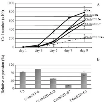
In order to assess the proliferation-altering potential of each shRNA vector, the parental C6 cell line (triplicate) and different clones of C6shE2f1-A (3 clones), -B (5 clo-nes), -C (3 clones) and C6shGFP (4 clones) were analyzed by a growth curve assay. At day zero, 5 x 10⁴cells from each of the different clones (C6shE2f1-A; -B; -C; -shGFP), as well as the parental C6 cell line, were each seeded into 10
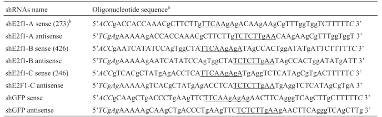
Table 1- Oligonucleotide sequences to construct pBS/hU6-1 encoding different shRNAs.
shRNAs name Oligonucleotide sequencea
shE2f1-A sense (273)b 5’ACCgACCACCAAACgCTTCTTgTTCAAgAgACAAgAAgCgTTTggTggTCTTTTTC3’
shE2f1-A antisense 5’TCgAgAAAAAgACCACCAAACgCTTCTTgTCTCTTgAACAAgAAgCgTTTggTggT 3’ shE2f1-B sense (426) 5’ACCgAATCATATCCAgTggCTATTCAAgAgATAgCCACTggATATgATTCTTTTTC3’ shE2f1-B antisense 5’TCgAgAAAAAgAATCATATCCAgTggCTATCTCTTgAATAgCCACTggATATgATT 3’ shE2f1-C sense (246) 5’ACCgTCACgCTATgAgACCTCATTCAAgAgATgAggTCTCATAgCgTgACTTTTTC3’ shE2F1-C antisense 5’TCgAgAAAAAgTCACgCTATgAgACCTCATCTCTTgAATgAggTCTCATAgCgTgA 3’ shGFP sense 5’ACCgCAAgCTgACCCTgAAgTTCTTCAAgAgAgAACTTCAgggTCAgCTTgCTTTTTC3’ shGFP antisense 5’TCgAgAAAAAgCAAgCTgACCCTgAAgTTCTCTCTTgAAgAACTTCAgggTCAgCTTg 3’ a: underlined, 9-nucleotide spacer sequence.
dishes, 35 mm in diameter, with DMEM supplemented with 5% FBS. At indicated times, each clone and the paren-tal cells were sampled in duplicate. The final results shown in Figure 1 represent the average among clones of each type. The culture medium was replaced every two days. The data presented indicate that construct pBSshE2f1-B significantly impaired the proliferation of C6 cells when compared to controls. A comparative analysis at day 9 showed that C6shE2f1 B cell proliferation was 3.5 to 4 times lower than that observed in the controls, these cells remaining with only 30% of the proliferative capacity ob-served in C6shGFP cells.
Total RNA was extracted from C6 (parental cell line), and clones C6shGFP-6 (control cells), C6shE2f1-A22, C6shE2f-C3 and 2 different clones from C6shE2f1-B (B7 and B11), by using Trizol (Invitrogen) according to manu-facturer’s protocol (see Figure 1). This was carried out on the 7thday of the growth curve, so as to ensure exponential growth and synchronized phases between the different cell-lines. This procedure was employed to minimize dif-ferences inE2f1expression due to the manner in which the cells were handled, and because E2f1 expression cycles during cell division. After RNA integrity was confirmed, each sample was treated with DNase I (Invitrogen) to avoid DNA contaminants, and purified by phenol/chloroform ex-traction before reverse transcription. An aliquot of 2mg of
RNA was used for first strand cDNA synthesis by priming with an oligo dT primer and using SuperScript II Reverse Transcriptase (Invitrogen) according to manufacturer’s in-structions. To control for DNA contamination of the cDNA samples, cDNA synthesis was performed in either the pres-ence or the abspres-ence of reverse transcriptase. Samples were used as template for real time PCR amplification, where each cDNA was sampled in triplicate to detectE2f1,E2f2, E2f3,Cyclin A and Cyclin E gene expression. Real time PCR was performed in an ABI Prism 7700 Sequence De-tection System (Applied Biosystems), according to manu-facturer’s guidelines. Expression ofb-actinwas assessed as an internal control, and used to calculate relative quantifi-cation as described by Pfaffl (2001). Each pair of primers was designed using Primer3 software, and their sequences are as follows: E2f1 F - 5’ TGTGCCCTGAGGAAAGTG 3’; E2f1 R 5’ AAGGTTGGGGATGTGGAG 3’; E2f2 F -5’ AGTTCCTGTCCCCAATCCT 3’; E2f2 R - -5’ GAGCCTGTCAATCTGTCTGTG 3’; E2f3 F - 5’ GCCCATTGAGGTTTACTTGTG 3’; E2f3 R - 5’ CCAGAGGAGAGAGGTTTGCT 3’ (designed using as a template GenBank database E2f3 LOC291105 - E2f3 pre-dicted from genome rat); Cyclin A F - 5’ TTTGCCA TCGCTTATTGCT 3’; Cyclin A R - 5’ TGTGGTGCTT TGAGGTAGGT 3’; Cyclin E F - 5’ CTCGCTGCTTCT GCTTTGT 3’; Cyclin E R - 5’ TGTGGGTCTGGATGTT GTG 3’;b-actin F 5’- ACCAACTgggACgATATggAgA AgA - 3’; andb-actin R 5’- TACgACCAgAggCATACA gggACAA - 3’ (Invitrogen).
Detection ofE2f1expression was performed in these samples by real time PCR to investigate shE2f1 efficiency. A comparative analysis of E2f1 expression between one clone of each construct C6shE2f1 (-A22; -B7; -3C), C6 pa-rental line and control clone C6shGFP-6 is presented in Figure 1b. The figure shows that E2f1 is more efficiently knocked down in C6shE2f1-B cells. These results are con-sistent with phenotypic observations.
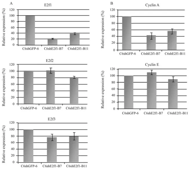
Based on these results, two clones, C6shE2f1-B7 and -B11, were chosen to test E2f1 expression by real time PCR. A comparative analysis ofE2f1expression between two C6shE2f1-B clones (B7 and B11) and control clone C6shGFP6 is presented in Figure 2a. The data show that E2f1 was knocked down by as much as 77% in C6shE2f1-B clones. The data are in accordance with inactivation by RNA interference (RNAi) of other genes, as described in the literature (Shi, 2003). Therefore, knockdown E2f1 ex-pression significantly impairs cell proliferation. These re-sults are in disagreement with those of Humbert et al. (2000) and Wuet al.(2001), who suggest that E2f1 does not play a key role in cell proliferation, since cell division in the E2F1 knockout mouse is maintained. However, the cells used in their experiments were not malignant, as is C6, and so there was no exacerbated proliferation or the accu-mulation of genetic alteration.
We showed that knockdown of E2F1 by RNAi is a promising approach to impair unwanted cell proliferation, but we have not yet explored the impact of reduced E2F1 on apoptosis. In E2f1 knockout mice, thymocytes revealed low levels of apoptosis and the animals had a high fre-quency of spontaneous tumor formation from different tis-sues (Fieldet al., 1996; Yamasakiet al., 1996). If designing a treatment strategy based on the induction of apoptosis, then shE2f1 may not be an appropriate option. Neverthe-less, E2F1 is not the only factor involved in controlling the apoptosis pathway, since tumors can undergo apoptosis in the absence of its expression (Baudinoet al., 2003). There-fore, the choice of treatment could depend on the back-ground of each tumor.
The E2F family includes 8 genes, most of which are involved in cell-cycle regulation. Only E2F1 to E2F3a are known to exert overlapping functions on inducing cell pro-liferation (Blais and Dynlacht, 2004). To test the influence
that the lack of E2f1 may exert over theE2f2,E2f3, genes, their expression levels were also assessed (Figure 2a). Their expression was not affected in C6shE2f1-B clones, when compared to control cells. These results suggest that: i) shE2f1-B does not disrupt E2f2 and E2f3 expression, thereby proving its specificity; ii) E2f1 is not responsible for controlling E2f2 and E3f3 expression; iii) E2f2 and E2f3 do not compensate for the absence of E2f1 in cell liferation, thus demonstrating that E2f1 was the major pro-mitotic effector under the present experimental conditions. Our data are in agreement with a recent study on HeLa cells, where the authors inhibited E2F1 by siRNA, and ob-served no effect on E2F2 expression (Gotoet al., 2006), and are also consistent with a study on double-knockout cells, where the authors found that E2F3 protein levels were unaffected by loss of E2F1/E2F2 (Liet al., 2003). How-ever, our findings are in contrast to those of Konget al. (2007), as these authors conclude that the long-term loss of
E2F activity leads to compensation by other family mem-bers. Nevertheless, our data were obtained from established clones and no compensatory effects were observed. A pos-sible explanation for these different observations may be due to the different cell types that were utilized. Knock-down of E2F1 in cells with normal E2F1 expression may release a compensatory response in E2F2 and E2F3 expres-sion, whereas in cancer cells, which usually over-express E2F1, knock down of this gene may not have a compensa-tory effect by other members of the family, thus being suffi-cient to impair proliferation.
Genes controlled by E2F1 have been described in a few studies where E2F1 was over-expressed. Cyclin A and Cyclin E were found to be over-expressed in response to E2F1, thereby demonstrating a direct correlationship be-tween E2F1 and its targets (Ohtaniet al., 1995; Inoshitaet al., 1999, Takahashiet al., 2000). The expression of these two genes was also assessed in a double-knockout model for E2f1/E2f2, where Cyclin A was down-regulated and Cyclin E was not significantly influenced (Liet al., 2003). In contrast to these findings, Gotoet al.(2006) revealed a different view by demonstrating that the lack of E2F1 does not negatively influence Cyclin A and Cyclin E. Because of the controversial function of E2F1 in controlling expres-sion of genes involved in cell-cycle progresexpres-sion, we also analyzed Cyclin A and Cyclin E gene expression in C6shE2f1-B cells by real time PCR (Figure 2b). In accor-dance to E2F1 overexpression studies, Cyclin A was down-regulated in our cells when compared to controls, thus accompanyingE2f1 knockdown. However, this was not the case forCyclin E, where expression was not signifi-cantly changed, when compared to control cells. This sug-gests that E2f1 does not controlCyclin Eexpression in C6 cells. However, continued expression of Cyclin E does not compromise the use of shE2F1-B in diminishing cell prolif-eration.
In conclusion, specific inactivation of E2f1 was suffi-cient to impair cell proliferation by 70%, and RNAi meth-odology seems to be an effective tool for targeting unwanted cell proliferation. Further investigation of these shRNAs in other cell-lines may provide additional infor-mation about this tool. Nevertheless, we have shown that shE2F1-B was capable of reducing the expression of E2f1, as well as impeding cell proliferation. With further devel-opment, shRNA against E2f1 may prove to be an interest-ing strategy in the treatment of proliferative diseases, such as cancer and other physiopathological conditions, includ-ing neointimal hyperplasia associated to cardiovascular de-rangements.
Acknowledgments
The authors thank Dr. David Baltimore (California Institute of Technology, CA- USA) for the pBS/hU6-1 plasmid, Dr. Carlos F. Menck and Dr. Eloísa S. Moreira for helpful comments, and Marcilene Floriano and Daniela
Jardim for expert technical assistance. This work was sup-ported by the Fundação de Amparo à Pesquisa do Estado de São Paulo (FAPESP, Brazil), and Conselho Nacional de Desenvolvimento Científico e Tecnológico (CNPq, Bra-zil). Dr. Luciana Vasques was a recipient of a Post-Doctoral Fellowship from FAPESP.
References
Bates S, Phillips AC, Clark PA, Stott F, Peters G, Ludwig RL and Vousden KH (1998) p14ARF links the tumour suppressors RB and p53. Nature. 395:124-1255.
Baudino T, Maclean KH, Brennan J, Parganas E, Yang C, Asla-nian A, Lees JA, Sherr CJ, Roussel MF and Cleveland JL (2003) Myc-mediated proliferation and lymphomagenesis, but not apoptosis, are compromised by E2f1 loss. Mol Cell 11:905-914.
Blais A and Dynlacht BD (2004) Hitting their targets: An emerg-ing picture of E2F and cell cycle control. Curr Opin Genet Dev 14:527-532.
DeGregori J, Kowalik T and Nevins JR (1995) Cellular targets for activation by the E2F1 transcription factor include DNA synthesis- and G1/S-regulatory genes. Mol Cell Biol 15:4215-4224. Erratum in: Mol Cell Biol 15:5846-5847. DeGregori J (2002) The genetics of the E2F family of
transcrip-tion factors: Shared functranscrip-tions and unique roles. Biochim Biophys Acta 1602:131-50.
DeGregori J and Johnson DG (2006) Distinct and overlapping roles for E2F family members in transcription, proliferation and apoptosis. Curr Mol Med 6:739-748.
Dyson N (1998) The regulation of E2F by pRB-family protein. Genes Dev 12:2245-2262.
Field SJ, Tsai FY, Kuo F, Zubiaga AM, Kaelin Jr WG, Livingston DM, Orkin SH and Greenberg ME (1996) E2F-1 functions in mice to promote apoptosis and suppress proliferation. Cell 85:549-561.
Frolov MV and Dyson NJ (2004) Molecular mechanisms of E2F-dependent activation and pRB-mediated repression. J Cell Sci 117:2173-2181.
Goto Y, Hayashi R, Kang D and Yoshida K (2006) Acute loss of transcription factor E2F1 induces mitochondrial biogenesis in HeLa cells. J Cell Physiol 209:923-934.
Halaban R (2005) Rb/E2F: A two-edged sword in the melanocytic system. Cancer Metastasis Rev 24:339-356.
Helin K, Lees JA, Vidal M, Dyson N, Harlow E and Fattaey A (1992) A cDNA encoding a pRB-binding protein with prop-erties of the transcription factor E2F. Cell 70:337-350. Humbert PO, Verona R, Trimarchi JM, Rogers C, Dandapani S
and Lees JA (2000) E2f3 is critical for normal cellular pro-liferation. Genes Dev 14:690-703.
Inoshita S, Terada Y, Nakashima O, Kuwahara M, Sasaki S and Marumo F (1999) Roles of E2F1 in mesangial cell prolifera-tionin vitro. Kidney Int 56:2085-2095.
Irwin M, Marin MC, Phillips AC, Seelan RS, Smith DI, Liu W, Flores ER, Tsai KY, Jacks T, Vousden KH,et al.(2000) Role for the p53 homologue p73 in E2F-1-induced apoptosis. Nature 407:645-648.
retino-blastoma-binding protein with E2F-like properties. Cell 70:351-364.
Kong LJ, Chang JT, Bild AH and Nevins JR (2007) Compensa-tion and specificity of funcCompensa-tion within the E2F family. Onco-gene 26:321-327.
Li FX, Zhu JW, Hogan CJ and DeGregori J (2003) Defective gene expression, S phase progression, and maturation during hematopoiesis in E2F1/E2F2 mutant mice. Mol Cell Biol 23:3607-3622.
Lissy NA, Davis PK, Irwin M, Kaelin Jr WG and Dowdy SF (2000) A common E2F-1 and p73 pathway mediates cell death induced by TCR activation. Nature 407:642-645. Moroni MC, Hickman ES, Lazzerini Denchi E, Caprara G, Colli
E, Cecconi F, Muller H and Helin K (2001) Apaf-1 is a transcriptional target for E2F and p53. Nat Cell Biol 3:552-558.
Mousses S, Caplen NJ, Cornelison R, Weaver D, Basik M, Hau-taniemi S, Elkahloun AG, Lotufo RA, Choudary A, Dou-gherty ER,et al.O (2003) RNAi microarray analysis in cul-tured mammalian cells. Genome Res 13:2341-2347. Ohtani K, DeGregori J and Nevins JR (1995) Regulation of the
cyclin E gene by transcription factor E2F1. Proc Natl Acad Sci USA 92:12146-12150.
Pfaffl MW (2001) A new mathematical model for relative quanti-fication in real-time RT-PCR. Nucleic Acids Res 29:2002-2007.
Pierce AM, Schneider-Broussard R, Gimenez-Conti IB, Russell JL, Conti CJ and Johnson DG (1999) E2F1 has both onco-genic and tumor-suppressive properties in a transonco-genic model. Mol Cell Biol 19:6408-6414.
Polyak K, Kato JY, Solomon MJ, Sherr CJ, Massaque J, Roberts JM and Koff A (1994) p27kip1, a cyclin-Cdk inhibitor, links transforming growth factor-beta and contact inhibition to cell cycle arrest. J Gene Dev 8:9-22.
Qin XF, An DS, Chen IS and Baltimore D (2003) Inhibiting HIV-1 infection in human T cells by lentiviral-mediated
de-livery of small interfering RNA against CCR5. Proc Natl Acad Sci USA 100:183-188.
Shan B, Zhu X, Chen PL, Durfee T, Yang Y, Sharp D and Lee WH (1992) Molecular cloning of cellular genes encoding retino-blastoma-associated proteins: Identification of a gene with properties of the transcription factor E2F. Mol Cell Biol 12:5620-5631.
Sherr CJ (1996) Cancer cell cycles. Science 274:1672-1677. Shi Y (2003) Mammalian RNAi for the masses. Trends Genet
19:9-12.
Takahashi Y, Rayman JB and Dynlacht BD (2000) Analysis of promoter binding by the E2F and pRB familiesin vivo: Dis-tinct E2F proteins mediate activation and repression. Genes Dev 14:804-816.
Tiscornia G, Singer O, Ikawa M and Verma IM (2003) A general method for gene knockdown in mice by using lentiviral vec-tors expressing small interfering RNA. Proc Natl Acad Sci USA 100:1844-1848.
Tsantoulis PK and Gorgoulis VG (2005) Involvement of E2F transcription factor family in cancer. Eur J Cancer 41:2403-2414.
Wells J, Yan PS, Cechvala M, Huang T and Farnham PJ (2003) Identification of novel pRb binding sites using CpG micro-arrays suggests that E2F recruits pRb to specific genomic sites during S phase. Oncogene 22:1445-1460.
Wu L, Timmers C, Maiti B, Saavedra HI, Sang L, Chong GT, Nuckolls F, Giangrande P, Wright FA, Field SJ,et al.(2001) The E2F1-3 transcription factors are essential for cellular proliferation. Nature 414:457-462.
Yamasaki L, Jacks T, Bronson R, Goillot E, Harlow E and Dyson NJ (1996) Tumor induction and tissue atrophy in mice lack-ing E2F-1. Cell 85:537-548.
Associate Editor: Carlos F.M. Menck