Fábio David Ferreira Rodrigues
Licenciado em Biologia Celular e Molecular
Functional genomics of
Rhipicephalus
bursa – Babesia ovis
interactions
towards disease control
Dissertação para obtenção do Grau de Mestre em Genética Molecular e Biomedicina
Orientador: Ana Isabel Amaro Gonçalves Domingos,
Investigadora Doutora, IHMT-UNL
Co-orientador: Sandra Isabel da Conceição Antunes,
Investigadora Pós-Doc, IHMT-UNL
Júri:
Presidente: Doutora Ilda Maria Barros dos Santos Gomes Sanches Arguente: Doutor Jacinto José Carneiro Gomes
Vogal: Doutora Sandra Isabel da Conceição Antunes
Functional genomics of Rhipicephalus bursa – Babesia ovis interactions towards disease control
Copyright Fábio David Ferreira Rodrigues, FCT/UNL, UNL
ii seria viável, porém, não posso deixar de demonstrar o meu profundo agradecimento para com algumas
pessoas que fazem e fizeram parte da minha vida e, portanto, gostaria de aproveitar esta oportunidade
para fazê-lo.
Às minhas orientadoras, Doutora Sandra Antunes e Investigadora Doutora Ana Domingos, por
poder sempre contar com o seu entusiasmo e alegria, bem como com a sua paciência e a sua palavra
amiga, de incentivo e tranquilidade, nos momentos em que as coisas corriam menos bem. O apoio e a
disponibilidade manifestados contribuíram de uma forma decisiva para que a conclusão deste trabalho
tenha sido possível. Agradeço, ainda, pela revisão científica deste manuscrito e pela enorme
disponibilidade com que sempre o fizeram.
Às minhas colegas e amigas Joana Couto, Joana Ferrolho, Samira d’Almeida, Catarina Rosa e Sara Pardal, por proporcionarem bons momentos, por me terem recebido tão bem no grupo, pelos
conselhos e pelos debates, e pela forma como sempre mostrou disponibilidade para ajudar em tarefas
no laboratório, o meu sincero obrigado Joana Couto.
Aos professores que me acompanharam ao longo deste percurso académico, sendo que alguns
deles vão ficar na minha memória pela forma como conseguiam transmitir o seu conhecimento.
O meu reconhecimento sincero aos funcionários do Instituto de Higiene e Medicina Tropical e
do Instituto Nacional de Investigação Agrária e Veterinária de Santarém, que colaboraram de forma
exemplar e com grande disponibilidade, tornando possível este estudo.
Aos colegas da Licenciatura em Biologia Celular e Molecular 2011/2014 e do Mestrado em
Genética Molecular e Biomedicina 20014/2016, com quem vivi um ambiente de aprendizagem
colaborativa e que com alguns dos quais partilhei momentos inesquecíveis e fiz boas amizades.
Aos meus amigos, alguns deles ouvintes de algumas dúvidas, inquietações, desânimos e
sucessos, pelo apoio e conforto, dando‐me, desta forma, coragem para ultrapassar os momentos
“menos bons” desta jornada.
À minha família em geral, uma palavra de agradecimento pela confiança que sempre
depositaram em mim, transmitindo-me sempre energias positivas e motivação para alcançar os meus
objetivos, sempre com um sorriso.
Uma chamada de reconhecimento muito especial para os meus pais, David e Luísa, por me
possibilitarem financeiramente este percurso académico e sempre me incentivarem perante os desafios
que a vida proporciona, a fazer mais e melhor. Portanto, quero partilhar convosco a alegria de
conseguir vencê-los continuamente!
iv Fábio David Ferreira Rodrigues
Palavras-chave: Babesia, Carraça, Vacina, RNA de interferência, Lachesina, Vitelogenina.
As carraças são ectoparasitas hematófagos obrigatórios de animais selvagens e domésticos,
sendo o Homem um hospedeiro acidental. Enquanto em saúde humana são consideradas o segundo
vetor mais importante de doenças, a seguir aos mosquitos, em saúde animal são os principais vetores.
Os protozoários do género Babesia, responsáveis pela doença denominada babesiose, são agentes patogénicos que afetam uma ampla variedade de animais, incluindo o Homem. Apesar de a babesiose
humana ser uma zoonose emergente, é na área animal que a babesiose se destaca, pois tem um grande
impacto na produção animal. Nomeadamente a espécie Babesia ovis, transmitida principalmente por
Rhipicephalus bursa, é altamente patogénica, particularmente em ovinos, podendo provocar febre, anemia, aborto, hemoglubinúria e até levar à morte. Atualmente, o principal método de controlo de
carraças e agentes a estas associados baseia-se no uso de acaricidas, mas a vacinação é um método
alternativo. O desenvolvimento de vacinas inicia-se com a identificação e caracterização de antigénios
com papel essencial no desenvolvimento da carraça. Assim, o objetivo do presente estudo foi clarificar
a função de genes de carraças diferenciadamente expressos em resposta à alimentação e infeção por B. ovis, visando o desenvolvimento de vacinas anti-carraças. Com base em estudos prévios foram identificados genes potencialmente envolvidos no processo de alimentação e infeção. Quatro destes
foram selecionados para caracterização funcional utilizando a metodologia de RNA de interferência. A
redução dos níveis de mRNA alvo nas carraças mostrou que a lachesina poderá estar envolvida no processo de infeção, uma vez que reduziu significativamente os níveis de infeção de B. ovis. Além disso, foi observado um efeito na fixação da carraça ao hospedeiro e aumento da mortalidade. O
silenciamento de vitelogenina-3 e do gene codificante para uma proteína de cimento demonstrou que ambos poderão estar associados ao processo de alimentação. Vários estudos têm caracterizado a
interface carraça-patogénio a nível molecular. Porém, este é o primeiro estudo de genómica funcional
vi
Functional genomics of Rhipicephalus bursa - Babesia ovis towards disease control.
Fábio David Ferreira Rodrigues
Keywords: Babesia, Tick, Vaccine, RNA interference, Lachesin, Vitellogenin.
Ticks are obligate hematophagous ectoparasites of wild and domestic animals, whereas is Man
an accidental host. While in human health are considered the second most important vector of
diseases, after mosquitoes, in animal health are the main vectors. Protozoans of the genus Babesia, responsible for the disease called babesiosis, are pathogens that affect a wide variety of animals,
including Man. Although human babesiosis is an emerging zoonosis, this disease has its greatest
impact on animal production. Namely Babesia ovis, mainly transmitted by Rhipicephalus bursa, is highly pathogenic, particularly in sheep, and can cause fever, anemia, abortion, hemoglobinuria and
even lead to death. Currently, the principal TTBD control method is based on the use of acaricides,
nevertheless vaccination is an alternative method. Vaccine development begins with the identification
and characterization of antigens that have an essential role in tick development. Therefore, the goal of
this study was to clarify the function of genes differentially expressed in response to blood-feeding
and infection by B. ovis, aiming the development of anti-tick vaccines. Based on previous studies, genes potentially involved in tick feeding and infection processes were identified. Four of these were
selected for functional characterization using the RNA interference methodology. Reduction of target
mRNA levels showed that lachesin may be involved in the infection process, since it significantly reduced the infection levels of B. ovis. Furthermore, it was observed an effect on tick attachment to the host and increased mortality. Silencing of vitellogenin-3 and the gene coding for a cement protein demonstrated that both may be associated to tick feeding. Several studies have characterized the
viii
μL Microliter
μM Micro molar
ºC Degree Celsius
64TRP 64 tick recombinant protein
AGE Agarose Gel Electrophoresis
AN Avogadro’s number
ATP Adenosine triphosphate
Ave Average
bp Base pair
cDNA Complementary DNA
Cq Quantification cycle
CRT Calreticulin
DDT Dichlorodiphenyltrichloroethane
DNA Deoxyribonucleic acid
dsRNA Double-stranded ribonucleic acid
EDTA Ethylene diamine tetra acetic acid
e.g. exempli gratia
ELF Elongation factor
ELI Expression library immunization
ELISA Enzyme-linked immunosorbent assay
EST Expressed sequence tag
g gram
g G-force
gDNA Genomic DNA
GRP Glycine-rich protein
ICT Immunochromatographic test
IFAT Immunoflorescence antibody test
kDa Kilo Dalton
ix
Lac Lachesin
Mg Milligram
min Minute
ml Millilitre
mm Millimetre
mM Millimolar
mRNA Messenger RNA
Mw Molecular weight
nl Nanolitre
LAMP Loop-Mediated Isothermal PCR
OTEs Off-target effects
OVs Ovaries
PBS Phosphate buffered saline
PCR Polymorphism chain reaction
qPCR Quantitative PCR
rCRT Recombinant calreticulin
rDNA Ribosomal DNA
RISC RNA-induced silencing complex
RLB Reverse line blot
RNA Ribonucleic acid
RNAi RNA interference
RNA-Seq RNA sequencing
RT-PCR Reverse transcriptase polymerase chain reaction
RTT Replication-Transcription-Translation
s Second
S.D. Standard deviation
SGs Salivary glands
siRNAs Small interfering RNAs
x
spp. Species (Plural)
s.s. sensu stricto
SSH Suppression-Subtractive Hybridization
TBD Tick borne diseases
TBE Tris/Borate/EDTA
TBEV Tick-borne encephalitis virus
TNF-α Tumor necrosis factor α
Tris-HCl Tris hydrochloride
TROSPA Tick receptor for outer surface protein A
TTBD Tick and tick borne diseases
UV Ultraviolet
V Volt
Vg Vitellogenin
VgR Vitellogenin receptor
Vn Vitellin
xii ACKNOWLEGMENTS ...I
RESUMO ...III
ABSTRACT ... V
ABBREVIATIONS ... VII
TABLE OF CONTENTS ... XI
INDEX OF FIGURES... XIV
INDEX OF TABLES ...XVII
INTRODUCTION ... 1
OVINE BABESIOSIS ... 2
1.1.1 BACKGROUND ... 2
1.1.2 HISTORICAL OVERVIEW ... 3
1.1.3 HOSTS ... 3
1.1.4 VECTORS ... 3
1.1.5 BABESIA SPP.LIFE CYCLE ... 4
1.1.5.1 Babesia spp.development in the vertebrate host ... 5
1.1.5.2 Babesia spp. development in the vector ... 5
1.1.6 PATHOGENESIS AND CLINICAL SIGNS ... 6
1.1.7 DIAGNOSIS ... 7
1.1.8 BABESIOSIS TREATMENT ... 8
1.1.9 ZOONOTIC RISK ... 9
TICK VECTOR ... 10
1.2.1 CLASSIFICATION ... 10
1.2.2 LIFE CYCLE... 11
1.2.3 TICK-HOST INTERACTIONS... 11
1.2.4 TICK ANATOMY AND PHYSIOLOGY ... 12
TICK CONTROL ... 14
1.3.1 CHEMICAL TICK CONTROL – ACARICIDES ... 14
1.3.2 ALTERNATIVE METHODS IN TICK CONTROL -VACCINES ... 15
1.3.3 IMMUNOLOGICAL TICK CONTROL ... 15
TICK AND TICK-BORNE DISEASES CONTROL ... 16
1.4.1 TICK ANTIGENS ... 16
1.4.2 FUNCTIONAL GENOMICS IN TICKS ... 18
AIMS OF THIS MASTER PROJECT... 19
2 MATERIALS AND METHODS ... 20
LAMB INFECTION WITH BABESIA OVIS ... 21
GENOMIC DNA EXTRACTION FROM LAMB BLOOD ... 21
DETECTION OF BABESIA OVIS IN BLOOD BY PCR... 21
AGAROSE GEL ELECTROPHORESIS ... 21
IDENTIFICATION OF DIFFERENTIALLY EXPRESSED GENES IN R. BURSA TICKS IN RESPONSE TO B. OVIS INFECTION... 22
2.5.1 SELECTION OF GENES ... 22
IN VIVO GENE SILENCING IN TICKS BY RNA INTERFERENCE ... 23
2.6.1 DSRNA SYNTHESIS ... 23
2.6.2 RHIPICEPHALUS BURSA COLONY ... 24
2.6.3 DSRNA INJECTION IN TICKS... 24
xiii
TICK DISSECTION ... 25
DNA AND TOTAL RNA EXTRACTION FROM TICK SGS AND OVS ... 25
GENE KNOCKDOWN ASSESSMENT BY QPCR ... 26
2.9.1 DETERMINATION OF TICK MRNA LEVELS BY QPCR ... 26
2.9.2 DETERMINATION OF BABESIA OVIS INFECTION BY QPCR ... 27
3 RESULTS ... 28
MONITORIZATION OF LAMB INFECTION ... 29
SEQUENCE ANALYSIS OF TICK GENES DIFFERENTIALLY EXPRESSED IN RESPONSE TO B. OVIS INFECTION 29 REFERENCE GENES SELECTION ... 29
EFFICIENCY OF GENE SILENCING IN R. BURSASGS AND OVS ... 30
FUNCTIONAL ANALYSIS OF TICK GENES DIFFERENTIALLY EXPRESSED IN RESPONSE TO B. OVIS INFECTION... 30
3.5.1 ANALYSIS OF TICK PHENOTYPE AFTER RNAI... 30
3.5.2 ANALYSIS OF BABESIA OVIS INFECTION LEVELS AFTER RNAI ... 31
4 DISCUSSION AND CONCLUSIONS ... 33
5 REFERENCES ... 39
6 APPENDIX ... 61
APPENDIX I ... 62
xv
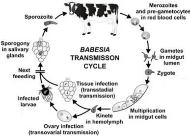
Figure 1.1: The transmission cycle of Babesia spp. in cattle ...5
Figure 1.2: Tick examples. ... 11
Figure 1.3: Characteristic Ixodid two-host life cycle. ... 12
Figure 1.4: Dissected SGs from Rhipicephalus annulatus. ... 13
Figure 1.5: Dissected ovaries from Rhipicephalus annulatus. ... 13
Figure 2.1: Injection of Rhipicephalus bursa female tick in the trochanter articulation. ... 25
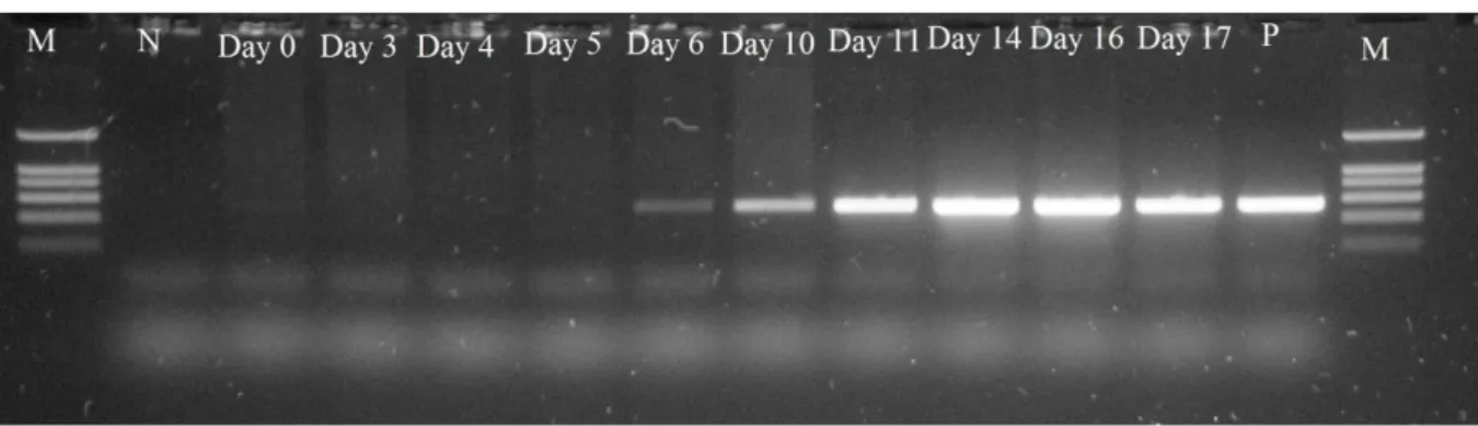
Figure 3.1: Detection of B. ovis in the lamb blood by AGE of PCR products. ... 29
Figure 3.2: Analysis of vitellogenin orthologue sequences. ... 29
Figure 3.3: Silenced and control female ticks recovered after feeding in B. ovis infected lamb. ... 31
Figure 6.1: Detection of B. ovis in the tick salivary glands by AGE of qPCR products in silenced groups st1 and mt5 ... 64
Figure 6.2: Detection of B. ovis in the tick salivary glands by AGE of qPCR products in the group Control and in the silenced groups cf2 and cf1+cf2... 64
Figure 6.3: Detection of B. ovis in the tick salivary glands by AGE of qPCR products in the group Control. ... 65
Figure 6.4: Detection of B. ovis in the tick salivary glands by AGE of qPCR products in the group Control. ... 65
Figure 6.5: Detection of B. ovis in the tick ovaries by AGE of qPCR products in the group Control and in the silenced groups cf1, cf2, st1, mt5 and cf1+cf2. ... 66
Figure 6.6: Detection of 16S tick gene in the tick ovaries by AGE of qPCR products in the group Control and in the silenced groups cf1, st1, mt5, cf2 and cf1+cf2. ... 66
Figure 6.7: Detection of 16S tick gene in the tick salivary glands by AGE of qPCR products in the group Control and in the silenced groups cf1, st1, cf2 and mt5. ... 67
Figure 6.8: Detection of 16S tick gene in the tick salivary glands by AGE of qPCR products in the silenced group cf1+cf2. ... 67
Figure 6.9: Detection of cf2 tick gene in the tick ovaries by AGE of qPCR products in the silenced group cf2. ... 68
Figure 6.10: Detection of st1, cf1 and β-tubulin tick genes in the tick ovaries by AGE of qPCR products in the silenced groups st1, cf1 and cf1+cf2. ... 68
Figure 6.11: Detection of st1, cf1 and β-tubulin tick genes in the tick ovaries by AGE of qPCR products in the silenced groups st1, cf1 and cf1+cf2. ... 69
Figure 6.12: Detection of mt5 tick gene in the tick salivary glands and ovaries by AGE of qPCR products in the group Control and in the silenced group mt5. ... 69
Figure 6.13: Detection of β-actin and cf2 tick genes in the tick ovaries by AGE of qPCR products in the silenced groups st1, cf1 and cf1+cf2. ... 70
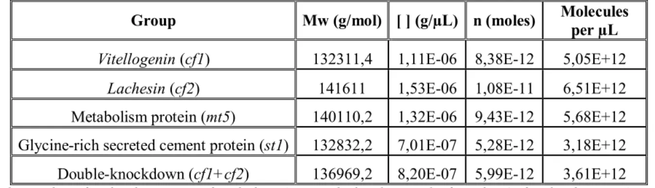
xviii Table 2.1: Number of molecules per µL of each dsRNA used to knockdown R. bursa genes in the present study. ... 24
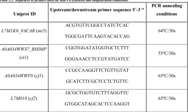
Table 2.2: Sequences of primers used for dsRNA synthesis and amplification conditions. ... 26
Table 2.3: Sequences of primers used for qPCR. ... 27
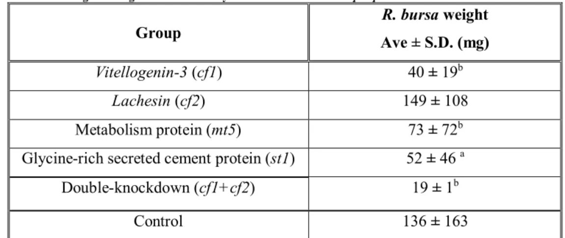
Table 3.1: Female tick weight after gene knockdown by RNA interference in Rhipicephalus bursa
ticks. ... 31
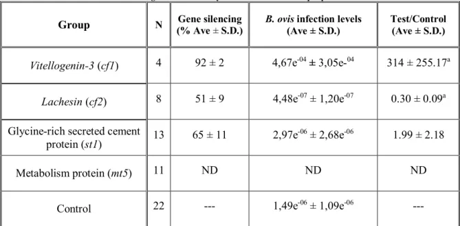
Table 3.2: Babesia ovis infection levels after gene knockdown by RNA interference in Rhipicephalus bursa ticks SGs. ... 32 Table 6.1: Sequence identity between different available vitellogenin nucleotide sequences and the obtained vitellogenin sequences. ... 62 Table 6.2: Sequence identity between different available genes encoding for secreted glycine-rich
cement proteins nucleotide sequences and the obtained sequence. ... 63
2
Ovine babesiosis
1.1.1 Background
Tick-borne protozoan parasites of the phylum Apicomplexa such as Babesia spp. are important pathogenic agents with a major impact in animal health leading to substantial financial
losses especially regarding livestock (Domingos et al., 2013; Guerrero et al., 2012a; Erster et al.,
2015b). In addition, some of these organisms can infect humans increasing the risks posed by ticks and
tick-borne diseases (TTBD). The first documented case of human babesiosis occurred in 1956 near
Zagreb, Croatia, when a splenectomized farmer was diagnosed with a B. divergens infection (Skrabalo & Deanovic, 1957) ever since, babesiosis came into view as a potentially life threatening zoonotic
infection in humans (Herwaldt et al., 2003; Homer et al., 2000; Hunfeld et al., 2008). Notably, Babesia
spp. can infect three host groups: domestic animals, humans and, most recently acknowledged, some
wildlife species make of this pathogen one of the most important protozoan transmitted by ticks
(Schnittger et al., 2012).
B. ovis is responsible for ovine babesiosis which as the name suggests is mainly associated with small ruminants. This protozoan species represents the principal ethiological agent of the disease,
although other species like B. motasi, B. crassa, B. taylori, B. foliata, among others have been described (Guan et al., 2008; Erster et al., 2015a; Ranjbar-Bahadori et al., 2012). Ovine babesiosis is
widespread in Eastern Asia, Iran, the Mediterranean basin and North Africa (Erster et al., 2015a;
Ranjbar-Bahadori et al., 2012; Rjeibi et al., 2014; Sevinc et al., 2013; Uilenberg, 2006), being
considered of great economic importance to the livestock industry, due to animal mortality, yield
losses and costs of treatment (Sevinc et al., 2015; Ranjbar-Bahadori et al., 2012). The principal vector
of B. ovis is the tick species Rhipicephalus bursa, whose distribution ranges from Asia to Africa, across the Mediterranean region (Erster et al., 2015a; Ranjbar-Bahadori et al., 2012). R. bursa is a two-host tick, feeding on a varied range of hosts, comprising hares, dogs and humans, as well as
several species of ungulates (Erster et al., 2015; Yeruham et al., 1996). Like other pathogens, B. ovis
displays transstadial and transovarial transmission, which means that the infection sustains tick
moulting and is successfully passed to progeny (Razmi & Nouroozi, 2010). Outbreaks of ovine
3
1.1.2 Historical overview
It was at the end of the 19th century that Babes discovered microorganisms in erythrocytes of
cattle in Rumania and associated them with bovine hemoglobinuria or red water fever (Babes, 1888).
Afterwards, the same author found similar organisms in sheep red blood cells. This seems to have
been the first report of the transmission of a protozoan parasite by an arthropod. In 1893, Starcovici
named these parasites as Babesia bovis, Babesia ovis and Babesia bigemina (Starcovici, 1893).
1.1.3 Hosts
As other protozoa pathogens, also Babesia spp. needs to interact with two different hosts to complete its life cycle: a vertebrate host and an arthropod, namely a tick. Initially, Babesia spp. was considered to be specific to a given vertebrate host but with the subsequent development of molecular
tools, some Babesia species have been shown to have a wider vertebrate host range than thought before. B. bovis and B. bigemina, primarily described as pathogens of cattle in tropical and sub-tropical areas, were both identified by specific serology and PCR in white-tailed deer (Odocoileus virginianus) in northern Mexico (Cantu et al., 2007; Chauvin et al., 2009; Mosqueda et al., 2012). B. divergens represents another parasite of cattle in temperate climates. Yet, it is capable to infect humans with a special impact in immunocompromised individuals (Cantu et al., 2007), primates
(chimpanzees and rhesus monkeys) (Garnham & Bray, 1959), ungulates (roe deer, fallow deer, red
deer, mouflon and sheep) (Penzhorn, 2006), and rodents (rat) (Ben Musa & Phillips, 1991) as well as
reindeer (Zintl et al., 2011), sheep (Malandrin et al., 2009) and gerbils (Lewis & Williams, 1979).
1.1.4 Vectors
The main experimental and biological vector of babesiosis in sheep is the tick Rhipicephalus bursa Canestrini and Fanzago, 1877 (Erster et al., 2015a; Erster et al., 2015b; Ferrolho et al., 2016).
R. bursa is a common ectoparasite of sheep and goat, although has also been documented in equines, cattle, dogs, gazelles and hares (Yeruham et al., 2000) and is widespread in the north
hemisphere, being particularly frequent in Mediterranean basin and central-western Asia (Erster et al.,
2015a; Ferrolho et al., 2016; Rjeibi et al., 2014; Sevinc et al., 2013; Uilenberg, 2006). This tick is also
known to act as vector of B. bigemina and B. bovis, the agents of bovine babesiosis, Theileria ovis, T. equi and T. annulata, etiological agents of theileriosis, Anaplasma marginale and A. ovis, agents of anaplasmosis, and Ehrlichia canis, responsible for canine monocytic ehrlichiosis (de la Fuente et al., 2008; Uilenberg, 2006; Dahmani et al., 2016; Ferrolho et al., 2016; Masala et al., 2012). Other
diseases have been associated with this tick, for example, Crimean Congo haemorrhagic fever virus
(Gargili et al., 2011; Papadopoulos & Koptopoulos, 1980).
4 but is mostly associated to the transmission of B. ovis. Like in all Babesial infections, once infected with B. ovis, R. bursa ticks remain infected during the course of their life cycle and transmit the parasite to the progeny, resulting in the emergence of infected larvae, nymphs and adults capable of
infect susceptible hosts during feeding. Besides this transstadial transmission, Babesia spp. also presents transovarial transmission (Erster et al., 2015a; Razmi & Nouroozi, 2010). Transovarial
transmission is considered a Babesia spp. adaptation for long-lasting persistence by the fact that some ticks remain infected and infective for many generations without needing to feed on infected animals
again, thus increasing transmission efficiency (Chauvin et al., 2009). Babesia spp. are usually divided into large and small forms such as B. ovis and B. microti, respectively. The large Babesia, also named as Babesia sensu stricto (s.s.), differ from small Babesia by their susceptibility to anti-Babesia drugs (Gray & Pudney, 1999) and by their life cycles, principally the occurrence of transovarial transmission
(Hunfeld et al., 2008; Uilenberg, 2006).
Using light microscopy, Weber and Friedhoff (1971) showed the development of B. ovis in R. bursa and could characterize the differentiated merozoites in the salivary glands (SGs) of female ticks (Weber & Friedhoff, 1971). Later, a study by Moltmann et al. (1982) using electron microscopy has
determined the development of B. ovis in the SGs of R. bursa. R. sanguineus and R. turanicus were reported that they could act as a vector of B. ovis as well (Moltmann et al., 1982; Razmi et al., 2002; Shayan et al., 2007). A study in Iran demonstrates that B. ovis DNA was found not just in R. bursa
ticks but also in R. sanguineus and R. turanicus (Shayan et al., 2007). Hyalomma marginatum is also known to act like a vector of B. ovis in cattle (Razmi & Nouroozi, 2010; Razmi et al., 2002; Taylor, 2015).
1.1.5 Babesia spp.life cycle
5 Figure 1.1: The transmission cycle of Babesia spp. in cattle (adapted from Hajdušek et al., 2013).
1.1.5.1 Babesia spp.development in the vertebrate host
Babesia spp. that belong to sensu stricto groups invade the host by injection of sporozoites with the saliva of the infected tick larvae, nymph or adult (Mehlhorn & Piekarski, 2002). Once in
circulation sporozoite penetrate erythrocyte’s cell membranes with the aid of a specialized apical complex forming a parasitophorous vacuole (Moltmann et al., 1982; Suarez & Noh, 2011; Yokoyama
et al., 2006). The parasite is left with the defining piroplasm feature of a single membrane by the
gradual disintegration of the vacuole membrane, contrasting to Plasmodium species that invade by a similar mode but retain the host membrane in addition to its own (Homer et al., 2000). Sporozoite
transforms into trophozoite by binary fission, from which two merozoites develop by merogony.
These last structures lyse the cell and continue to infect more erythrocytes. Rapid reproduction
destroys the host cell and results in hemoglobinuria in the host. Different divisional stages can be
observed in the bloodstream at the same time due to the asynchronous multiplication of the parasite
(Chauvin et al., 2009). Some trophozoites develop into a diploid ovoid type of merozoite, named
gamont precursor, which do not develop further until they are taken up by the tick in the blood meal
later on, when in the tick gut, even before to leaving the erythrocytes, these precursors develop into
gametocytes (Chauvin et al., 2009; Hildebrandt et al., 2013; Homer et al., 2000; Hunfeld et al., 2008;
Mackenstedt et al., 1995).
1.1.5.2 Babesia spp. development in the vector
Once in the arthropod vector, most of the parasites degenerate and are destroyed when
Babesia-infected erythrocytes are ingested by ticks. Nevertheless, in the passage from host blood to the midgut of the tick there are environmental changes that stimulate the development of “pre
6 1995; Schnittger et al., 2012). The ray bodies go through further multiplication inside the infected
erythrocyte, leading to the formation of large aggregations of multinucleated ray bodies. Once
gametogenesis is accomplished and after digestion of the consumed erythrocyte, single-nucleated and
haploid gametes emerge from the aggregates (Mackenstedt et al., 1995). The elongated zygote is
formed by the fusion of gametes in the lumen of tick’s digestive tract. When the zygote reaches the midgut cell membrane invaginates at the point of contact, apparently due to the action of enzymes
released by the invading parasite and no parasitophorous membrane is formed (Chauvin et al., 2009).
At some moment, the zygote undergoes one-step meiosis to form a haploid zygote (Mackenstedt et al.,
1995) and motile kinetes are formed (primary schizogony). These organisms escape the midgut
epithelium into the haemolymph and infect a variety of cell types and tissues, including ovaries (OVs)
where successive cycles of secondary schizogony occur (Chauvin et al., 2009; Homer et al., 2000;
Mackenstedt et al., 1995). Hence, transovarial transmission succeeds with further development taking
place in tick larvae. Sporogony occurs at each tick stage and the Babesia spp. infection acquired during one life stage is passed on to the next (transtadial transmission). Kinetes are transformed into
multinucleated stages in the SGs and break up to form sporozoites (Mackenstedt et al., 1995). A recent
study showed an inefficient transmission of the parasite by immature tick stages that indicates that the
transmission of B. ovis by R. bursa occurs mainly by the adult stage (Erster et al., 2015a), which is in agreement with previous reports on the seasonality of ovine babesiosis, describing that the outbreak of
the disease corresponds to the emergence of adult R. bursa (Erster et al., 2015a; Yeruham et al., 1998a).
1.1.6 Pathogenesis and clinical signs
After Babesia spp. transmission to the vertebrate host, the pathogenesis of babesiosis consists of an incubation period of between 1 week and 6 weeks followed sequentially by acute, subclinical,
and in some cases chronic phases (Conrad et al., 1991; Figueroa et al., 1992; Homer et al., 2000).
A few studies suggest that the pathogenesis is possibly related to an excessive immunological
reaction of the vertebrate host to the Babesia agent (Hemmer et al., 2000; Hunfeld et al., 2008; Telford III & Maguire, 2006). Studies in a mouse model of B. microti infection demonstrated that T-cell receptor-deficient mice are easily infected in comparing with B-cell receptor-deficient mice (Hunfeld
et al., 2008; Telford III & Maguire, 2006). Immunological studies on mice also reveal an important
role of CD4+ T cells in controlling parasitemia (Hemmer et al., 2000; Hunfeld et al., 2008). These data
are in agreement with the known difficulties of depressed cellular immunity individuals to control
persistent parasitemia (Telford III & Maguire, 2006; Haselbarth et al., 2007; Hunfeld et al., 2008;
Hildebrandt et al., 2008). In the same way, reduction of host macrophages and natural killer cells
increases susceptibility to infection (Hunfeld et al., 2008). A devastating production of
7 human cases, symptoms occur at parasitemias of less than 1% and experiments on several Babesia
spp. suggest that an excessive host immune response is an important pathogenetic cofactor for severe
babesiosis (Hunfeld et al., 2008; Gray & Weiss, 2008).The acute phase generally runs a course of one
week, in which mild and non-specific signs are described, such as fever, loss of appetite, tachycardia,
dyspnea, icterus, hemoglobinuria and hemolytic anemia, with lymphadenopathy, splenomegaly and
hemorrhagic tendencies in worst cases, which eventually might lead to death (Conrad et al., 1991;
Yeruham et al., 1998a; Yeruham et al., 1998b). Mortality rates in susceptible hosts range from 30% to
50% after field infections with B. ovis (Aktas et al., 2005; Hashemi-Fesharki, 1997). Whereas B. ovis
infections of young animals are not usually followed by clinical signs, primary exposure of adult sheep
and goats to this parasite may lead to clinical symptoms of the disease (Yeruham et al., 1998b; Carletti
et al., 2015; Suarez & Noh, 2011). The pathogenicity of B. ovis strains are directly related to erythrocyte destruction. In the case of strains of B. bovis, the ethiological agent of bovine babesiosis, hemolysis involves the release of many pharmacologically active agents like proteolytic enzymes,
which affect microcirculation by vasodilatation and increased permeability, leading to hypotension
and edema, and affect blood viscosity, coagulation and cytoadherence, resulting in ischemia. Central
nervous system complications due to adhesion of parasitized erythrocytes in brain capillaries can
occur with B. bovis infections (Mosqueda et al., 2012; Seifert, 1996). Afterwards, babesial infections may continue after spontaneous clinical recovery or ineffective treatment, and such animals may enter
the subclinical phase of babesiosis with no clinical signs. Consequently, clinically healthy sheep in the
subclinical phase of babesiosis are carriers of the parasite for years without developing clinical
disease, during which time tick vectors could still acquire and spread the pathogen to other hosts
(Buling et al., 2007; Conrad et al., 1991; Homer et al., 2000). For an unknown reason, certain animals
will progress to the chronic phase of babesiosis, which can be absent of clinical signs for years due to
complete cure or more often associated with the persistence of small numbers of parasites, being
consequently considered natural reservoirs of Babesia spp. (Homer et al., 2000). Chronically infected animals maintain elevated antibody titers, and some can develop signs of other chronic diseases, such
as liver disease (Conrad et al., 1991; Homer et al., 2000).
Babesiosis is a multisystemic disease and several factors may contribute to its severity, such as
pathogenicity of different strains, host age, immunocompetence and co-infections with other
pathogenic agents (Homer et al., 2000; Marathe et al., 2005).
1.1.7 Diagnosis
The diagnosis of babesiosis should begin with a descriptive history, which might include
clinical manifestations, history of travel to an area where it is endemic, tick bite, or exposure to a
tick-infested area, recent blood transfusion and splenectomy (Homer et al., 2000).
Clinical cases of babesiosis can be detected by microscopy, immunological assays or using
8 be dyed by staining with Giemsa or acridine orange. Thin blood films are prepared from capillary
blood, since blood of general circulation may contain fewer parasites due to sequestration of infected
erythrocytes in capillaries of brain or other organs (Böse et al., 1995). For low levels of parasitemia,
diagnosis is carried on by thick smears of infected blood stained with Giemsa (Mosqueda et al., 2012).
The advantage of the thick smear consists in a large amount of erythrocytes analyzed in a reduced
space. Hence, the probability of finding infected cells is higher than in a thin smear. Such methods are
inexpensive and portable, nevertheless, accuracy of diagnosis depend on the skills of the microscopist
(Mosqueda et al., 2012).
Some immunological tests have been described for Babesia spp. detection, as the indirect immunofluorescence antibody test (IFAT), the enzyme-linked immunosorbent assay (ELISA) and the
immunochromatographic test (ICT), being all based on the recognition of parasite antigens by serum
antibodies in the blood of the tested animal. ELISA includes the use of recombinant antigens and
monoclonal antibodies, increasing specificity and decreasing unspecific binding and signal (Goff et
al., 2008; Mosqueda et al., 2012). The ICT is a quick diagnostic device that detects antibodies against
a specific antigen in a small amount of serum by means of specific antibody and a recombinant
antigen both imbued on a nitrocellulose membrane-based test strip (Weigl et al., 2008). Since it is very
easy to perform and read, does not require a trained technician, can be implemented in the field and is
inexpensive (Mosqueda et al., 2012). The main disadvantage of the immunological tests consists in the
relying on the presence of specific antibodies against parasites and that may take days or weeks to
develop in an infected animal (Mosqueda et al., 2012).
The molecular diagnosis methods can distinguish active infections by detection and
amplification of pathogen DNA (PCR based assays). Since the improvement of the sensitivity of PCR
based techniques, many methods for the detection and differentiation of babesiosis infections have
been described, among them nested PCR (Figueroa et al., 1993), reverse line blot (RLB) hybridization
(Schouls et al., 1999), LAMP (Loop-Mediated Isothermal PCR) (Iseki et al., 2007) and real time PCR
(Buling et al., 2007; Criado-Fornelio et al., 2009). Due to factors like costs, contaminations and
validation, none of these methods is globally used, in spite of the advantages of these techniques
concerning to sensitivity. There are some studies that used PCR to diagnose B. ovis and this technique demonstrates to be specific and sensitive in detecting the pathogen (Aktaş et al., 2005; Shahzad et al., 2013).
1.1.8 Babesiosis treatment
Due to its implications in animal production and in public health, babesiosis control is crucial
(Bock et al., 2004). Currently, due to the introduction of exotic breeds, which typically do not display
9 Chemotherapy is usually effective against ovine babesiosis and several chemical compounds
have been reported to be active against Babesia parasites (Vial & Gorenflot, 2006). An early diagnosis and the rapid administration of drugs are factors that contribute to a successful treatment. Present
treatments afford protection from disease but normally permit an appropriate level of infection (low
level parasitemias) in order to develop immunity which is important in babesiosis endemic areas. Only
a few Babesiacides are available commercially, being diminazene aceturate and imidocarb dipropionate the most used:
Diminazene aceturate – In Pakistan, chemotherapy against babesiosis was studied (Rashid et
al., 2010). Diminazene® was administered to a group of sheep at the dose of 3.5 mg/kg body weight
and showed 80% efficacy at day 10 post-medication. These results agree with the study of Baby and
his colleagues, who treated simultaneous babesiosis and anaplasmosis in goat with diminazene (Baby
et al., 2001). Equivalent results were also found by Cordoves & Polanco (1983), Simitch et al. (1956),
Aliu & Odegaard (1985), Mohamed & Yagoub (1990) and Manget (1983), who obtained an
acceptable effect of diminazene against babesiosis.
Imidocarb dipropianate –used subcutaneously at a dose of 1.2 mg/kg for treatment or at a
dose of 3 mg/kg for chemoprophylactic use will prevent babesiosis (Vial & Gorenflot, 2006). Several
studies have presented that imidocarb is retained in comestible tissues of ruminants for long periods
after treatment (McHardy et al., 1986; Mosqueda et al., 2012; Suarez & Noh, 2011). High doses of this
drug completely eliminate parasites, leaving the animals susceptible to reinfection and for this motive
reduced drug levels are sometimes designated (Bock et al., 2004; Vial & Gorenflot, 2006), particularly
in endemic areas where the development of protective immunity is desired. In other hand, the use of
reduced drug doses increases the risk of resistance acquisition against the drug by the extensive use
(Rodriguez & Trees, 1996). Rashid et al. (2010) also studied the effect of imidocarb dipropianate in
treatment of babesiosis in sheep. Imizol® was administered to a group of sheep at the dose rate of 2
mg/kg body weight. The efficacy was 60% at day 3, 90% at day 7 and 100% at day 10
post-medication. Ramin (2000)and McHardy et al. (1986)have found similar results, by recording 97.28%
and 100% imidocarb efficacy.
1.1.9 Zoonotic risk
Although recognized as an animal disease, more attention is being given to babesiosis as a
worldwide emerging zoonosis due to the increase of reports of human cases. The rodent parasite B. microti and the cattle parasite B. divergens are the most commonly implicated species in North America and Europe, respectively. Cases reported in splenectomized or otherwise
immunocompromised individuals are often fatal (Herwaldt et al., 2003, 2004).
The first human case of babesiosis was identified in 1957 near Zagreb, Croatia (Skrabalo &
Deanovic, 1957). A young farmer had been grazing cattle on tick-infested pastures and presented with
10 week of illness. Firstly, described as B. bovis, the agent most likely was B. divergens. In 1968, B. divergens was confirmed as the etiologic agent in a splenectomized person infected while vacationing in the Irish countryside (Fitzpatrick et al., 1968; Vannier et al., 2008). Primarily detected in Europe
and North America, human babesiosis is now described worldwide.
Over the past 50 years, the epidemiology of the human babesiosis has changed from a few
isolated cases to the establishment of endemic areas in southern New England, New York, and the
north central Midwest. Human babesiosis due to B. microti has been reported in Connecticut, Massachusetts, Minnesota, New Jersey, New York, Rhode Island, and Wisconsin (Esernio-Jenssen et
al., 1987; Meldrum et al., 1992; Spielman, 1988; Spielman et al., 1981; Spielman et al., 1979; Steketee
et al., 1985; Western et al., 1970; Spielman et al., 1985; Eskow et al., 1999; Herwaldt et al., 2002;
Krause et al., 1991). Moderately severe illness caused by B. duncani occurred in Washington state and California (Conrad et al., 2006; Persing et al., 1995). Cases of B. divergens-like infection have been reported from Missouri (Herwaldt et al., 1996), Kentucky (Beattie et al., 2002), and Washington state
(Herwaldt et al., 2004). In Europe, B. divergens, B. microti, and B. EU1, an etiological agent of babesiosis found in ticks from Slovenia (Duh et al., 2005), have been reported to cause babesiosis in
humans and are thought to be transmitted by Ixodes ricinus (Herwaldt et al., 2003; Hildebrandt et al., 2007). In Asia, babesiosis has been reported in Japan (B. microti-like) (Wei et al., 2001), Korea (KO1) (Kim et al., 2007), Taiwan (TW1) (Shih et al., 1997), and India (Marathe et al., 2005). Human
babesiosis also has been reported in Africa (Bush et al., 1990) and South America (Ríos et al., 2003).
The disease manifestations are similar to the other types of babesiosis (Benach & Habicht,
1981; Persing et al., 1995). The cases due to B. divergens infections seen in Europe are usually more severe than those caused by B. microti. Onset of disease symptoms usually occurs within 1 to 3 weeks of the infecting tick bite (Homer et al., 2000; Hunfeld et al., 2008; Leiby, 2006). In splenectomized
patients, illness appears suddenly, with hemoglobinuria followed by jaundice due to severe hemolysis.
In the most severe cases, patients show renal failure and pulmonary edema (Homer et al., 2000; Vial &
Gorenflot, 2006).
Tick vector
1.2.1 Classification
Ticks belong to phylum Arthropoda, subphylum Chelicerata, class Arachnida, subclass Acari,
superorder Parasitiformes, order Ixodida and superfamily Ixodoidea. There are three families of ticks,
in which Argasidae or soft ticks with 193 species, Ixodidae or hard ticks with 702 species and, with
only one species, Nuttalliellidae (Brites-Neto et al., 2015). The most remarkable difference between
the two most representative tick families is the presence of a hard sclerotized shield or scutum on the
anterior dorsal surface of hard ticks, which is absent in soft ticks. There are other dissimilarities, like
11 grooves in hard ticks, and the position of mouthparts, which are located ventrally in soft ticks and
anterior in hard ticks, making them visible from a dorsal view. Nuttalliellidae family is considered the
most ancestral lineage of ticks, sharing features characteristic of both Argasidae and Ixodidae
(Klompen et al., 2007; Mans et al., 2011). The largest family of ticks can be divided in Prostriata,
which is considered as the most basal line and can copulate either on or off the host, aggregating only
the Ixodes genus, in contrast with Metastriata that can mate only on the host (Barker & Murrell, 2008).
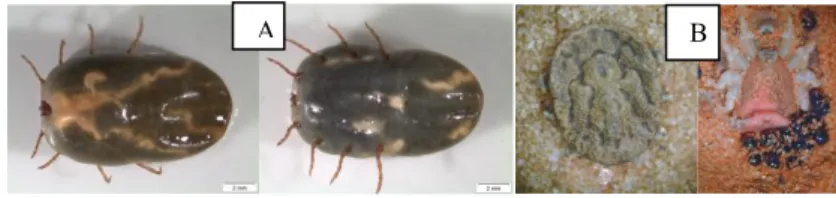
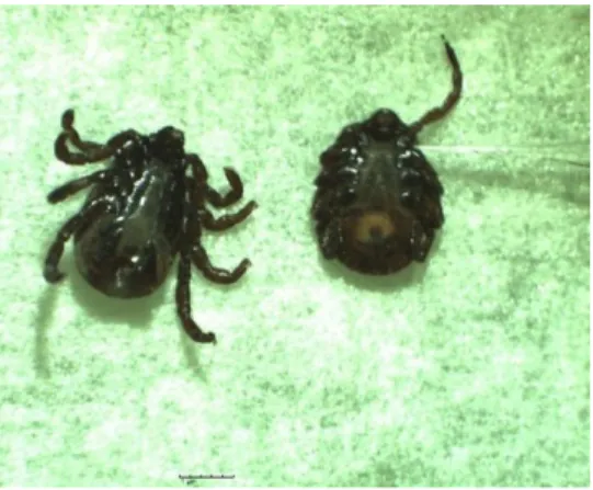
Figure 1.2: Tick examples. (A) dorsal (left) and ventral (right) view of an Rhipicephalus annulatus female, representative of a hard tick species (original and authorized from Sandra Antunes). (B) dorsal (left) and ventral (right) view of an Ornithodoros savignyi with eggs,
representative of a soft tick species (original and authorized from Ard Nijhof).
1.2.2 Life cycle
Ticks go through four stages, specifically egg, larvae, nymph and adult (Oliver, 1989;
Sonenshine & Roe, 2014). Hard ticks only have one nymph instar, differing to the several nymphal
instars of soft ticks (Oliver, 1989). Ixodid ticks require some days to feed and after the female is
engorged falls from the host to lay thousands of eggs and then dies. Argasid ticks may feed for several
times and intermittently in their lifetime and lay few hundreds of eggs in batches on different hosts
because these parasites don’t remain attached to the hosts. These last have a huge longevity living for
many years and may tolerate long periods of starvation (Sonenshine & Roe, 2014). Relatively to
Ixodid ticks, larval, nymphal and adult feeding normally requires 3-7, 4-8 and 7-9 days, respectively.
Through this time, occurs the growth of gut and cuticle in order to accommodate the blood meal,
mostly acquired in the last 24 hours of engorgement. Male hard ticks feed intermittently, since small
quantities of blood are enough to mature reproductive organs. As soon as genus Ixodes male ticks moult from the nymphal stage, they have already active reproductive organs and do not need to feed.
Resulting of many factors of nature such as photoperiods, temperature, humidity and availability of
appropriate hosts, the length of life cycles is variable. In colder regions, ticks can take until three years
to complete their life cycle, being one generation a year the usual pattern for most ticks in warmer
regions (Oliver, 1989; Sonenshine & Roe, 2014).
1.2.3 Tick-host interactions
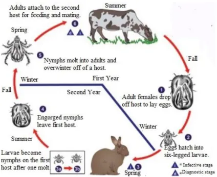
Ixodid ticks can be three-, two-, or one-host arthropods. Regarding the two-host ticks, larvae
attach to the host and when full of blood they hatch and nymphs reattach feeding again until repletion.
Nymphs drop from the host and, after some days, adults hatch and search for a new host to complete
the life cycle. Under certain conditions, ticks can use one or two hosts or use two instead of three
(Oliver, 1989), demonstrating some flexibility in feeding behavior. There are ticks that accept an
12 extensive variety of host species, other might be more selective and other attach to only one host
species.
Figure 1.3: Characteristic Ixodid two-host life cycle.(Adapted from: http://www.cdc.gov/ accessed in 20 July 2016).
1.2.4 Tick anatomy and physiology
Ticks body is externally divided in two main parts: the anterior capitulum or gnathosoma
containing the head and mouthparts and the posterior idiosoma that contains the legs, digestive tract
and reproductive organs. Whole tick body is covered by cuticle that works as an exoskeleton, like in
other arthropods. The exterior part of cuticle, termed procuticle, is sclerotized in certain parts and
forms sclerites. The biggest sclerite, scutum, covers the anterior part of the body and protects the
dorsal side of it. Cuticle major components are proteins and chitin, whereas lipids represent a minor
part (Sonenshine & Roe, 2014).
Within ticks body there are different organs surrounded by hemolymph, including the midgut,
SGs and the ovary, which are organs that can be easily detected upon dissection of engorged female
ticks.
The midgut is the most notable organ in the tick body and is divided into an anterior and a
post-ventricular region, lined by a simple pseudo-statified epithelium composed of cells with diverse
classifications and functions (Coons & Alberti, 1999). Throughout feeding, almost all body cavity of
the tick is occupied by it and his branches are for storage. Unlike in insects, the digestion in ticks is an
intracellular process, except the intraluminal digestion of erythrocytes (Coons & Alberti, 1999;
Sonenshine & Roe, 2014). At a structural level, midgut’s cells of Ixodidae ticks are complex by having different organelles and many cytoplasmic inclusions and that reflects the multifunctional
activity of the midgut (Caperucci et al., 2010).
13 the development and transmission of tick-borne pathogens, designed transstadial transmission
(Sonenshine & Roe, 2014). The pair of SGs is located in the lateral regions of the body cavity in both
Argasid and Ixodid ticks, and was described as grape-like (alveolar structures) clusters composed of
the granular and agranular acini. The saliva is drained by a system of small secondary ducts to the
main duct towards the opening in the mouthpart (Sonenshine & Roe, 2014). Previously to feeding,
SGs are crucial in water balance regulation, during attachment and feeding are responsible for cement
proteins secretion as well as other molecules transported by saliva (Sonenshine & Roe, 2014). During
feeding SGs expand several times and once females fully engorge suffer degeneration and
transformation processes that are under hormonal regulation (L’Amoreaux et al., 2003).
Female reproductive system consists of a single U-shaped ovary, which is found in the
posterior region of the body and is responsible for transovarial transmission in some pathogens. In the
unfed females the ovary is thin and small otherwise in fed females it’s a big organ with a tube-like structure of luminal epithelium and developing oocytes connected with an epithelium by a short
hollow stalk called funiculus (Sonenshine & Roe, 2014).
The sequential life cycle stages of Babesia spp. occur in different sections of ixodid ticks. So, these pathogens have to cross barriers like midgut and salivary gland epithelium, and, in ticks that
transmit these parasites transovarially, also need to cross ovary epithelium (Florin-Christensen &
Schnittger, 2009).
Figure 1.4: Dissected SGs from Rhipicephalus annulatus. (original and authorized from Sandra Antunes).
14 Ticks represents one of the most important groups of arthropod vectors of pathogens
worldwide and are considered obligate, bloodsucking, nonpermanent ectoparasitic arthropods that feed
on all animals except fish (Schwan, 2011).
Although ticks are considered zoophilic, several species can be related to the transmission of
agents to humans, making these last accidental hosts (Silva et al., 2006). In order to qualify as a
vector, a tick must feed on infectious vertebrates, acquire the pathogen during the blood meal, keep the
pathogen through one or more stages of life-cycle and has to be able to transmit the pathogen to other
unexposed hosts while feeding again (Estrada-Peña et al., 2013; Jongejan & Uilenberg, 2004).
Tick control
Tick control is fundamental to reduce impact on livestock productivity and also to contract
tick-borne diseases occurrence, including control measures predominantly based on the application of
acaricides, however, other methods such as vaccination has been applied (Willadsen, 2006).
1.3.1 Chemical tick control – acaricides
Up to now, the use of acaricides has been a main factor of an integrate tick control measures.
There are several acaricides that can be used against ticks: pyrethroids as flumethrin and deltamethrin;
organochlorines, as dichlorodiphenyltrichloroethane (DDT); organophosphates, as diazinon and
coumaphos; carbamates, as carbaril; formamidines, as amitraz; cicloamidines as, clenpirin and
macrocyclic lactones (avermectins and milbemycins), among others (George et al., 2004; Latif &
Walker, 2004). Owing to limitations like contamination problems, ineffective issues or resistance
arising, some acaricides were withdrawn from the market unless others are still accessible. Acaricides
can lead to residual effects in milk and meat products, as well as in the environment. Acaricides
application strategies are frequently hard to preserve and consequently, tend to be improperly used,
being responsible for acaricide-resistant ticks increasing (George et al., 2004; Graf et al., 2004). This
resistance is associated with mutations in genes related to drug susceptibility, like detoxificating
enzymes, like esterases, glutathione-S-transferases and mono-oxidases, and due to genetic drift
(Guerrero et al., 2012a). Combinations of acaricides have been used globally, which products combine
active components, in order to exploit a diverse number of mechanisms of action, aiming the reduction
of insecticide resistance (Veiga et al., 2012).
The public awareness of the damaging effects of pesticides on the environment and increasing
concerns about resistance of insecticides, demands the need of discovering new methodologies in tick
control. Besides, the introduction in the market of a new acaricide is time-consuming and has a
15
1.3.2 Alternative methods in tick control - Vaccines
Ticks have relatively few natural enemies, although the use of predators, parasites and
pathogens has been studied aiming tick control (Miranda-Miranda et al., 2011). Tick control strategies
could be based on interference with tick bacteria endosymbionts, which are essential to arthropod
survival (Ghosh et al., 2007). Other approach is the application of entomopathogenic fungi that have
been reported to attack and kill ticks. These organisms have been applied in field trials with moderate
success and commercial products have been developed (Samish et al., 2004; Stafford & Allan, 2010).
Other measure in controlling the tick vector is the genetic control, in which consists in the
release of sterilized ticks into the environment, identical to sterile insect techniques developed for the
control of pests. Ticks can be sterilized through hybridization (Hilburn et al., 1991), treatment with
chemicals (Hayes & Oliver, 1981) or by RNA interference (RNAi) (de la Fuente et al., 2006b; Merino
et al., 2011a).
1.3.3 Immunological tick control
Alternatives to acaricide treatments have been developed and anti-tick vaccines are among the
most significant developments. The identification of antigens capable of induce animal protection to
ticks is critical and during the decade of 1980´s several midgut protein combinations were tested until
an antigen termed Bm86, a membrane-bound glycoprotein in the cell surface, was discovered, which
conferred significant protection of cattle against R. microplus infestations (Willadsen et al., 1989). This protein of unknown function in tick biology is localized on the microvilli of the midgut digest
cells, and tick ingestion of antigen specific antibodies leads to lysis of these cells, resulting in
mortality and a deleterious effect on the reproductive performance of tick (de la Fuente et al., 1998c;
Willadsen, 2004). The discovery of this protective antigen was a revolutionary moment in the
development of anti-tick vaccines. From this breakthrough, two commercial vaccines containing the
Bm86 recombinant protein emerged in the early 1990’s, Gavac in Cuba and TickGARD in Australia (Willadsen, 2004; Ghosh et al., 2007). In spite of the effectiveness of these commercial Bm86-based
vaccines for cattle tick infestations control, they show strain-to-strain variation in efficacy being
predominantly effective against Rhipicephalus tick species (de la Fuente & Kocan, 2003; Guerrero et al., 2012b; Willadsen, 2006).
Since the commercialization of these vaccines no other has become available but research
focusing this alternative tick control method has ascended. In the last decade new molecular tools such
as next generation sequencing, proteomics or RNA interference (RNAi) have brought to light some
potential vaccine candidates allowing a rapid, systematic and comprehensive approach to tick vaccine
discovery (de la Fuente & Kocan, 2006c; Domingos et al., 2013). Nevertheless, the availability of
these techniques, the identification and characterization of effective antigens remains a noteworthy
16 This approach is based on recombinant protein as antigens to immunize animals and
demonstrate to be a good-looking alternative for the control of tick plagues, since they exhibit several
advantages, such as prevention or reduction of pathogens transmission (Almazán et al., 2005; de la
Fuente et al., 1998c; de la Fuente et al., 2007a, 2011; Merino et al., 2011b), environmental safety, low
cost production (Kiss et al., 2012), avoidance of drug-resistant selection (Parizi et al., 2012) and
inclusion of multiple antigens that are able to target many tick species (de la Fuente et al., 2000; de la
Fuente & Kocan, 2006c; Parizi et al., 2012; Willadsen, 2008; Willadsen, 2004).
A study from Rodriguez-Mallon focused on Rhipicephalus ribosomal proteins (Rodríguez-Mallon et al., 2012) identified a unique immunogenic region of protein P0. This protein seems to be
important in the assembly of 60S ribosomal subunit (Rodríguez-Mallon et al., 2012). Silencing effects
of tick protective antigens 4D8 and Rs86, homologues of Bm86, were evaluated in R. sanguineus (de la Fuente et al., 2006a). Silencing of 4D8 alone had effect on tick feeding, attachment and oviposition and silencing of Rs86 had an effect on tick weight and oviposition. Silencing of expression of both genes had substantial effect on R. sanguineus survival, attachment, feeding, weight and oviposition (de la Fuente et al., 2006a). The authors of this study suggested the development of multi-antigenic
vaccines, in order to prevent infestation from R. sanguineus (de la Fuente et al., 2006a).
Tick and tick-borne diseases control
The aim of anti-arthropod vaccines is not only the control of vector infestations but also the
agents harbored by them. The effect of such vaccines could be achieved by a) reducing vector
populations and therefore the exposure of hosts to vector-borne pathogens, b) reducing the arthropod
vector capacity for pathogen transmission, and, ideally, c) a combination of these factors (Merino et
al., 2013). As it is more and more clear that disease transmission can implicate complex interactions
between host, vector and disease organism, it is accepted that by disturbing the tick the vaccine will
also have impact on the disease (Willadsen, 2004).
1.4.1 Tick antigens
Current molecular techniques are supporting in the identification of potencial tick-protective
antigens. Bioinformatic tools and high throughput DNA sequencing technologies development enable
undertake of provisional function to expressed sequence tags (ESTs). ESTs are fragments of mRNA sequences of approximately 200-800 base pairs (bp) derived from single sequencing reactions
performed on randomly selected cDNA clones and show to be really useful for gene identification and
verification of gene predictions, since they represent the expressed portion of a genome and offer a
low-cost alternative to full genome sequencing, particularly for eukaryote organisms, whose genomes
tend to be larger and less gene-dense than prokaryotes (Parkinson & Blaxter, 2009). The first study
that reports the use of ESTs was in 1983 (Putney et al., 1983). An alternative approach for
high-17 throughput technology that uses the immune system to screen the entire genome of a pathogen, in
combination with sequence analysis of EST’s, resulting in the expressed genes without prior knowledge of the antigens encoded by the cDNAs (Almazán et al., 2003; Barry et al., 2004; Ghosh et
al., 2007). Also, suppression subtractive hybridization (SSH) is a broadly used technique for
separating DNA molecules that discriminate two closely related DNA samples of either cDNA or
genomic DNA (Diatchenko et al., 1999). Both these methods assure antigen identification without
introducing prior criteria to manage the selection of candidate genes and thereby may result in the
finding of new and unexpected antigens.
Several studies successfully identified antigens related to tick feeding and pathogen infection
using SSH technique (Antunes et al., 2012; Heekin et al., 2012, 2013; McNally et al., 2012). A study
performed by Antunes et al. (2012) applied this technique to identify R. microplus and R. annulatus
genes induced by infection with B. bigemina (e.g. TROSPA, calreticulin, serum amyloid A, subolesin). Posterior studies perfomed in vitro and in vivo supported the inclusion of TROSPA in the development of new anti-TTBD vaccine (Antunes et al., 2014, 2015; Merino et al., 2013).
Direct RNA sequencing (RNA-Seq) offers the possibility to obtain both sequence and
frequency of RNA molecules that are present at any particular time in a particular cell type, tissue or
organ. Briefly, a population of RNA is converted to a library of cDNA fragments with adaptors
attached to one or both ends. After this, fragments are sequenced in a high-throughput manner to
obtain short sequences, typically 30–400 bp. After sequencing, the resulting reads are either aligned to a reference genome or reference transcripts, or assembled de novo without the genomic sequence to produce a genome-scale transcription map that consists of both the transcriptional structure and/or
level of expression for each gene (Wang et al., 2009).
Although RNA-Seq is a technology under development, it presents several advantages over
existing technologies. First, contrasting with hybridization-based approaches like microarrays, based
on the use of probes to simultaneously analyze the expression of thousands of genes in a certain point
in time, this method is not limited to detecting transcripts that correspond to existing genomic
sequence (Nagalakshmi et al., 2008; Wang et al., 2009). For example, 454-based RNA-Seq has been
used to sequence the transcriptome of the Glanville fritillary butterfly (Melitaea cinxia) (Vera et al., 2008). For non-model organisms, whose genomic sequences that are yet to be determined, this
technique shows to be particularly attractive. RNA-Seq has very low, if any, background signal
because DNA sequences can been unambiguously mapped to unique regions of the genome, unlike
microarrays that have high background signal and cannot distinguish two closely related sequences
due to cross-hybridization of probes (Nagalakshmi et al., 2008; Wang et al., 2009). This technique can
also reveal sequence variations, as single nucleotide polymorphisms (SNPs), in the transcribed regions
(Cloonan et al., 2008). The results of RNA-Seq also demonstrate high levels of reproducibility for
both technical and biological replicates, as well as high accuracy in quantifying expression levels, as
18 (Cloonan et al., 2008; Mortazavi et al., 2008). Finally, because there are no cloning steps, RNA-Seq
requires less RNA sample. These factors make RNA-Seq useful for studying complex transcriptomes.
There are reports that use RNA-Seq in order to get new insights into the sialotranscriptome of
A. americanum tick (Karim & Ribeiro, 2015), as well for A. parvum, A. cajennense and A. triste
(Garcia et al., 2014), aiming the identification of antigens that might confer anti-tick immunity.
1.4.2 Functional genomics in ticks
RNAi or post transcriptional gene silencing is a conserved and natural process that cells use to
turn down or silence specific genes (Fire, 1999; Montgomery et al., 1998). Though RNAi mechanism
in ticks has not been fully elucidated, it has been well studied in the nematode Caenorhabditis elegans
and the fruit fly Drosophila melanogaster (Hannon, 2002; Mello & Conte, 2004). RNAi begins with the uptake of dsRNA by the cell, followed by its cleavage, which is assessed by an RNAse III called
Dicer, producing small interfering RNAs (siRNAs). The siRNAs are then incorporated into
RNA-induced silencing complex (RISC), which results in a huge sequence-specific degradation of
cytoplasmic mRNAs containing the same sequence as dsRNA trigger, leading to gene silencing (de la
Fuente et al., 2007b; Mello & Conte, 2004). This silencing signal may spread among the cells and
different tissues, generating a systemic gene silencing in the organism (Whangbo & Hunter, 2008). A
study from Kurscheid et al. (Kurscheid et al., 2009) shown that some components of RNAi machinery
in other invertebrates are also present in the ticks.
The key challenge is to find easy and trustworthy methods for delivering dsRNA. There are
three main strategies to delivery dsRNA, being microinjection the most commonly used method for
dsRNA delivery to arthropods and insects (Antunes et al., 2012). Ingestion of dsRNA through oral
feeding of diet mixed with dsRNA or transgenic plants expressing dsRNA could be performed (Baum
et al., 2007). In some cases, dsRNA could be delivered by soak organisms in dsRNA solution (Galay
et al., 2016; Whyard et al., 2009) but is mainly used for cell culture work. Prior experiments proposed
that dsRNAs could be designed as specific pesticides due to its high specificity (Baum et al., 2007;
Whyard et al., 2009).
Since the first report of RNAi application in Amblyommaamericanum (Aljamali et al., 2002), in which ticks injected with histamine binding protein (HBP) dsRNA presented a decrease in feeding
compared to control ticks, several studies have been used RNAi in ticks to evaluate tick gene function
in response to pathogen infections, aiming the development of vaccines against tick specific antigens
(Antunes et al., 2012; Galay et al., 2013, 2016; Hajdušek et al., 2016; Lu et al., 2016). TROSPA and
serum amyloid A knockdowns using RNAi reduced B. bigemina infection in R. annulatus whereas in
R. microplus, knockdown of TROSPA, serum amyloid A and calreticulin reduced pathogen infection as well, comparing with controls (Antunes et al., 2012). Recently, knockdown of subolesin, a transcription factor that regulates gene expression (Naranjo et al., 2013), by RNAi decreased the