1D e p a rtment of Pathology, State University of Health Sciences of Alagoas - UNCISAL, Maceió AL - Brazil;2D e p a rtment of Pathology,
Paulista School of Medicine of Federal University of São Paulo - UNIFESP/EPM, São Paulo SP - Brazil;3D e p a rtment of Pathology,
Federal University Fluminense - UFF, Niterói RJ - Brazil.
Received 20 February 2006, received in final form 21 June 2006. Accepted 28 July 2006.
D r. Henrique de Oliveira Costa - Departamento de Patologia / EPM/UNIFESP - Rua Botucatu 740 - 04023-900 São Paulo SP - Brasil. E-mail: lapac@uol.com.br
PRIMARY LYMPHOMA OF THE
CENTRAL NERVOUS SYSTEM
A clinical-pathological and immunohistochemical
study of ten autopsy cases
Henrique Costa
1, Marcello Franco
2, Myriam Dumas Hahn
3ABSTRACT - Context:P r i m a ry central nervous system lymphomas (PCNSL) are a rare subgroup of lymphomas generally associated with HIV and EBV. Objective:To study ten autopsy cases of PCNSL, to describe the n e u ropathological findings, to characterize the phenotype of the neoplastic cells, to detect EBV in the lesion and to compare the findings with the clinical and laboratory data of the patients. Method:T h e clinical, histological and immunohistochemical data of ten cases of PCNSL, eight cases from patients with AIDS, identified among 265 autopsies of these patients were analyzed. Results:Seven patients were males and the mean age was 40.9 years. The most frequent symptomatology was focal neurologic deficit (70%). Six patients presented with only one lesion. Histologically, densely cellular and polymorphous neoplasms with angiocentrism were observed, in 90% of cases. An association with other diseases was observed in four cases. Most patients had diffuse large B cell non-Hodgkin’s lymphoma. EBV was detected by immuno-histochemistry in only one case. The lack of detection of the virus might have been due to the long time of fixation of the brain which might have inactivate epitopes there f o re compromising the testing. Conclusion:In the present series, PCNSL presented with focal symptoms, with unifocal or multifocal lesions, with a predominant B-cell CD20 positive phenotype, rarely associated with EBV.
KEY WORDS: primary central nervous system lymphoma, neuro p a t h o l o g y, immunohistochemistry, acquire d immunodeficiency syndrome.
Linfoma primário do sistema nervoso central: estudo clínico-patológico e imuno-histoquímico de dez casos de necropsia
RESUMO -Contexto:Linfoma primário do sistema nervoso central (LP-SNC) é raro subgrupo de linfomas relacionado à AIDS, geralmente associado EBV. Objetivo:Identificar os achados clínico-patológicos dos pacientes com LP-SNC. Método:Foram analisados dados clínicos, histológicos e imuno-histoquímicos de dez necrópsias de LP-SNC, oito deles de pacientes com AIDS, identificados entre 265 autopsias destes. Resultados:Sete pacientes foram masculinos e a idade média foi 40,9 anos. A sintomatologia neurológi-ca mais freqüente era foneurológi-cal (70%). Seis exibiram lesão únineurológi-ca. Histologineurológi-camente, eram neoplasias densa-mente celulares e polimorfas, com angiocentrismo em 90% dos casos. Em quatro casos, houve associação com outras afecções. A maioria dos casos foi de linfoma não-Hodgkin difuso de grandes células B. A pesquisa para EBV foi positiva em um caso. Conclusão:P redominaram os LP-SNC associados à AIDS, com sintoma-tologia focal, lesão em massa ou multifocal, com predominância de células B CD-20.
PA L AV R A S - C H AVE: linfoma primário do sistema nervoso central, achados neuropatológicos, imuno-histo-química, síndrome da imunodeficiência adquirida.
P r i m a ry central nervous system lymphomas (PCN S L ) a re a rare group of neoplasms, accounting for about 0.3 to 1.5% of all intracranial tumors and for 0.7 to 2% of lymphomas1. These tumors are generally asso-ciated with immunosuppression due to either the use
i-culties in the classification according to the Wo r k i n g F o rmulation, the Kiel criteria and the revised Euro-pean-American Lymphoma (REAL) system. In view of their restriction to a non-lymphoid organ, among other reasons, PCNSL have been considered a sepa-rate entity whose prognosis is generally worse than that of systemic lymphomas6. With the advent of AIDS and the introduction of organ transplants, the inci-dence of PCNSL has been increasing. These tumors c u rrently re p resent an important subgroup of lym-phomas2,4. In patients with AIDS, PCNSL are the sec-ond most common lesion with a mass eff e c t4and the most frequent noninfectious cause of death7. The tumor re p resents the fourth cause of death in this g roup of patients8. PCNSL show a male/female ratio of 1.5 to 1, whereas the T subtype presents a ratio of 7 : 19. The clinical manifestations are indistinguishable f rom cerebral toxoplasmosis and include loss of con-sciousness and focal neurologic deficits1 0. In the case of meningeal involvement, the tumor manifests as a meningeal syndro m e1 1 , 1 2. Most PCNSL involve the ba-sal nuclei or the frontal and parietal lobes, and pre s-ent as homogenous mass lesions with ring-like zone of contrast enhancement and perilesional edema. The neuroimaging diff e rential diagnosis with cere-bral toxoplasmosis is difficult10. The lesions are mul-tifocal, bilateral or periventricular and may extend to the subarachnoid space or ventricular cavity1 3. The m a c roscopic findings are variable1 4. Some tumors pre-sent as well-defined masses with a homogenous cut s u rface. Other lesions are poorly demarcated, dif-fusely infiltrating, showing areas of softening, hem-o rrhage and necrhem-o s i s1 5. In immunocompetent pa-tients, PCNSL are more superficial and located in the white and/or gray matter1 6, or exclusively in the sub-arachnoid space17.
The histopathological findings of PCNSL are sim-ilar to those of systemic non-Hodgkin’s lymphomas and include densely cellular lesions comprised by atypical lymphoid cells1 3with an angiocentric pat-tern at the margins of the infiltrate and a rich retic-ulin network dissociating the vascular wall1 8. In volu-minous tumors, the areas of necrosis can be exten-sive. Mitotic activity is high and hemorrhage is com-mon. Neoplastic cells may invade normal adjacent s t ru c t u res. There f o re subependymal infiltration ex-tending into the ventricular cavity and subarachnoid space are not uncommon1 4. The oncogenesis of PCNSL is still controversial. The site of origin of these lym-phomas and the oncogenetic mechanisms are still u n c l e a r, especially because the tumor develops fro m and is restricted to a non-lymphoid organ without
lymphatic vessels19. It might occur due to the development of a monoclonal proliferation from a pre -existing inflammatory lesion, similar to the mecha-nism underlying the origin of MALT lymphoma fro m
Helicobacter pylori-induced gastritis. This hypothe-sis is supported by the demonstration of inflamma-t o ry lesions preceding inflamma-the developmeninflamma-t of PCNSL2 0 , 2 1. Other authors suggest that non-neoplastic lympho-cytes might be targeted to the CNS by a viral infec-tion, more notably HTLV-1 or EBV, with the possible later occurrence of clonal neoplastic transform a t i o n of these cells12. In the case of immunodepressed pa-tients, one may speculate that the lack of control of T cells over EBV present in patients with AIDS pro-motes the reactivation of EBV infection, leading to the proliferation of B cells, induction of oncogenes and gene re a rrangements, and finally to the devel-opment of PCNSL2 2. There f o re in patients with AIDS, infection with EBV is suggested to be the most like-ly oncogenetic mechanisms since the EBV genome is found in more than 95% of these tumors and in less of 20% of PCNSL not associated with AIDS2 3 - 2 5. Herpes v i rus genomes such as HHV-6 or HHV-8 have been identified in some cases, suggesting that these or other herpes viruses may predispose to the develop-ment of primary lymphoma, even in immunocompe-tent patients2 3. HIV-infected subjects show a 60 to 100 times higher risk of developing malignant lym-phoma, which re p resents a diagnostic criterion for AIDS5.
Based on the above observations, we studied ten autopsy cases of PCNSL to more accurately describe the neuropathological findings and to characterize the phenotype of the neoplastic cells, to test the pre s-ence of EBV and to compare the morphological find-ings with the clinical and laboratory data of the patients.
METHOD
A total of 265 autops ies of patients perf o rmed at the S e rvice of Pathological Anatomy, Department of Pathology, Antônio Pedro University Hospital, Federal University Fluminense, between 1987 and 1996 were revised and 10 cases of PCNSL were selected (3.7%).
P reparation of the material and fixation – The autop-sy was complete in all cases and the CNS was fixed in 30% f o rmalin for 21 days and then examined. The cerebral hemi-s p h e rehemi-s were hemi-studied in approximately 1.0-cm thick coro-nal sections and in 0.5- to 1.0-cm perpendicular sections cut from the cerebral trunk and cerebellar hemispheres.
I m m u n o h i s t o c h e m i s t ry–5 -µm thick CNS sections were mounted on slides previously treated with 3-aminopropy-ltriethoxysilane (Sigma, A3648). After incubation in an oven at 60ºC for about 18 h, the sections were deparaffinized in xylene and hydrated in decreasing ethanol concentrations. Next, the specimens were incubated in two baths of 3% oxygenated water in methanol for 15 min each to inhibit endogenous peroxidase and washed with Tris buff e r, pH 7.4. For antigen retrieval, the sections were immersed in citrate buff e r, pH 6.0, and heated in a microwave oven for 5 min at maximum potency. Pre s s u re chamber was used to CD3, CD20, CD15, KAPPA, LAMBDA e EBV antibodies. To inhibit nonspecific binding, the sections were incubated in a Tr i s - b u ff e red solution of 1% skim milk (w/v) and 1% bovine serum albumin (w/v) in a humid chamber for 1 h at room temperature. Next, the sections were incubated over-night in a humid chamber at 4ºC with the following pri-m a ry antibodies: rabbit anti-hupri-man CD3 polyclonal (Dako, A 0452, diluted 1/200), mouse anti-human CD20 monoclon-al (Dako, M 0755, diluted 1/250), mouse anti-human CD15 (Dako, M 733, diluted 1/150), mouse anti-human Kappa monoclonal (Sigma, K-4377, diluted 1/4000), mouse anti-human Lambda monoclonal (Sigma, L-6522, diluted 1/4000), and mouse anti-EBV monoclonal (Dako, M 0897, diluted 1/2500).
For the anti-CD30, anti-CMV and anti-GFAP antibodies, the sections were trypsinized, nonspecific binding of the antibodies was blocked and the specimens w ere incubat-ed in a humid chamber at 37ºC with the following primary antibodies: mouse anti-human CD30 monoclonal antibody (Dako, M 751, diluted 1/100), mouse anti-CMV monoclon-al antibody (Dako, M 854, diluted 1/100), and rabbit anti-GFAP polyclonal antibody (Dako, Z 0334, diluted 1/400).
Next, the sections were washed three times in Tris buff e r for 10 min each and incubated with the biotinylated sec-o n d a ry antibsec-odies: gsec-oat anti-rabbit immunsec-oglsec-obulins (Daksec-o, E 432, diluted 1/200) or rabbit anti-mouse immunoglobu-lins (Dako, E 0354, diluted 1/200) for 1 h in a humid cham-ber at 37ºC. The primary and secondary antibodies were diluted in Tr i s - b u ff e red 1% bovine serum albumin (w/v). After three washes in Tris buffer for 10 min each, the
sec-tions were incubated with the stre p t a v i d i n - b i o t i n - p e ro x i-dase complex (Dako, K 0377) for 45 min in a humid cham-ber at room temperature, washed again three times in Tr i s b u ff e r, and the peroxidase reaction was developed with diaminobenzidine (Sigma, 5637) and oxygenated water. The sections were counterstained with Mayer’s hematoxylin. The antibodies used are shown in Table 1.
RESULTS
General clinical data and laboratory exams –T h e g e n d e r, age, risk behavior, clinical data and comple-m e n t a ry exacomple-ms of the patients are sucomple-mcomple-marized in Ta-ble 2. Seven patients were males and three were females, with age ranging from 24 to 64 years (mean
±s t a n d a rddeviation: 40.9±11.8 years). Neuro l o g i c a l signs or symptoms were observed in all but one case, with a predominance of focal neurological deficit in seven patients. The time between the discovery of HIV positivity and the diagnosis of PCNSL ranged fro m 6 months to 3 years.
G ross findings –B rown-grayish, poorly demarc a t-ed lesions with areas of necrosis, comprist-ed by soft, fleshy to friable tissue, were identified in the basal nuclei (Fig 1) and thalamus of six, in the cere b r a l t runk of four, in the cerebellar hemisphere of thre e , and in the cortex and corpus callosum of two pa-tients. Multiple lesions were observed in six cases, with the number of lesions ranging from 2 to 4. In three cases, only one lesion was identified.
In one case, the gross findings diff e red from those observed in other cases. The tumor lesion was char-acterized by a whitish color and homogenous surf a-ce with a fish-flesh appearana-ce, and involved stru c-t u res of c-the midline and deep gray mac-tc-ter, exc-tend- extend-ing to the corpus callosum and adjacent white mat-ter (Fig 2). Another case was characmat-terized by the e n l a rgement of the nerve roots and cranial nerv e s , and opacity of the leptomeninges. Analysis of the Table 1. Panel of antibodies.
Primary Diluition Source Code Origin Secundary Antigen retrieval
antibody antibodies procedure
CD30 1/100 Dako M 751 Mouse R x M * Trypsin
CD15 1/150 Dako M 733 Mouse R x M * MW/PC*
CD3 1/200 Dako A 0452 Rabbit G x R*** MW/PC*
CD20 1/250 Dako M 0755 Mouse R x M * MW/PC*
Kappa 1/4000 Sigma K-4377 Mouse R x M * MW/PC*
Lambda 1/4000 Sigma L-6522 Mouse R x M * MW/PC*
EBV 1/2500 Dako M 0897 Mouse R x M * MW/PC*
CMV 1/100 Dako M 854 Mouse R x M * Trypsin
GFAP 1/400 Dako Z 0334 Rabbit G x R*** Trypsin
Table 2. Identification, clinical data, risk behavior and complementary exams of 10 autopsy cases of CNS primary lymphoma diag -nosed.
Case Age Gender Risk behavior HIV Time of Clinical data Neurological signs
(Y/O) evolution
(months)
1 64 F Hemotransfusion + 10 Loss weight, fever, Focal neurologic deficit,
N87-297 diarrhea, hemorrhage convulsion, coma
2 35 M NE + NE Loss weight, fever, No neurological signs.
N88-422 diarrhea, pulmonary
infection
3 46 M Promiscuous + 12 Loss weight, fever, Paraplegy, dysfunction
N89-009 diarrhea, pneumocystosis of sphincter
4 28 M NE + 12 Loss weight, fever, Meningeal syndrome,
N89-224 diarrhea, pneumocystosis, coma
Kaposi’s sarcoma
5 38 M NE + 8 Loss weight, fever, Focal neurologic deficit,
N91-013 diarrhea, pneumocystosis meningeal syndrome
6 42 M Hemotransfusion + 13 Loss weight, fever, Focal neurologic deficit
N91-113 Homosexualism diarrhea, pulmonary
infection, Kaposi’s sarcom
7 24 M Hemotransfusion + 24 Loss weight, fever, Focal neurologic deficit,
N93-041 diarrhea,pneumocystosis, convulsion
digestive hemorrhage
8 33 F Hemotransfusion + 36 Loss weight, fever, Focal neurologic deficits,
N95-163 Promiscuous diarrhea convulsion, headache,
vomiting, meningeal syndrome, coma
9 50 M – – 4 – Focal neurologic deficit,
N95-205 headache, vomiting,
cerebellar syndrome
10 49 F – – 4 – Focal neurologic deficit,
N96-326 p a r a p l e g y, vomiting, pain
in inferior extremities, visual hallucinations, dysfunction of sphincter
NE, not especificated; +, positive; –, absent/negative.
Fig 1. Tumoral involvement of basal nuclei (arrow), with hyper -emic halo and compression of the lateral ventricle (LV) and deviation of the central structures.
various sections did not reveal any pare n c h y m a l abnormalities.
Other important associated macroscopic findings w e re cortical atrophy in four cases and cerebral ede-ma accompanied by uncal and tonsillar herniation in two (Table 3).
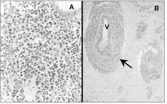
M i c roscopic findings –M i c roscopic examination revealed densely cellular parenchymatous lesions in nine cases (Fig 3A). These lesions were characterized by atypical lymphoid cells, a central area of necrosis and an angiocentric pattern at the periphery of the infiltrate (Fig 3B), with a rich reticulin network
dis-sociating the vascular wall. Only one case did not pre-sent the angiocentric arrangement.
In most cases, infiltration was diffuse and consist-ed of a polymorphic infiltrate comprisconsist-ed pre d o m i n a n-tly by large, sometimes bi- or multinucleated, immuno-blast-like cells containing large, pleomorphic, vesicu-lar nuclei, clearly visible nucleoli and scarce cytoplasm. The mitotic count was high. In two cases the cellular infiltration was made up by small lymphoid cells with inconspicuous mitotic activity. Plasma cell diff e re n t i-ation was also observed in one case (Table 4).
Gliosis was identified in all cases, especially at the Fig 3. Histopathology of the non-Hodgkin large B-cell lymphoma. (A) Note dense sheet of typical
lymphocytes (HE; 40x). (B) Angiocentric pattern (arrow; V) of the tumoral involvement (HE; 5x). Table 3. Gross findings at of 10 autopsy cases of CNS primary lymphomas.
Case External exam Nº tumoral Lobar Basal Cerebral Cerebellar Size lesions lesion nuclei trunk hemisphere (major lesion)
1 Cortical atrophy / old infarct 3 – 1 1 1 1.5 x 1.0 cm
2 Congestion 1 1 – – – < 1.0 cm
3 NL 1 – – – – < 1.0 cm
4 Oedema / cerebral hernia 2 – 1 1 – 4.0 x 3.5 cm
5 Oedema / cerebral hernia 1 – 1 – – 6.0 x 4.0 cm
6 Cortical atrophy 2 – 2 – – 2.0 x 2.0 cm
7 Cortical atrophy 2 2 – – – 2.0 x 1.5 cm
Cavitary lesion
8 Cortical atrophy 4 – – – 2 3.0 x 2.5 cm
9 Mild oedema 4 – 2 1 1 5.0 x 4.0 cm
10* Mild oedema – – – – – –
p e r i p h e ry of the tumoral mass, and reactive astro c y-tes were eventually observed within the tumor tissue.
Associated disorders –O p p o rtunistic infections w e re observed in six cases: HIV-encephalitis (n=2), CMV - encephalitis (n=2), cerebral toxoplasmosis (n=1) and cerebral cryptococcosis (n=1) (Table 4).
Immunohistochemical findings –All cases but one showed a B phenotype (CD20 positive); the remain-ing case, from a HIV negative patient, was positive for T-lymphocytes (CD3 positive). In addition, just one
case was CD30 positive and one case EBV positive (Fig 4).
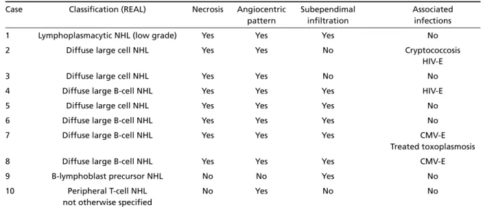
According to the REAL classification26, three cas-es were classified as diffuse large B-cell non-Hodgkin’s lymphoma, one case as lymphoplasmacytic non-Hod-g k i n ’s lymphoma, one case as B-lymphoblast pre c u r-sor non-Hodgkin’s lymphoma, and one case as periph-eral T-cell non-Hodgkin’s lymphoma, not otherw i s e specified. The three cases, in which no immunohis-t o c h e m i s immunohis-t ry could be perf o rmed, were classified as probably diffuse large B-cell lymphoma.
Table 4. Classification and histopathologic findings at of 10 autopsy cases of CNS primary limphoma.
Case Classification (REAL) Necrosis Angiocentric Subependimal Associated pattern infiltration infections
1 Lymphoplasmacytic NHL (low grade) Yes Yes Yes No
2 Diffuse large cell NHL Yes Yes No Cryptococcosis
HIV-E
3 Diffuse large cell NHL Yes Yes No No
4 Diffuse large B-cell NHL Yes Yes Yes HIV-E
5 Diffuse large cell NHL Yes Yes Yes No
6 Diffuse large B-cell NHL Yes Yes Yes No
7 Diffuse large B-cell NHL Yes Yes Yes CMV-E
Treated toxoplasmosis
8 Diffuse large B-cell NHL Yes Yes Yes CMV-E
9 B-lymphoblast precursor NHL No No Yes No
10 Peripheral T-cell NHL No Yes No No
not otherwise specified
NHL, non-Hodgkin’s lymphoma; HIV, human immunodeficiency virus; CMV, cytomegalovirus; E, encephalitis.
DISCUSSION
In the present study, all tumors were only identi-fied in the CNS, in agreement with the criteria for the diagnosis of PCNSL which re q u i re that no lympho-mas are detected in organs other than the CNS or posterior region of the retina at the onset of neuro-logical symptoms11,14. By definition, extranodal lym-phomas are recognized as lymphoid tumors arising f rom lymphoid cell-containing sites other than the lymph nodes27.
M a c ro s c o p i c a l l y, the lesions were similar to those of toxoplasmosis, a finding also re p o rted by Cirilo and Rosenblum1 5. Similarly, neuroimaging does not p e rmit a diff e rential diagnosis with cerebral toxo-p l a s m o s i s4and other methods are necessary for diag-nosis conclusion2 8. Thus, in the present study, the diagnosis of PCNSL was made only by microscopy.
In the present study, immunohistochemistry for EBV revealed only one positive case in an AIDS pa-tient. The lack of detection of the virus might have been due to the long time of fixation of the brain which might have inactivate epitopes there f o re com-promising the testing.
Association with other CNS disorders was a fre-quent finding in the present series. It might be spec-ulated as proposed by Aderson et al.2 0and Aozasa et al.2 1that the presence of HIV- or CMV-induced en-cephalitis is responsible for B-cell hyperreactivity.
PCNSL continue to be a challenge in terms of their morphology and oncogenesis. Molecular studies of oncogenes and suppressor genes are still inconclu-sive. These lymphomas develop in non-lymphoid or-gans without lymphatic vessels, and as such re p sent one of the main areas of neuro-oncological re-search.
Acknowledgment –Thanks to Maria C. Aparecida do Nascimento for secretarial assistance. The investigation was c a rried out at the Depar tment of Pathology, Fluminense Federal University (FFU), University Hospital Antônio Pedro . It was financed by CAPES and based on a master disserta-tion from the Inter-institudisserta-tional Post-Graduate Course of FFU and State University of Health Sciences of Alagoas.
REFERENCES
1. Mead GM, Bleehen NM, Gregor A, et al. A medical re s e a rch council randomized trial in patients with primary cerebral non-Hodgkin lym-phoma: cerebral radiotherapy with and without cyclophosphamide, d o x o rubicin, vincristine and prednisone chemotherapy. Cancer 2000; 89:1359-1370.
2. Castellano-Sanchez AA, Li S, Qian J, Lagoo A, Weir E, Brat DJ. Primary central nervous system posttransplant lymphoproliferative disord e r s . Am J Clin Pathol 2004;121:246-253.
3. Bacchi CE, Bazan R, Padovani EG, et al. Central nervous system lym-phoma: association with Epstein-Barr virus. J Bras Patol 1996;32:103-109.
4. B e rger JR. Mass lesions of the brain in AIDS: the dilemmas of distin-guishing toxoplasmosis from primary CNS lymphoma. AJNR 2003;24: 554-555.
5. Okano M, Gross TG. A review of Epstein-Barr virus infection in pati-ents with immunodeficiency disorders. Am J Med Sci 2000;319:392-396. 6. M o rgello, S. Pathogenesis and classification of primary central
nerv-ous system lymphoma: an update. Brain Pathol 1995;5:383-393. 7. Levy JA, Bredesen DE, Rosenblum ML. Neurological manifestations
of AIDS: experience at UCSF and review of the literature. J Neuro s u rg 1985;62:475-495.
8. O’neill BP, Illig JJ. Primary central nervous system lymphoma. Mayo Clin Proc 1989;64:1005-1020.
9. Murray K, Kun L, Cox J. Primary malignant lymphoma of the central nervous system: results of treatment of 11 cases and review of the lit-eratura. J Neurosurg 1986;65:600-607.
10. B e rger JR. Mass lesions of the brain in AIDS: the dilemmas of distin-guishing toxoplasmosis from primary CNS lymphoma. AJNR 2003; 24: 554-555.
11. H o c h b e rg FH, Miller DC. Primary central nervous system lymphoma. J Neurosurg 1988;68:835-853.
12. Julien J, Vital C, Rivel J, et al. Primary meningeal B lymphoma pre s e n t-ing as a subacute ascendt-ing polyradiculoneuro p a t h y. J Neurol Neuro-surg Psychiatry 1991;54:610-613.
13. Lantos PL, Va n d e r b e rg SR, Kleihues P. Tumours of the central nerv-ous. In Graham D, Lantos PI (eds). Greenfield’s Neuro p a t h o l o g y. 6. ed. Hodder Headline Group. Londres, 1997:583-879.
14. Grant JW, Isaacson P. Primary central nervous system lymphoma. Brain Pathol 1992;2:97-102.
15. Ciricillo SF, Rosenblum ML. Use of CT and MR imaging to distinguish intracranial lesions and to define the need for biopsy in AIDS patients. J Neurosurg 1990;73:720-724.
16. Isaacson PG, Norton AJ. Lymphomas of the nervous system. In Isaacson PG, Norton AJ (eds). Extranodal lymphomas. New York: Churchill Li-vingstone, 1994:217-227.
17. Lachance DH, O’neill BP, Macdonald DR, et al. Primary leptomeningeal lymphoma: report of 9 cases, diagnosis with immunocytochemical analysis, and review of the literature. Neurology 1991;4:95-100. 18. Kalimo H, Lehto M, Näntö-Salonen K, et al. Characterization of the
pe-rivascular reticulin network in a case of primary brain lymphoma. Immunohistochemical demonstration of collagen types I, III, IV and V; laminin; and fibronectin. Acta Neuropathol (Berl) 1985;66:299-305. 19. Hoang-Xuan, Khe A, Camilleri-Broet Sophie B, Soussain Carole C. Re-cent advances in primary CNS lymphoma. Curr Op Oncol 2004; 16:601-606.
20. Alderson L, Fetell MR, Sisti M, Hochberg F, Cohen M, Louis DN. Sen-tinel lesions of primary CNS lymphoma. J Neurol Neuro s u rg Psychiatry 1996;60:102-105.
21. Aozasa K, Saeki K, Horiuchi K, et al. Primary lymphoma of the brain developing in a boy after a 5-year history of encephalitis: polymerase chain reaction and in situ hybridization analyses for Epstein-Barr viru s . Hum Pathol 1993;24:802-805.
22. Rosenblum ML, Levy RM, Bredesen DE, et al. Primary central lym-phomas in patients with AIDS. Ann Neurol 1988;23(Suppl):S13-S16. 23. Paulus W, Jellinger K, Hallas C, Ott G, Müller-Hermelink HK. Human
h e r p e s v i rus-6 and Epstein–Barr virus genome in primary cerebral lym-phomas. Neurology 1993;43:1591-1593.
24. AboodyGuterman K, Hair L, Morgello S. EpsteinBarr virus and A I D S -related primary central nervous system lymphoma: viral detection by i m m u n o h i s t o c h e m i s t r y, RNAin situhybridization, and polymerase chain reaction. Clin Neuropathol 1996;15:79-86.
25. C a m i l l e r i - B roet S, Davi F, Feuillard J, et al. and The French Study gro u p for HIV-Associated Tumors. A I D S related primary brain lymphomas: histopathologic and immunohistochemical study of 51 cases. The Fre n c h Study Group for HIV-Associated Tumors. Hum Pathol 1997;28:367-374. 26. Harris N, Jaffe E, Stein H, et al. A revised European-American classi-fication of lymphoid neoplasms: a proposal from the international lym-phoma study group. Blood 1994;84:1361-1392.
27. Knowles DM. The extranodal lymphoid infiltrate: a diagnostic dilem-ma. Sem Diagn Pathol 1985;2:147-151.