39
ANATOMY OF
GYNURA AURANTUACA (BLUME) SCH.BIP. EX
DC. (ASTERACEAE)
Rodica BERCU
Faculty of Natural and Agricultural Sciences,”Ovidius” University, Constantza University Alley, No. 1, B, 900470, Constantza
Corresponding author e-mail:rodicabercu@yahoo.com
Received 21 April 2014; accepted 20 May 2014
ABSTRACT
The paper presents a detailed histoanatomical description, of the vegetative organs (root, stem and leaf) and photographs as well of Gynura aurantiaca (Blume) Sch.Bip. ex DC. It was observed that the root have typical primary dictos structure. The stem has a differentiated in two regions cortex and the stele comprise one ring of open collateral vascular bundles with secondary xylem due to the cambium activity. The petiole anatomy is quite similar in its basic structure with the stem. The blade presents a heterogenous and hypostomatic mesophyll and a number of vascular bundles in the midrib zone. Remarkable is the presence of the filamentous, uniseriate non-glandular hairs in the stem, petiole and leaf blade. The mechanical tissue is present in the stem, petiole and blade as well.
KEY WORDS: anatomy, vegetative organs, Gynura aurantiaca
INTRODUCTION
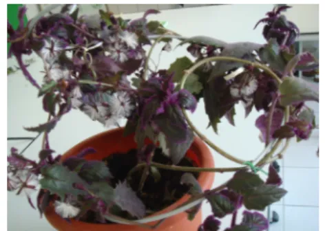
Gynura aurantiaca (Blume) Sch.Bip. ex DC native from and South- East Asia, Java, Indonezia is commonly known as the purple passion plant or purple velvet plant. It is a perennial vine species that grows up to 60-90 cm, bearing oval leaves with large teeth along the margins. The leaves and stems are covered with short, bright-purple hairs that become more colorful if the plant is grown in bright light. It grows rapidly, but is cultivated primarily for those attractive purple leaves (Fig. 1). Annually during the spring, this plant produces prominent flowers both in appearance and smell, gaudy and malodorous. Parts of plant are poisonous if ingested(Chen et al., 2000; Mioulane, 2004; Sadovsky,1996).
40
Few data are known about this species anatomy. Most of the studies refer to the plant organogenesis, physiological studies concerning the flowers productivity growth and development, the effect of leaf pubescence on light refletance (Chen et al., 2000; Gausman and Cardenas, 1969; LI Luan et al., 2009) or phytopatological studies (Conejero and Semanicik, 1977). In the Romanian literature data on this species structure lack.
The aims of the present study were to analysis the vegetative organs of Gynura aurantiaca and to contribute to the knowledge of this species.
MATERIALS AND METHODS
The plant leaves were collected form S.C. “Iris International” S.R.L. greenhouse. Small pieces of root, stem and leaves were fixed in FAA (formalin: glacial acetic acid: alcohol 5:5:90). Cross sections of the vegetative organs were performed by the freehand made technique (Bercu and Jianu, 2003; Ianovici, 2009). The cross sections samples were stained with alum carmine and iodine green. Histological observations and micrographs were performed with a BIOROM–T bright field microscope, equipped with a TOPICA 6001A digital camera attachment.
RESULTS AND DISCUSSION
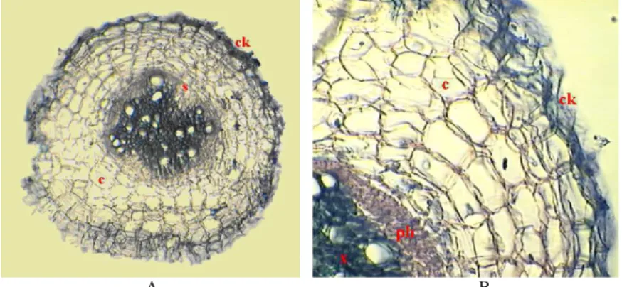
Transection of the root reveals a few layers of suberized cells to the exterior (the cork) and 5-6 layers of phelloderm inside, produced by phellogen. Just below the cortex is the centrally located stele consisting of secondary phloem and xylem arranged in a compact mass. The xylem vessels are placed into a sclerencymatous parenchyma (Fig. 2, A, B).
A B
FIG. 2. Cross section of the root – ensamble (A, x 120). Portion with cork and cortex (B, x 322): c- cortex, ck- cork, ph- phloem, st- stele, x- xylem.
41
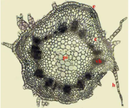
composed of small isodiametric radial elongated cells, without intercellular spaces interrupted by the presence of stomata and long, uniseriate - one cell layer thick, filamentous and sharp-typed non-glandular hairs (Bavaru and Bercu, 2002; Esau, 1977). The epidermal cells are covered by a thin cuticle. Just below the epidermis is the cortex, differentiated into two zones, one zone consisting of 2-3 layers of collenchyma another area made up of several layers of parenchyma cells (Fig. 3, 4, A).
FIG. 3. Cross section of the stem - ensemble (x 50): c- cortex, e- epidermis, h- hair, pi- pith, vb- vascular bundles.
A B
Fig. 4. Cross section of the stem. Portion with epidermis and cortex (A, x 180). A stele vascular bundle - detail – (B, x 190): cb- cambium, co- collenchyma, e- epidermis, ic- inner cortex, ph- phloem, pfb- periphloemic fibers, x- xylem.
42
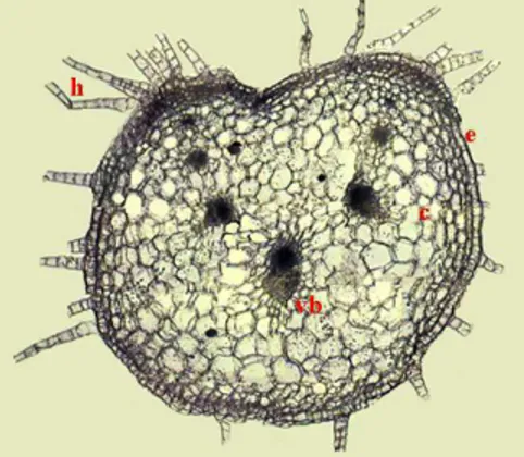
Cross section of the petiole discloses a one-layered epidermis, covered by cuticle and broken by the presence of stomata and non-glandular hairs the same as those of the stem (Fig. 5; 6, A).
FIG. 5. Cross section of the stem -ensemble (x 30): c- cortex, e- epidermis, h- hair, vb- vascular bundle.
A B
FIG. Cross section of the stem. Portion with epidermis and cortex. A vascular bundle - detail (A, B, x 107): co- collenchyma, e- epidermis, h- hair, ic- inner cortex, vb- vascular bundle, ph- phloem, x- xylem.
Just below epidermis is the hypodermis composed of 1-2 layers of collenchymatous cells, followed by a number o parenchyma cells, forming the inner cortex. The vascular bundles, three larger than the rest of four, are the same as those of the stem (Fig. 5; 6, B)
43
that the epidermal cells and hairs possess reddish purple anthocyanins in the vacuolar systems.
A B
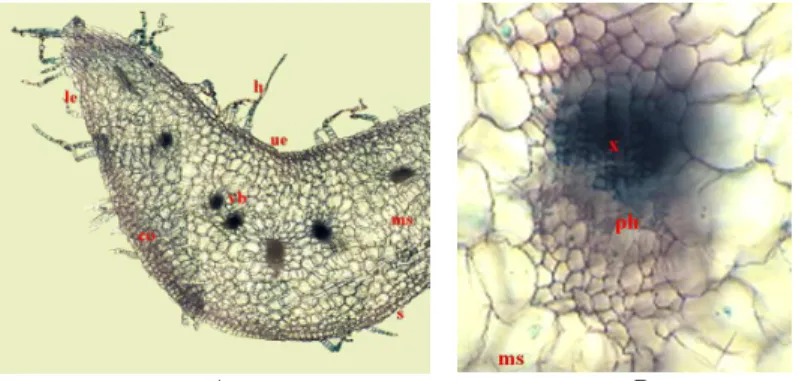
FIG. 6. Cross section of the blade – ensemble (A, x 70). A midrib vascular bundle (B, x 110): co- collenchyma, h- hair, le- lower epidermis, ms- mesophyll, ph- phloem, s- stoma, ue- upper epidermis, vb- vascular bundle, x- xylem.
The mesophyll is composed of palisade tissue and spongy tissues as well, in witch vascular bundles are embedded (Fig. 6, A).
The midrib collateral 4 vascular bundles, present the typical foliar arrangement of the conductive elements, unprotected by sclerenchyma or parenchyma sheaths. In the midrib aria, just bellow the upper and lower epidermis, 1-2 layers of collenchyma are present (Fig. 6, B).
CONCLUSIONS
The root has a secondary structure. The stem cortex is differentiated into two zones. The stem stele is represented by a circular ring of collateral vascular bundles with secondary xylem due to the cambium activity.
The petiole anatomy is quite similar in its basic structure with the stem but differences appear concerning the petiole shape and vascular bundles arrangement. The blade is bifacial and hypostomatic with a heterogenous mesophyll. Uniseriate filamentous non glandular hairs are present in stem, petiole and blade.
The mechanical tissue, present in the stem and less in the leaf, is poorly developed and represented by collenchyma and some vascular bundles sclerenchymatous elements as well.
ACKNOWLEEDGEMENTS
44
REFERENCES
• Bavaru A., Bercu R. 2002. Morphology and anatomy of plants, Ed. Ex Ponto, Constanta.
• Bercu R.; Jianu D.L. 2003. Practicum de Morfologia si anatomia plantelor, Ed. by “Ovidius” University Press, Constanta.
• Chen J., Russell D.C., Robinson C.A. 2000. Suppressing Purple Passion (Gynura aurantiaca) Flowering Using Selected Plant Growth Regulators. Hort. Science, 35(3): 416.
• Conejero, V., Semanicik, J.S. 1977. Analysis of the proteins in crude plant extract of polyacrylamide.
Phytopathology, 67: 1424-1426.
• Esau K. 1977. Anatomy of Seed Plants, 2nd ed., John Wiley & Sons, New York.
• Foster P. I., Thongpukdee A. 1988. Variation in Gynura drymophyla (F. Muell.). F.G. Davies Asteraceae Senecioneae. Austrobaileya, 2(5): 557-566.
• Gausman H.W., Cardenas R. 1969. Effect of Leaf Pubescence of Gynura aurantiaca on Light Refletance, Bot. Gaz., 130(3): 158-162.
• Ianovici N. 2009. Biologie vegetală - lucrări practice de citohistologie şi organografie, Ed. Mirton, Timişoara: 205 p.
• Mioulane P. 2004. Enciclopedia Truffault. Grădini şi plante de interior. Ed. Grupul Editorial RAO, Bucuresti.
• Sadovsky, E. 1996. Plante de apartament şi decoraţiuni florale, vol I, Ed. Academia Romana, Bucuresti.
• Toma C., Rugina, R. 1998. Anatomia plantelor medicinale, Ed. Academia Romana. Bucuresti.