Abstract
The present study deals with the leaf anatomy and leaf surface of
Posoqueria acutifolia
Mart.,
P. latifolia
Mart.,
P.
longiflora
Aublet,
P. macropus
Mart.,
P. palustris
(Rudge) Roem. and
Posoqueria
sp., collected in fragments of
Atlantic rain forest, Rio de Janeiro, Brazil. The epicuticular wax may occur in the form of filaments, granules or crusts.
The leaves are covered by a thick cuticular layer that may be smooth or striated. Paracytic stomata, and non-glandular
trichomes are limited to the abaxial surface; the latter are numerous in
P. palustris
, and rare in
P. longiflora
and
P.
latifolia
. Leaves have a dorsiventral structure, with only one layer of palisade parenchyma and varied amounts of
spongy parenchyma. Idioblasts containing crystalliferous sand were observed, and were more abundant in
P. latifolia
.
The leaf blade vascular system is formed by collateral bundles with a parenchymatous sheath, associated with fibers.
The vascular system of the petiole and the leaf blade forms an arch. Some of the anatomical features observed can be
used to distinguish the species studied. Anatomical leaf characters could be used in the recognition of six species of
Posoqueria
studied, such as anticlinal wall of epidermal cells, wax deposition, trichomes and shape of the leaf margin.
Key words
: leaf anatomy, scanning electron microscopy, taxonomy, Cinchonoideae.
Resumo
São apresentadas informações sobre a anatomia e a superfície foliar de
Posoqueria
acutifolia
Mart.,
P.
latifolia
Mart.,
P
.
longiflora
Aublet,
P. macropus
Mart.,
P. palustris
(Rudge) Roem
e
Posoqueria
sp., espécies ocorrentes em fragmentos da
Floresta Pluvial Atlântica, Rio de Janeiro, Brasil. A cera epicuticular pode ocorrer na forma de filamentos, grânulos ou
crostas. As folhas são recobertas por espessa camada cuticular, que pode ser lisa ou estriada. Na face abaxial a cutícula
apresenta-se estriada em
P. latifolia
e
P. palustris
. Os estômatos estão restritos à face abaxial e são do tipo paracítico.
Tricomas tectores ocorrem apenas nesta face sendo numerosos em
P. palustris
, raros em
P. longiflora
e
P. latifolia
. As
folhas têm estrutura dorsiventral, com apenas uma camada de parênquima paliçádico e variado número no parênquima
esponjoso. No mesofilo foram observados idioblastos contendo areia cristalífera, mais abundantes em
P. latifolia
. Na
lâmina foliar o sistema vascular é formado por feixes do tipo colateral, envolvidos por bainha parenquimática, e
apresentam fibras associadas. No pecíolo e na nervura principal o sistema vascular apresenta-se em arco. Características
anatômicas foliares podem ser usadas na distinção das seis espécies de
Posoqueria
estudadas, tais como: a parede
anticlinal das células epidérmicas, deposição de cera, tricomas e a forma da margem da folha.
Palavra-chave
: anatomia foliar, microscopia eletrônica de varredura, taxonomia, Cinchonoideae.
Leaf anatomy and micromorphology of six
Leaf anatomy and micromorphology of six
Leaf anatomy and micromorphology of six
Leaf anatomy and micromorphology of six
Leaf anatomy and micromorphology of six
Posoqueria
Posoqueria
Posoqueria
Posoqueria
Posoqueria Aublet species (Rubiaceae)
Aublet species (Rubiaceae)
Aublet species (Rubiaceae)
Aublet species (Rubiaceae)
Aublet species (Rubiaceae)
Anatomia e micromorfologia foliar de seis espécies de Posoqueria Aublet (Rubiaceae)
Rosani do Carmo de Oliveira Arruda
¹
, Doria Maria Saiter Gomes
²
Aline Carvalho de Azevedo
1,3, Michelle Lima Magalhães
1,3& Mario Gomes
4 http://rodriguesia.jbrj.gov.br1
Universidade Federal do Estado do Rio de Janeiro (UNIRIO), Instituto de Biociências, Depto. Botânica, Av. Pasteur 458, 22290-540, Rio de Janeiro, RJ. Autora para correspondência: rosaniarruda@gmail.com
2Universidade Federal Rural do Rio de Janeiro (UFRRJ), Instituto de Biologia, Depto. Botânica, Rod. 465, km 7, 23890-000, Seropédica, RJ. 3 Iniciação Científica.
4Extracta Moléculas Naturais S.A., Av. Carlos Chagas Filho 791, Cidade Universitária, 21941-904, Rio de Janeiro, RJ.
Introduction
Rubiaceae (Gentianales) is one of the four largest families of angiosperms, with representatives widely distributed worldwide, presenting diverse habits and life forms. The family is monophyletic and includes about 10.700 species, which were
recently divided into two subfamilies, the Cinchonoideae and the Rubioideae (Robbrecht & Manen 2006).
Belonging to Cinchonoideae subfamily,
Posoqueria Aublet is found in the Neotropic region
among other compounds, have been identified as perfume sources in Posoqueria latifolia (Rudge)
Roem., popularly known as “açucena-do-mato”. For
P. acutifolia Mart., laboratory tests have identified
analgesic and anti-inflammatory activities of the methanolic extracts, which may be related to this species use in popular medicine (Souza et al. 2007).
Among the organs that can be useful to taxonomic propositions, the leaf is the most examined one because of its internal architecture, which is a classic source of useful information in the systematics, specially the epidermis and the cuticle (Stuessy 1990; Judd et al. 2009).
About micromorphology the studies of Mantovani & Vieira (1993/1997) and Kocsis et al.
(2004) stand out and are source of important information about the leaf surface of diverse species of this family. Descriptive anatomical studies of the vegetative or reproductive parts also have been carried out with other Brazilian rubioid species (Mantovani et al. 1995; Gomes et al. 2000; Arruda
& Gomes 1996. De Toni & Mariath 2008).
This study describes the leaf anatomy and micromorphology of six Brazilian Posoqueria
species: P. acutifolia Mart., P. latifolia (Rudge)
Roem. & Schult., P. longiflora Aubl., P. macropus
Mart., P.palustris Mart., and Posoqueria sp. aiming
to identify features that may be useful in recognizing these species.
Materials and Methods
Adult leaves of Posoqueria latifolia and P. longiflora were collected from plants found at the
Parque Nacional da Floresta da Tijuca (22º57’S and 43º18’W), in Rio de Janeiro, (RJ). In this region, the annual avarage temperature varies between 19.3ºC in the winter and 25.5°C in the summer, and the rainfall is about 2000 mm annually (Vieira 1994). Posoqueria palustris was collected in Rio das Ostras (RJ,
22º31S’-41º55’W) where the annual average temperature is 22ºC and the annual rainfall between 1500 and 2000 mm
Botânico do Rio de Janeiro (RB), were rehydrated according to Gomes (2002).
The fresh leaves were fixed in F.A.A. (formalin, acetic acid, ethanol 70%) for 48h and preserved in 70% ethanol (Johansen 1940). For light microscopy, sections of the leaf blade and petiole were dehydrated in graded ethanol-butanol series, embedded in paraffin, sectioned transversely and longitudinally in rotatory microtome at a thickness of 12 mm. The sections were stained with 1% safranin - 1% astra blue in tartaric acid (Luque et al. 1996).
Leaf anatomy and micromorphology of Posoqueria Aublet (Rubiaceae)
Results
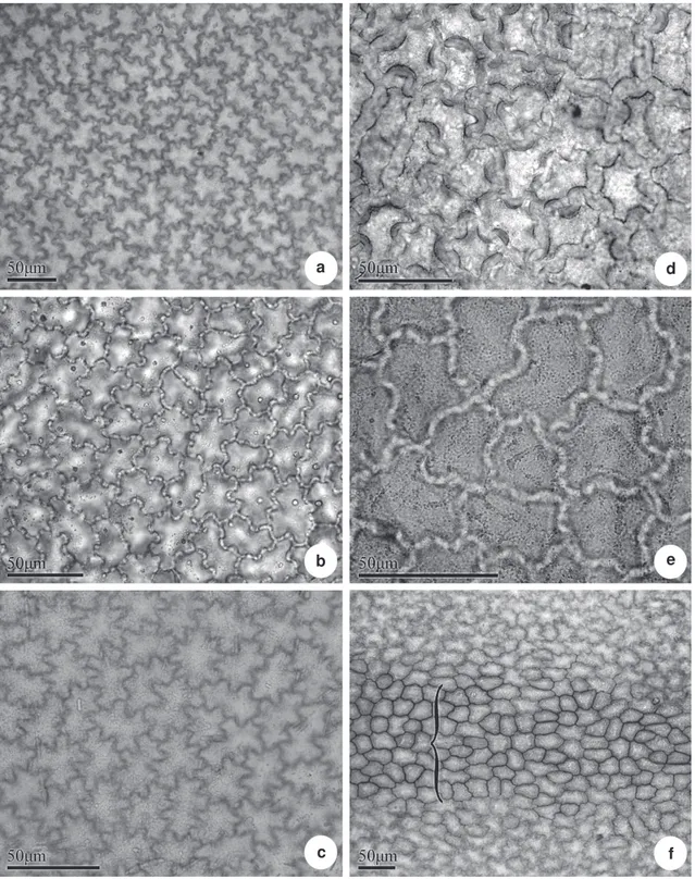
In most of the studied species, the epidermis, in adaxial surface is composed of ordinary epidermal cells, with sinuous anticlinal walls (Fig. 1 a-f), and thick, notably in Posoqueria macropus (Fig. 1 d).
Over vascular bundles, such as in the abaxial surface of Posoqueria sp. the anticlinal walls can present
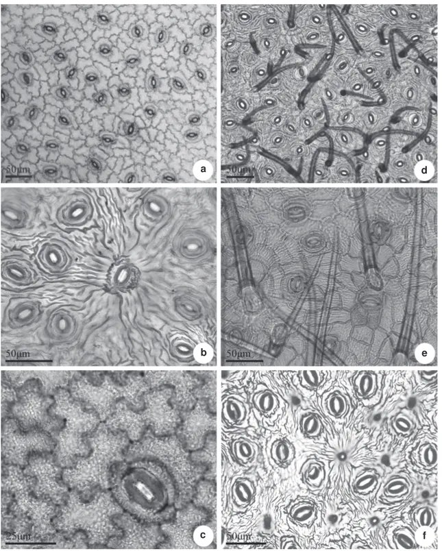
themselves straight or curved (Fig. 1 f). The abaxial surface is composed by ordinary epidermal cells, stomata and trichomes (Fig. 2 a-f). The epidermal anticlinal cells walls are sinuous in all species (Fig. 2 a-e), with the exception of Posoqueria sp in which
they are straight. P. acutifolia has glabrous leaves
(Fig. 2 a), in P. macropus numerous, non-glandular,
unicellular trichomes, with thick lignified walls were observed (Fig. 2 d). In P. palustris there are
pluricellular uniseriated trichomes, long, non lignified (Fig. 2 e). Posoqueria sp is marked by presence of
numerous papillae (Fig. 2 f). Rare short unicellular non-glandular trichomes are observed on the abaxial surface of the leaf of P. latifolia and P. longiflora,
restricted to midvein; in P. palustris, numerous
pluricellular trichomes are observed.
The stomata belong to the paracytic type and are arranged randomly (Fig 2). The subsidiary cells may be of different sizes; some stomata may be flanked in parallel by more than one subsidiary cell. The six species investigated present some large stomata, around which the epidermal cells are organized radially, and the cuticle layer forms a radiate pattern (Fig. 2 b). In P.macropus and P. palustris the stomata are partly covered by numerous trichomes (Fig. 2 d-e).
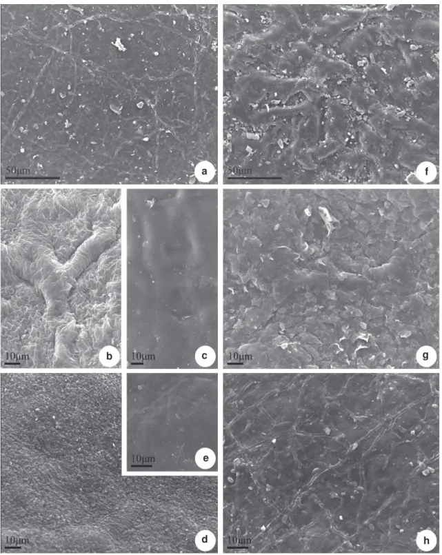
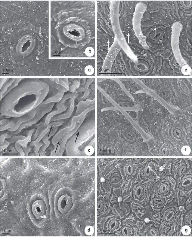
The epicuticular wax can occur without defined ornamentation, in the form of filaments, granules and in crusts (Figs. 3 a-h, 4 a-g). The granules can be of varied sizes and are present in Posoqueria acutifolia
(Fig. 4a), P. longiflora (Fig. 3 d, 4d), P. macropus
(Fig. 4 e), P. palustris (Fig. 4 f) and Posoqueria sp
(Fig. 4 g), including over the stomata (Fig. 4 d, g) and trichomes (Fig. 4 e). The crusts occur in P. macropus
(Fig. 3 f), P. palustris (Fig. 3 g) and are thick in P. latifolia (Fig. 3 b); the filaments occur in abaxial surface
of P. latifolia (Fig. 4 c). The cuticle is smooth on
the adaxial surface of all species (Fig. 3 a-h) and on the abaxial surface of P. longiflora (Fig. 4 d), and
striated on the abaxial surface of the other species (Fig. 4 b, e-g), specially in P. latifolia (Fig. 4 c) and
in Posoqueria sp (Fig. 4 g).
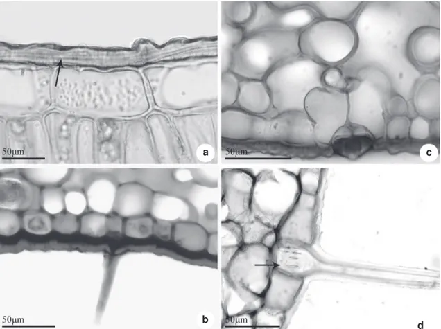
In all species investigated the leaf epidermis is one-layred formed by thick-walled cells and covered by a layer of cuticle and well-developed
cuticular-wax strata (Fig 5 a-d). On the abaxial surface, above the stomatal pore, the cuticle layer may form conspicuous ledges (Fig. 5 c). The stomata are located on the same level or slightly below the other epidermal cell and the subsidiary cells can form projections in the region of the substomatal chamber (Fig. 5 c). The trichomes have thick walls (Fig. 5 b), and in Posoqueria palustris, the basal
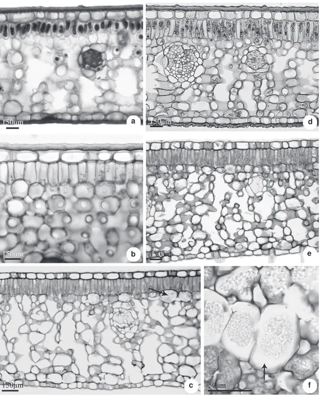
region of the septate trichome is pitted (Fig. 5 d). All the species studied show dorsiventral mesophyll, with one layer of cells in the palisade parenchyma and about 10 to 14 layers in the spongy parenchyma (Fig. 6 a-e). The palisade cells are wider and shorter in Posoqueria acutifolia and P. latifolia
(Fig. 6 a-b) and longer and narrower in the remaining species (Fig. 6 c-e). Idioblasts containing tiny polyhedral crystals of calcium oxalate (crystalliferous sand) are observed on the entire mesophyll in all species studied (Fig. 6 c, f). They are more abundant in P. latifolia
and P. palustris. These cells occur in a greater
frequency beneath the palisade parenchyma, or near the epidermis of the abaxial surface, isolated or in groups, especially in P. latifolia and P. palustris
(Fig. 6 e-f). The histochemical tests showed that the crystalliferous cells also accumulate phenolic compounds.
The vascular system of the leaf blade is formed by bundles of the collateral type, of varied calibers (Fig. 6 a, d). The vascular bundles are surrounded by a parenchymatous sheath that may contain chloroplasts, crystalliferous sand, and phenolic compounds. Internally to this sheath lignified fibers occur in variable proportions. In
Posoqueria acutifolia, P. latifolia and P. macropus
the sclerenchyma occurs in a larger proportion than the vascular tissues (Fig. 6 a, d).
The cross section showed that the outline of leaf margin is truncate-revolute in Posoqueria longiflora and slightly revolute in the others
species (Fig. 7 a-d). In the epidermis, characteristics similar to those of the rest of the leaf blade can be observed, noticing that the cuticular layer is strongly thickened, forming conspicious cuticular flanges. The common epidermal cells are smaller then in the rest of the leaf blade and in P. longiflora
have a papillose appearance. Internally, the margin is filled with parenchymatous cells with thickened walls; among them idioblasts with crystals occur. Lignified fibers were observed associated to the vascular system.
Figure 1
– Leaf epidermis of
Posoqueria
Aublet in frontal view, adaxial surface – a.
P. acutifolia
; b.
P. latifolia;
c.
P. longiflora
;
d.
P. macropus
; e.
P. palustris
; f.
P. longiflora
, in the midvein region (
{
).
c f
e b
Leaf anatomy and micromorphology of Posoqueria Aublet (Rubiaceae)
Figure 2
– Leaf epidermis of
Posoqueria
Aublet in frontal view, abaxial surface – a.
P. acutifolia
; b.
P. latifolia;
c.
P.longiflora
; d.
P. macropus
; e.
P. palustris
; f.
Posoqueria
sp. showing the the arrangement of stomata,
trichomes (d, e) and papillae (f).
f e
c b
Figure 3
– Scanning electron microscopy of leaf blade of
Posoqueria
Aublet – a.
P. acutifolia
; b-c.
P. latifolia
;
d-e.
P. longiflora
; f.
P. macropus
; g.
P. palustris
; h.
Posoqueria
sp. Adaxial surface showing wax in granules
(d), in crusts (f) and in thick crusts (b).
h g
d e
f
c b
Leaf anatomy and micromorphology of Posoqueria Aublet (Rubiaceae)
Figure 4
– Scanning electron microscopy of leaf blade of
Posoqueria
Aublet – a.
P. acutifolia;
b.
P. acutifolia
boiled;
c.
P. latifolia;
d.
P. longiflora;
e.
P. macropus;
f.
P. palustris;
g.
Posoqueria
sp. Abaxial surface showing stomata (a-g),
trichomes in
P. macropus
(e) and
P. palustris
(f) and papillae in
Posoqueria
sp. (g). The surface can be smooth (d) or
striated (b-c, e-g). Epicuticular wax (arrow) in filaments (c) and granules (e).
g f e
Figure 5
– Cross sections of
Posoqueria
Aublet leaf, details of epidermis – a.
P. latifolia
with a thick cuticular layer
(arrow); b.
P. longiflora
, trichome in midvein region; c.
P. longiflora
, stomata with developed rims and subsidiry cells
in the substomatal chamber; d.
P. palustris
, lignified trichome with pitted wall (arrow).
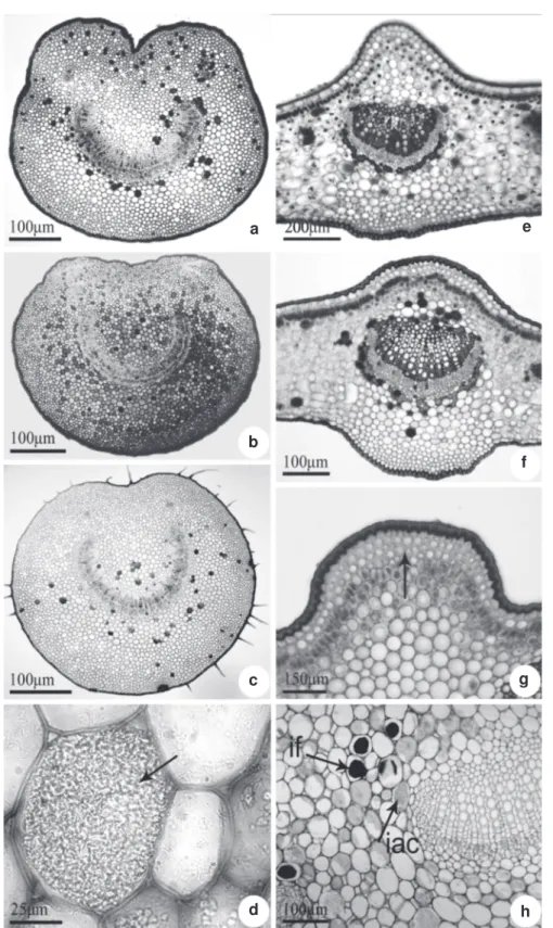
both surfaces of the leaf, the epidermal cells are organized in rows and have thicker, nearly straight walls. Stomata can be found in this region of the leaf blade. In the cortical region of the petiole there are two to five layers of angular collenchyma beneath the epidermis. A fundamental parenchyma containing idioblasts with crystals, phenols, or some sclereids are located internally (Fig. 8 d, h). In all species, the vascular system is arranged in an arch (Fig. 8 a-c, e), with the xylem cells arranged in rows and the phloem in the external position, like the collateral type. Additional bundles are observed at the extremities of the vascular system.
In the region of the midvein, beneath the epidermis, the cortical region is occupied by three to five layers of collenchyma that varies between the angular and lamellar types, and underneath there is a palisade parenchyma connecting the two parts of the leaf blade (Fig. 8 f-g). In this portion, the cells of the
palisade parenchyma are shorter and wider than the remaining parts of the leaf blade. In the cortical portion below the abaxial surface there is a parenchyma which shows progressively more-voluminous cells with thinner walls towards the vascular system.
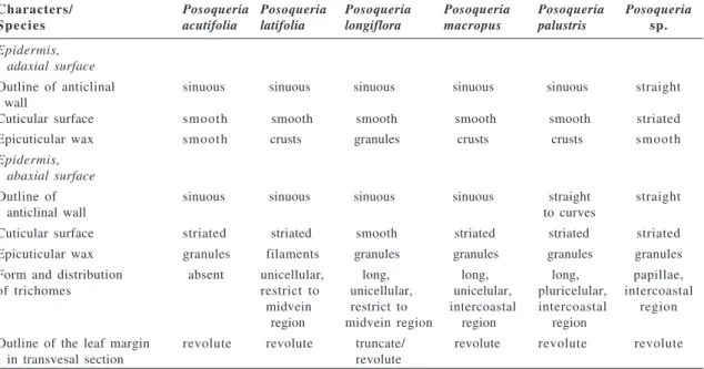
In this study the most relevant leaf characteristics to the distinguishing of the species are related in the Table 1.
Discussion
From all the anatomical features examined, the ones that showed a higher potencial of utilization to identify the studied species were: cuticular and wax sculpturing, aspect of the anticlinal walls of epidermal cells and presence and types of trichomes. The variation on the thickness em sinuosity from the anticlinal walls is quoted fot the Rubiaceae by Metcalfe & Chalk (1950) and Gomes
et al. (2000). Harberlandt (1928) believes that the
d c
Leaf anatomy and micromorphology of Posoqueria Aublet (Rubiaceae)
Figure 6
– Cross sections of
Posoqueria
Aublet leaf showing the dorsiventral mesophyll structure – a.
P. acutifolia
;
b.
P. latifolia
; c.
P. longiflora
; d.
P. macropus
; e.
P. palustris
; f.
P. palustris
with crystal sand in idioblasts (arrow).
f ce b
Figure 7
– Cross sections of
Posoqueria
Aublet showing variation outline of the leaf margin – a.
P. acutifolia
;
b.
P. latifolia
; c.
P. longiflora
;
d.
P. palustris
.
Table 1
– Distinctive characteres to the identification of six
Posoqueria
Aublet (Rubiaceae) species.
Characters/ Posoqueria Posoqueria Posoqueria Posoqueria Posoqueria Posoqueria
S p e c i e s acutifolia latifolia longiflora macropus palustris sp.
Epidermis, adaxial surface
Outline of anticlinal sinuous sinuous sinuous sinuous sinuous straight wall
Cuticular surface smooth smooth smooth smooth smooth striated Epicuticular wax smooth crusts granules crusts crusts smooth
Epidermis, abaxial surface
Outline of sinuous sinuous sinuous sinuous straight straight
anticlinal wall to curves
Cuticular surface striated striated smooth striated striated striated Epicuticular wax granules filaments granules granules granules granules Form and distribution absent unicellular, long, long, long, papillae, of trichomes restrict to unicellular, unicelular, pluricelular, intercoastal
midvein restrict to intercoastal intercoastal region region midvein region region region
Outline of the leaf margin revolute revolute truncate/ revolute revolute revolute
in transvesal section revolute
d c
Leaf anatomy and micromorphology of Posoqueria Aublet (Rubiaceae)
Figure 8
– Petiole and midvein of
Posoqueria
Aublet in cross section – a.
P. latifolia
; b.
P. longiflora
; c.
P. palustris
;
d.
P. palustris
showing idioblast with crystal sand in detail (arrow). e.
P. acutifolia;
f.
P. latifolia
g. and h.
P. palustris
with
collenchyma and palisade tissue under adaxial epidermis (g, arrow), idioblasts with phenols (if) and crystal sand (iac),
on cortical region of petiole (h).
h g f e
For the plants studied here, which were collected in the interior of the Atlantic Rain Forest, the thick cuticular layer, besides protecting the plants from dehydration and from invasion by fungi and bacteria, also protects them from the heavy rain that is common in the neotropics (Juniper & Jeffree 1983). The different aspects of the cuticular layer of the abaxial surfaces and the ornamentation of epicuticular wax could be used in the characterization of these species, confirming the potencial value of this anatomical characteristics for the taxonomists (Wilkinson 1979).
The presence of hypostomatous leaves appears to be the most common case among the Rubiaceae (Metcalfe & Chalk 1950). In all cases, paracytic stomata predominate, although other patterns such as the anomocytic or anisocytic are reported in the family (Metcalfe & Chalk 1950). Stomata with two or more subsidiary cells arranged parallel to the stomata cells can be classified as parallelocytic, as in the species of Posoqueria
studied here and also previously described by several members of Rubiaceae (Mantovani et al.
1995; Kocsis et al. 2004). According to Carpenter
(2005), the anomocitic type is an ancestral condition between the basal angiosperms; besides, the paracytic stomatal architecture has been derived independently some families in this group.
Trichomes have great value in taxonomical, ecological, and evolutionary studies (Gomes & Neves 2009). In the Rubiaceae, the leaf trichomes are of the non-glandular type, uni-or multicellular, in variable densities and sizes, from papillae to longer ones that form a dense indumentum (Metcalfe & Chalk 1979; Gomes et al. 2000; Kocsis et al. 2004). Robbrect & Manen (2006) proposed
that whereas the Rubioideae are distinguished by having generally articulated trichomes, those of the Cinchonoideae are distinguished by the cylindrical type (unicellular), as observed in Posoqueria latifolia and P. longiflora. In P. palustris, however,
Psychotria nuda Wawra, P. leiocarpa Mart., Bathysa gymnocarpa K. Schum, B. mendonçaei K.
Schum, B. cuspidata (A.St.-Hil.) Hook.f. and B. australis (A.St.-Hil.) Benth. & Hook.f. among others
(Vieira et al. 1992; Gomes et al. 2000). Rubiaceae in
open areas, such as the coastal dune forests (restingas), has thicker leaves (Arruda & Gomes 1996). The greater leaf thickness may be a result of accentuated development of the palisade or even the spongy parenchyma, under intense irradiation (Dickison 2006). The species of Posoqueria studied
even though collected in areas where they are relatively protected by the crowns of the canopy plants, have proportionately thicker leaves as a result of the larger number of spongy parenchyma layers. Considering the plastic variation of this character, it has no taxonomic value to the identification of the analized Posoqueria species.
Calcium oxalate crystals are an important source of information for defining the subfamilies, tribes, and subtribes of Rubiaceae. Few studies about the development of these idioblasts or ontogenetic and evolutionary relationships of these cells for the subfamilies of Rubiaceae are available in the literature, except the work of Horner & Whitmoyer (1972) with Psychotria punctata.
Crystals are abundant in the family, and include crystal-sand, raphides, clustered, styloids and other acicular forms. According to Metcalfe & Chalk (1950) the crystal distribution is helpful in genera delimitation. Recently the raphides-type crystals were said to be related to the subfamily Rubioideae, and the absence of this type and the presence of druses or crystalliferous sand, with some exceptions, characterizes the Cinchonoideae (Jansen et al. 2003; Andersson & Antonelli 2005;
Robbrecht & Manen 2006). In the six species of
Posoqueria examined, only idioblasts containing
Leaf anatomy and micromorphology of Posoqueria Aublet (Rubiaceae)
P. latifolia the amount of idioblasts in the leaf blade
is comparatively much higher than in the leaf blade of the other two species.
The arrangement of the vascular system in the petiole and the midvein can be useful in the diagnosis of some plant species. In this sense, morphological patterns have been established for taxonomic purposes dealing with several plant families. In the leaves of many Rubiaceae, the vascular tissues may be arranged in a U, O, or V-shape (Metcalfe & Chalk 1950; Kocsis et al. 2004). The organization of the
vascular system can be used in superior taxonomic levels in Rubiaceae (Martinez-Cabrera et al. 2009).
In all species analyzed, the vascular system is organized in an arch from the petiole to the principal vein, where it begins to be accompanied by some fibers. For the Posoqueria analyzed, a small diagnostic
value was attributed to the shape of vascular system in transversal section at species level.
Although the species of Posoqueria are
considered to be typical shade plants (Macias 1988), some xeromorphic elements were identified in the leaf blade, specially in P. macropus e P. palustris.
These species are eventually affected by the dry and flood periods, a trademark of the flooded region in which they were found, and show, besides the thick cuticular layer, a high amount of fibers associated to the vascular system. The xeromorphic features observed in the studied plants, especially in P. macropus and P. palustris, can be related to
the coastal environment where they are found, marked by a deficiency of nitrogen, phosphorus, calcium and elevated acidity (Henriques et al. 1986).
The sclerification is considered an important strategy for the survival in poor resourced places, because it gives the plants an augmentation of the longetivity and a higher efficiency in the use of available nutrients (van Arendonk & Poorter 1994). This study show that anatomical leaf characters could be allied to morphological ones in the recognition of six species of Posoqueria evaluated,
such as, outline of anticlinal wall of epidermal cells in frontal view, the patterns of the wax and cuticle deposition, the presence and type of trichomes, and the cross-sectional shape of the leaf margin.
Acknowledgments
We want to thank the Curator of the Herbarium of Instituto de Pesquisas Jardim Botânico do Rio de Janeiro (RB) for the permission of using his collection. We want to thank Rosilene Gonçalves, Anna Carina Defaveri, Jefferson Andrade Ferrão
and Ana Angélica M. de Barros for their assistance with field and laboratory work. We want to express our gratitude also to Alice Sato, Alcides Guarino and Edwin Azzero for the many laboratories facilities. To Flora Gomes Elias for English revision.
References
Andersson, L. & Antonelli, A. 2005. Phylogeny of the tree Cinchoneae (Rubiaceae), its position in Cinchonoideae, and discription of a new genus,
Liliosemina. Taxon 54: 17-28.
Ariza, O.A.; Parra, E.D.R.; Archila, J.A.; Morales, J.M. & Stashenko, E. E. 2007. Determinación mediante HS-SPNE / GC-MS, de la composición química de la fragancia y el absoluto de las flores de Posoqueria
latifolia. Scientia et Technica 33: 59-61.
Arruda, R.C.O. & Gomes, D.M.S. 1996. Anatomia Foliar
de Mitracarpus frigidus (Wild). K. Shum. var
Salzmannianus (D.C) K. Shum. e Mitracarpus
lhotzkianus Cham. (Rubiaceae). Boletim do
Herbarium Bradeanum. 6: 431-444.
Bidegain, P. & Michael, C. 2003. Bacias hidrográficas dos Rios São João e das Ostras. Águas, Terra e Conservação Ambiental. Consórcio Intermunicipal Lagos São João (CILSJ). Available in <http:// www.lagossaojoao.org.br/index-cilsj.html>. Access on 10 August 2006.
Carpenter, K.J. 2005. Stomatal architecture and evolution in basal angiosperms. American Journal of Botany 92: 1595-1615.
De Toni, K.L.G. & Mariath, J.E.A. 2008. Ovule ontogeny in Rubiaceae (Juss): Chomelia obtusa (Cinchonoideae
– Guettardeae) and Ixora coccinea (Ixoroideae-Ixorea).
Plant Systematics and Evolution 272: 39-48. Delprete, P.G. 2009. Taxonomic history, morphology
and reproductive biology of the tribe Posoquerieae (Rubiaceae,Ixoroideae). Annals of the Missouri Botanical Garden 96: 79-89.
Dickson, W.C. 2006. Integrative plant anatomy. San Diego. HP Harcourt. Academic Press. 533p. Fahn, A. & Cuttler, D.F. 1992. Xerophytes. Ed.
Gerbrüder Borntraeger, Berlin, 176p.
Foster, A.S. 1949. Practical plant anatomy. Princeton, D. Van Nostrand Company Inc. 228p.
Franklin, G.L. 1945. Preparation of thin sections of synthetic resins and wood-resin composites, and a new macerating method for wood. Nature 155: 51. Gomes, D.M.S. & Neves, L.J. 2009. Scanning electron microscopy of the leaf epidermis of Merostachys
Spreng. (Poaceae: Bambusoideae). Acta Botanica Brasilica 23: 516-525.
Gomes, D.M.S. 2002. Anatomia foliar de espécies de
Merostachys Spreng. (Poaceae:Bambusoideae) no
Horner, H.T.J. & Whitmoyer, R.E. 1972. Raphide crystal cell development in leaves of Psychotria punctata
(Rubiaceae). Journal of Cell Science 11: 339-355. Jansen, S.; Watanabe, T.; Dessein, S.; Smets, E. &
Robbrecht, E. 2003. A comparative study of metal levels in leaves of some Al-accumulating Rubiaceae. Annals of Botany 91: 657-663.
Jensen, W.A. 1962. Botanical histochemistry principles and pratice. W.H. Freeman and Co., San Francisco. 408p.
Johansen, D.A. 1940. Plant microtechnique. McGrow-Hill Book Co. Inc, New York. 523p.
Judd, W.S.; Campbell, C.S.; Kellogg, E.A.; Stevens, P.F. & Donoghue, M.J. 2009. Sistemática vegetal. Um enfoque filogenético. Armed Ed., Porto Alegre. 612p. Juniper, B.E. & Jeffre, C. 1983. Plant surfaces. Edward
Arnold Pub., London. 93p.
Kocsis, M.; Dárok, J. & Borhidi, A. 2004. Comparative leaf anatomy and morphology of some neotropical
Rondeletia (Rubiaceae) species. Plant Systematics
and Evolution 248: 205-218.
Kraus, J.E. & Arduin, M. 1997. Manual básico de métodos em morfologia vegetal. EDUR, Seropédica, 198p. Leonhardt, C.; Bueno, O.L.; Busnello A.; Calil, A.C. &
Rosa, R. 2008. Morfologia e desenvolvimento de plântulas de 29 espécies arbóreas nativas da área da bacia hidrográfica do Guaíba, Rio Grande do Sul, Brasil. Iheringia, Série Botânica 63: 5-14.
Luque, R.; Sousa, H.C. & Kraus, J.E. 1996. Métodos de coloração de azul de Roeser (1972) – modificado – e Kropp (1972) visando a substituição do azul de astra por azul de alcião 8GS ou 8GX. Acta Botanica Brasilica 10:199-212.
Macias, L.F.N. 1988. Revisão taxonômica do gênero Posoqueria
Aublet (Rubiaceae). Dissertação de Mestrado. Universidade de Campinas, Campinas. 197p. Mantovani, A. & Vieira, R.C. 1993/1997. Leaf surface
of two understorey shrubs. Rodriguesia 45/46: 7-13.
Metcalfe, C.R. & Chalk, L. 1979. Anatomy of the Dicotyledons: systematic anatomy of the leaf and stem. 2 ed. Vol. 1. Oxford University Press, New York. 276p.
Robbrecht, E. & Manen, J.F. 2006. The major evolutionary lineages of the coffee family (Rubiaceae, angiosperms).Combined analyses(nDNA an cpDNA) to infer the position of Coptosapelta and
Luculia, and supertree construction based on rbcL,
rps 16, trnL-trnF and atpB rbcL data. A new classification in two subfamilies, Cinchonoideae and Rubioideae. Systematics and Geography of Plants National Botanic Garden 76: 85-146.
Sousa, O.V.; Del Vechio-Vieira, G.; Almeida, B.I.; Miranda, M.A.; Filgueiras, R.C.; Campos, A.C. & Silvério, M.S. 2007. Efeitos farmacológicos e toxicológicos do extrato de Posoqueria acutifolia
Mart (Rubiaceae) em roedores. Revista de Ciências Farmacêuticas Básicas e Aplicadas 28: 51- 56. Stuessy, T.F. 1990. Plant Taxonomy: the systematic
evaluation of comparative data. Columbia University Press, New York. 514p.
van Arendonk, J.J.C.M. & Poorter, H. 1994. The chemical composition and anatomical structure of leaves of grass species differing in relative growth rate. Plant, Cell and Environment 17: 963-970. Vieira, R.C. 1994. Considerações sobre o clima e solo da
floresta da Tijuca e de Búzios. Cadernos de Geociências 12: 45-50.
Vieira, R.C.; Gomes, D.M. & Ferraz, C.L.A. 1992. Anatomia foliar de Psychotria nuda Wawra e
Psychotria leiocarpa Mart. (Rubiaceae). Hoehnea
19: 185-195.
Wilkinson, H.P. 1979. The plant surface (mainly leaf) Part VII: Epicuticular wax and its morphology. In:
Metcalfe, C.R. & Chalk, L. 1979. Anatomy of the dicotyledons: systematic anatomy of the leaf and stem. 2 ed. Vol. 1. Oxford University Press, New York. 276p.