Original Paper
Cell Physiol Biochem 2010;26:523-530 Accepted: September 22, 2010
Cellular Physiology
Cellular Physiology
Cellular Physiology
Cellular Physiology
Cellular Physiology
and Biochemistr
and Biochemistr
and Biochemistr
and Biochemistr
and Biochemistry
yy
yy
Copyright © 2010 S. Karger AG, Basel
Tissue Vitamin A Insufficiency Results in Adverse
Ventricular Remodeling after Experimental
Myocardial Infarction
Marcos F. Minicucci
1, Paula S. Azevedo
1, Silvio A. Oliveira Jr.
1, Paula
F. Martinez
1, Fernanda Chiuso-Minicucci
2, Bertha F. Polegato
1, Luis
A. Justulin Jr.
3, Luiz S. Matsubara
1, Beatriz B. Matsubara
1, Sergio A.
R. Paiva
1and Leonardo A. M. Zornoff
11Internal Medicine Department, Botucatu Medical School, UNESP - São Paulo State University, Botucatu, 2Department of Microbiology and Immunology, Institute of Biosciences, São Paulo State University,
Botucatu, 3Department of Morphology, Institute of Biosciences, São Paulo State University, Botucatu
Key Words
Ventricular unction • Fibrosis • Ventricular ilatation
Abstract
Background/Aims: The role of tissue vitamin-A insufficiency on post-infarction ventricular remodeling is unknown. We tested the hypothesis that cardiac vitamin A insufficiency on post-infarction is associated with adverse myocardial remodeling. Methods: After infarction, rats were allocated into two groups: C (controls, n=25); VA (dietary vitamin A restriction, n= 26). After 3 months, the animals were submitted to echocardiogram, morphometric and biochemical analysis. Results: Rats fed the vitamin-A-deficient diet had lower heart and liver retinol concentration and normal plasma retinol. There were no differences in infarct size between the groups. VA showed higher diastolic left ventricular area normalised by body weight (C= 1.81 ± 0.4 cm2/kg, VA= 2.15 ± 0.3 cm2/ kg; p=0.03), left ventricular diameter (C= 9.4 ± 1.4 mm, VA= 10.5 ± 1.2 mm; p=0.04), but similar systolic ventricular fractional area change (C= 33.0 ± 10.0 %, VA= 32.1 ± 8.7 %; p=0.82). VA showed decreased isovolumetric relaxation time normalised by heart rate (C= 68.8 ± 11.4 ms, VA= 56.3 ± 16.8 ms; p=0.04). VA
showed higher interstitial collagen fraction (C= 2.8 ± 0.9 %, VA= 3.7 ± 1.1 %; p=0.05). There were no differences in myosin heavy chain expression, metalloproteinase 2 and 9 activation, or IFN-γ and TNF-αcardiac levels. Conclusion: Local tissue vitamin A insufficiency intensified ventricular remodeling after MI, worsening diastolic dysfunction.
Introduction
Soon after myocardial infarction (MI), left ventricular enlargement can occur as a result of infarct expansion, which increases the surface of the infarcted area by stretching and thinning of the damaged region. This regional alteration in cavity size produces left-ventricular-chamber enlargement, thus increasing wall stress on the remaining normal regions. This, in turn, stimulates expression of altered contractile proteins, myocyte hypertrophy, metalloproteinase activation, and myocardial fibrosis. This overall process – which can be detected by alterations in ventricular function, size, composition, and
mass – is known as ventricular remodeling [1-3]. Ventricular remodeling is associated with cardiac rupture, ventricular aneurysm, increased risk for progressive ventricular dysfunction, and cardiovascular death after MI. Therefore, several strategies have been used to attenuate the remodeling process [4-8]. On the other hand, strategies that increase ventricular remodeling should be avoided.
Vitamin A is a dietary compound that modulates cardiac structure and function throughout life. In recent years, vitamin A supplementation was recognised as an effective means to prevent ventricular remodeling. Indeed, experimental studies suggested that retinoic acid (RA), a derivative of vitamin A, suppresses morphological, functional, and gene expression alterations induced by several cardiac injuries [9-13]. In addition, a previous study showed that retinoic acid supplementation attenuated ventricular remodeling after MI [14].
Considering vitamin A deficiency, in the embryonic period, vitamin A deficit is associated with several cardiac malformations [15]. On the other hand, the cardiac consequences of post-embryonic vitamin A deficiency are less clear. A recent study demonstrated that an adequate concentration of plasma vitamin A alone can be insufficient to maintain tissue vitamin levels under circumstances in which vitamin A intake is low [16]. Additionally, our group demonstrated that tissue vitamin A insufficiency stimulated cardiac remodeling and ventricular dysfunction in adult normal rats [17]. However, both the morphological and functional consequences of this phenomenon following a coronary occlusion are unknown. Thus, to address this issue, the present study aimed to test the hypothesis that a cardiac vitamin A insufficiency would aggravate the ventricular remodeling after MI in the adult rat model.
Materials and Methods
Groups and Treatment
All experiments and procedures were performed in concordance with the National Institute of Health’s Guide for the Care and Use of Laboratory Animals and were approved by the Animal Ethics Committee of our Institution.
Our protocol was adapted from Gardner and Ross [18]. Considering that tissue vitamin A concentration is high in normal situation, this protocol reduces the transfer of vitamin A in milk to the nursling pups and shortens the onset of local vitamin A deficiency. Male Wistar rats were utilised. Control animals were born from dams fed an AIN-93 diet sufficient in vitamin A (4 retinol equivalent (RE)/g diet) during pregnancy and lactation. Post weaning, the animals were fed the same
diet. Dietary vitamin A-deficient rats were born to dams fed an AIN-93 vitamin A-free diet during pregnancy and lactation. After weaning, rats were fed AIN-93 containing 0.18 RE /g diet. After birth, 24 pups from each group were sacrificed. Liver and heart samples were collected and total vitamin A was determined in 3 pooled samples (n= 6 neonates/treatment in each pool). When the animals achieved a weight of 200-250g, at the time of MI, liver and heart total vitamin A, and plasma retinol were measured again in each group. Additional rats were submitted to MI. After 24 hours of MI, two groups were constituted: 1) Control (C, n=25) animals, fed an AIN-93 diet sufficient in vitamin A (4 retinol equivalent (RE)/g diet) before and after surgery; 2) Dietary vitamin A-deficient animals (VA, n=26), fed AIN-93 containing 0.18 RE /g diet before and after surgery. The C and VA groups were constituted after 24 hours given that within the first 24 hours, bleeding, pneumothorax, and anaesthetic effects can occur and are not related to infarct size or different treatments. Food and water were supplied ad libitum. The planned observation period was 90 days, when morphological, biochemical and functional analyses were performed.
Coronary artery ligation
When the animals achieved 200-250 g body weight, myocardial infarction was produced as previously described [19]. In brief, the rats were anesthetized with ether, and after a left thoracotomy, the heart was exteriorised. The left atrium was retracted to facilitate ligation of the left coronary artery with 5-0 mononylon between the pulmonary outflow tract and the left atrium. The heart was then replaced in the thorax, the lungs inflated by positive pressure, as the thoracotomy was closed. The rats were housed in a temperature controlled room (24ºC) with a 12-hour light:dark cycle.
Echocardiographic Study
endocardial borders were traced in both short-axis and long-axis views. The end-systolic and end-diastolic cavity areas were calculated as the sum of the areas from both the short-and long-axis views in diastole (SumD) short-and systole (SumS), respectively. Fractional area change (FAC) was calculated from the composite cavity areas as: FAC = (SumD-SumS)/SumD [21]. The velocities of transmitral diastolic flow (E and A velocities) were obtained from the apical four-chamber view. The E/A ratio, the isovolumetric relaxation time, and the isovolumetric relaxation time normalised by heart rate (TRIV/RR0,5) were used as indices of LV diastolic function.
Morphometric analysis
At the completion of the functional study, the right and left ventricles (including the interventricular septum) were dissected, separated, and weighed.
The morphometric analysis of the myocardium was performed as previously described [22]. Transverse 3 mm-thick sections of LV were fixed in 10% buffered formalin and embedded in paraffin. Five-micron-thick sections were stained with hematoxylin-eosin and with the collagen-specific stain picrosirius red (Sirius red F3BA in aqueous saturated picric acid). Myocyte cross sectional area (CSA) was determined for at least 100 myocytes per slide stained with hematoxylin-eosin. The measurements were performed using a Leica microscope (lens magnification 400X) attached to a video camera and connected to a personal computer equipped with image analyser software (Image-Pro Plus 3.0, Media Cybernetics, Silver Spring, MD). CSA was measured with a digitising pad, and the selected cells were transversely cut with the nucleus clearly identified in the centre of the myocyte. Interstitial collagen volume fraction (IC) was determined for the entire picrosirius red stained cardiac section using an automated image analyser (Image-Pro Plus 3.0, Media Cybernetics). The components of the cardiac tissue were identified according to the colour level: red for collagen fibres, yellow for myocytes, and white for interstitial space. The digitised profiles were sent to a computer that calculated collagen volume fraction as the sum of all connective tissue areas divided by the sum of all connective tissue and myocyte areas. An average of 35 microscopic fields were analysed with a 40X lens. Perivascular collagen was excluded from this analysis. The lengths of the infarcted and viable muscle for both the endocardial and epicardial circumferences were determined by planimetry. Infarct size was calculated by dividing the endocardial and epicardial circumferences of the infarcted area by total epicardial and endocardial ventricular circumferences. Measurements were performed on midventricular slices (5-6 mm from the apex), under the assumption that the left midventricular slice showed a close linear relation with the sum of the area measurements from all heart slices [23, 24].
Vitamin A analysis
Serum retinol was extracted by using a modified version of the extraction method reported by Tang et al. [25] Total vitamin A in cardiac and liver tissue was also assayed by an HPLC system after saponification [26, 27]. In the pups, liver and heart total vitamin A were determined in 3 pooled samples after
saponification, extraction, and reverse-phase HPLC.
Myosin isoform distribution
Electrophoretic separation of myosin heavy chain (MHC) isoforms α and β were determined as previously reported by Vescovo et al. [28-30]. Electrophoresis conditions: The upper buffer reservoir had 600 ml of 0.05 M Tris-base, 0.384M glycine, 0.2% (w/v) SDS, and 546 µl 2-mercaptoethanol. The lower buffer chamber had 4 L of the same buffer without 2-mercaptoethanol. Gels were run using 4-16 mA constant current for 36h with constant cooling to 20°C using a circulating water bath. After the electrophoresis was complete, the gels were stained using Coomassie blue R-250. MHC isoforms were identified by molecular mass, and their relative percentages were quantified by densitometry. α and β-MHC isoforms were separated according to relative mobility in relation to rat soleus muscle used as standard control.
Metalloproteinase-2 and -9 activity
The metalloproteinase (MMP)-2 and -9 activity was determined as reported by Tyagi et al. [31]. In brief, samples for analysis were prepared by dilution in extraction sample buffer consisting of 50mM Tris, pH 7.4; 0.2 M NaCl; 0.1% Triton X and 10 mM CaCl2. Then they were diluted in application sample buffer consisting of 0.5 M Tris, pH 6.8; 100% glycerol, and 0.05% bromophenol blue. The samples were loaded into the wells of 8% SDS-polyacrylamide containing 1% gelatin. Electrophoresis carried out in a Bio-Rad apparatus at 80V for 2 hours, when bromophenol blue reaches the bottom of the gel. The gel was removed and washed 2 times with 2.5% Triton-X-100 and then washed with 50mM Tris pH 8.4. The gel was then incubated at 37o C overnight in activation solution consisting of 50 mM Tris pH 8.4; 5 mM CaCl2 and Zn Cl2. The staining was performed for 2 hours with 0.5% comassie blue and destaining in 30% methanol and 10% acetic acid until clear bands over a dark background were observed. Staining and destaining were performed at room temperature on a rotatory shaker. The gels were photographed and the intensity of gelatinolytic action (clear bands) analyzed in UVP,UV, White Darkhon image analyzer.
Evaluation of cytokine production
Briefly 60 mg of cardiac tissue samples were homogenised and solubilised in 50 mM potassium phosphate buffer, pH 7.4; 0.3 M sucrose; 0.5 mM DTT; 1mM EDTA, pH 8.0; 0.3 mM PMSF; 10 mM NaF, and 1:100 protease inhibitor. Cytokine levels in cardiac homogenate were evaluated by ELISA according to manufacturer instructions (R & D Systems, Minneapolis, MN, USA). Sensitivity of ELISA for IFN-γ and TNF-α were 19 and 31 pg/mL respectively.
Statistical analysis
out with SigmaStat for Windows v2.03 (SPSS Inc, Chicago, IL). The significance level was considered 5%.
Results
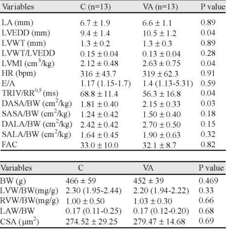
Table 1 summarises vitamin A status in plasma, liver and heart after birth and at the moment of MI. Both groups had normal liver and heart retinol values at birth. At the time of experimental MI, the rats fed vitamin A-deficient diet had lower heart and liver retinol concentrations even with normal plasma retinol concentrations. In addition, at
the time of MI, VA rats showed reduced retinoic acid concentration in the heart (C = 0.193 ± 0.054 µmol/kg of tissue and VA =0.063 ± 0.019 µmol/kg of tissue, P = 0.004). There was no difference in the body weight (BW) at infarction moment (C=210 (190 - 227) g, VA=220 (206 – 230) g; p=0.28) or in infarct size (C=36.9 ± 9.6 %, VA=36.3 ± 8.9 %; p=0.88) between the two groups. Mortality during the 3-month period after infarction did not differ between the groups (C=48%, VA=50%; p=0.89).
Table 2 summarises echocardiographic data. The VA group showed higher aorta diameter (C=3.4 (3.1-3.6) mm, VA=3.5 (3.4-3.9) mm; p=0.04), left ventricle diastolic
Table 3. Morphological data. C: infarcted
control animals; VA: infarcted dietary vitamin A restriction animals; LV: left ventricle; BW: body weight; LVW: left ventricular weight; RVW: right ventricular weight; LAW: left atrium weight; CSA: myocyte cross sectional area; Data are expressed as mean ± SD or medians (including the lower quartile and upper quartile).
Table 2.Echocardiographic data. C: infarcted control animals; VA: infarcted dietary vitamin A restriction animals; LV: left ventricle; LA: left atrium; LVEDD: LV end-diastolic dimension; LVWT: LV posterior wall thickness; LVMI: mass index; HR: heart rate; E: peak velocity of early ventricular filling; A: peak velocity of transmitral flow during atrial contraction; TRIV/RR0,5 : isovolumetric relaxation time normalised by heart rate; DASA/BW : diastolic area in short axis/body weight; SASA/BW: systolic area in short axis/body weight; DALA/BW: diastolic area in long axis/body weight; SALA/BW: systolic area in long axis/body weight; FAC: fractional area change. Data are expressed as mean ± SD or medians (including the lower quartile and upper quartile).
diameter (C=9.4 ± 1.4 mm, VA=10.5 ± 1.2 mm; p=0.04) and diastolic area in short axis /body weight (C=1.8 ± 0.4 cm2/kg, VA=2.1 ± 0.3 cm2/kg; p=0.03) than C group. Considering variables that represent diastolic function, VA group showed lower TRIV/RR0,5 than C group (C=68.8 ± 11.4 ms, VA=56.3 ± 16.8 ms; p=0.04). No differences were observed in the E/A ratio. Systolic function did not differ between the groups.
The morphometric data are shown in Table 3. The groups did not differ in myocyte cross-sectional area, BW, LVW/BW or RVW/BW. On the other hand, VA group showed higher collagen percentage than C group (C=2.8 ± 0.9 %, VA=3.7 ± 1.1 %, p=0.05; Figure 1).
There was no difference in the relative amount of myosin heavy chain β (C=47.3 ± 6.6 %, VA= 46.6 ± 5.8 %; p=0.85; Figure 2), and MMP-2 (C=37.0 (30.8-43.9)%, VA= 35.4 (30.2-36.7)%; p=0.730) or MMP-9 (C=1.12 ± 0.46%, VA= 1.73 ± 0.69%; p=0.173) activation (Figure 3) between the groups.
Considering the cytokine production, there were no differences in IFN-γ (C=321 ± 87 pg/ml, VA= 301 ± 54; p=0.69) and TNF-α (C=356 ± 107 pg/ml, VA= 336 ± 71 pg/ml; p=0.73) cardiac levels between the groups.
Discussion
The present study aimed to evaluate the effect of a tissue vitamin A insufficiency on post-infarction ventricular remodeling. The main finding of this study is that a vitamin A-deficient diet, even when serum retinol is within the normal range, may not provide enough retinol to the heart. Additionally, differences in heart retinol levels were associated with morphological and functional abnormalities. Indeed, tissue vitamin A insufficiency intensified ventricular remodeling after myocardial
Fig. 1. Interstitial
collagen volume fraction in picrosirius red stained cardiac section – red colour for collagen fibres, yellow for myocytes. C: control infarcted rats; VA: infarcted dietary vitamin A-deficient animals. VA group presented higher collagen percentage than C group (C=2.8 ± 0.9 %, VA=3.7 ± 1.1 %, p=0.05). Data are expressed as mean ± SD.
Fig. 2. MHC: myosin heavy chain; C: control infarcted rats; VA: infarcted dietary vitamin A-deficient animals. There were no differences between the groups (p>0.05).
Fig. 3.Metalloproteinase-2 (MMP-2) and
infarction characterised by increased left chamber size, collagen accumulation, and diastolic dysfunction. Therefore, our data strongly suggest that the heart appears to be susceptible functionally to vitamin A insufficiency induced by diminished vitamin A intake and declining tissue vitamin A stores.
Considering the effects of vitamin A-deficient diet, a recent study showed that normal plasma retinol concentration alone is not sufficient to maintain lung vitamin levels when vitamin A intake is low. A diet deficient in Vitamin A may lead to lower delivery of this vitamin to target tissues via chylomicrons [16]. In addition, our group demonstrated that, in adult normal rats, differences in heart retinol levels induced by low intake of vitamin A were associated with physiological differences. Indeed, the tissue vitamin A deficiency induced ventricular remodeling characterized by increased left chamber size and ventricular dysfunction [17]. However, until now, the potential morphological and functional consequences of this event after MI were unknown.
In the present study, an important issue to be considered is related to vitamin A status. Our data showed that both groups had similar normal liver and heart retinol concentration at birth. Thus, this model of vitamin deficient diet did not induce embryonic vitamin A deficiency. Likewise, our protocol did not induce vitamin A deficiency in the rat after birth, since that the restricted group had serum retinol values similar to those of the control group at the moment of MI. On the other hand, a vitamin A-deficient diet results in lower liver and heart vitamin A concentration, despite adequate levels of plasma retinol, a finding that indicates the importance of the dietary vitamin A to provide enough retinol to vitamin A requiring target tissues. Importantly, this insufficiency of tissue vitamin A concentration induced adverse ventricular remodeling after myocardial infarction, characterised by increased left chamber size and mass, collagen accumulation, and diastolic dysfunction.
Regardless of the complexity of the remodeling process, after MI the term is frequently used as a synonym for ventricular dilation [1-3]. Consequently, our data indicate that a tissue vitamin A insufficiency intensifies this deleterious process after MI. The left ventricular enlargement after MI could be explained by eccentric remodeling due to myocyte hypertrophy and/or myocyte slippage due, in turn, to MMP activation. In our study, hypertrophy was assessed by mass index, left ventricular weight adjusted for body weight, and myocyte cross sectional area. However, the left ventricular weight is
not a reliable variable of hypertrophy in this model, due to reabsorption of the necrotic tissue and collagen accumulation in viable myocardium. Likewise, there was no difference between the groups in relation to myocyte cross sectional area. Nonetheless, in this model, we must to consider that myocyte increases preferentially in series. On the other hand, the analyses of variables showed increased mass index induced by cardiac vitamin A deficiency. Therefore, our study suggests the presence of eccentric hypertrophy induced by tissue vitamin A insufficiency.
The myocardial extracellular matrix surrounds and interconnects muscle fibres, cardiac myocytes, and myofibrils. Extracellular matrix degradation by MMP has been associated with slippage of myocyte fascicles and left ventricular wall thinning [32]. Extracellular matrix degradation paralleled by an abnormal collagen accumulation has been reported after MI, where left ventricular enlargement also occurs [32]. Importantly, this abnormal collagen accumulation is associated with myocardial dysfunction, beginning with diastolic dysfunction followed by abnormalities in systolic function [33-35]. However, in our study, despite left ventricular enlargement, we did not find evidence of MMP-2 and MMP-9 activation in the VA group. Importantly, we cannot comment on the participation of other MMPs in ventricular remodeling induced by tissue vitamin A insufficiency.
In some heart failure models, cytokine production can modulates left ventricular remodeling. Indeed, increased IFN-γ, and mainly TNF-α levels is associated to left ventricular dysfunction, cachexia, activation of fetal genes program, apoptosis, hypertrophy, and fibrosis [36]. However, in this model, the remodeling intensification was not associated to altered IFN-γ and TNF-α levels. Therefore, at this point, our data suggest that other mechanisms not related to IFN-γ and TNF-α stimulation might be involved in this remodeling process. An important issue is that, in normal rats with the same protocol of vitamin A dietetic restriction, tissue vitamin A insufficiency resulted in cardiac remodeling associated with increased levels of cardiac IFN-γ and TNF-α [17]. Therefore, our data suggest that the mechanisms involved in the remodeling process induced by tissue vitamin A insufficiency in normal rats and after MI might be different.
References
1 Pfeffer MA, Braunwald E: Ventricular
remodeling after myocardial infarction: experimental observations and clinical implications. Circulation 1990;81:1161-1172.
2 Zornoff LAM, Paiva SAR, Duarte DR,
Sparado J: Ventricular remodeling after myocardial infarction: concepts and clinical implications. Arq Bras Cardiol 2009;92:157-164.
3 Cohn JN, Ferrari R, Sharpe N: Cardiac
remodeling- concepts and clinical implications: a consensus paper from an international forum on cardiac remodeling. J Am Coll Cardiol 2000;35:569-582.
4 Pfeffer JM, Pfeffer MA, Braunwald E:
Influence of chronic captopril therapy on the infarcted left ventricle of the rat. Circ Res 2002;57:84-95.
5 Auricchio A, Spinelli JC, Trautmann SI,
Kloss M: Effect of cardiac resynchronization therapy on ventricular remodeling. J Card Fail 2002;8:S549-555.
6 Abdulla J, Barlera S, Latini R,
Kjoller-Hansen L, Sogaard P, Christensen E, Kober L, Torp-Pedersen C: A systematic review: Effect of angiotensin converting enzyme inhibition on left ventricular volumes and ejection fraction in patients with a myocardial infarction and in patients with left ventricular dysfunction. Eur J Heart Fail 2007;9:129-135.
7 Oie E, Bjonerheim R, Grogaard HK,
Kongshaug H, Smiseth OA, Attramadal H: ET-receptor antagonism, myocardial gene expression, and ventricular remodeling during CHF in rats. Am J Physiol 1998;275:H868-877.
8 Bristow MR: Beta-adrenergic blockade in
chronic heart failure. Circulation 2000;101:558-569.
9 Wang HJ, Zhu YC, Yao T: Effects of
all-trans retinoic acid on angiotensin II-induced myocyte hypertrophy. J Appl Physiol 2002;92:2162-2168.
1 0 De Paiva SA, Zornoff LAM, Okoshi MP,
Okoshi K, Matsubara LS, Matsubara BB, Cicogna AC, Campana AO: Ventricular remodeling induced by retinoic acid supplementation in adult rats. Am J Physiol Heart Circ Physiol 2003;284:H2242-2246.
1 1 Zhou MD, Sucov HM, Evans RM, Chien
KR: Retinoid-dependent pathways suppress myocardial-cell hypertrophy. Proc Natl Acad Sci U S A 1995;92:7391-7395.
1 2 Choudhard R, Baker KM, Pan J: All-trans
retinoic acid prevents angiotensin II- and mechanical stretch-induced reactive oxygen species generation and cardiomyocyte apoptosis. J Cell Physiol 2008;215:172-181.
1 3 Choudhard R, Palm-Leis A, Scott RC 3rd,
Guleria RS, Rachut E, Baker KM, Pan J: All-trans retinoic acid prevents development of cardiac remodeling in aortic banded rats by inhibiting the renin-angiotensin system. Am J Physiol Heart Circ Physiol 2008;294:H633-644.
dysfunction. Although no difference was observed in E/ A ratio, TRIV/RR0,5 was reduced, suggesting a restrictive pattern in the VA group. Therefore, our data suggest that diastolic dysfunction already presented in the MI model, when infarct size is greater than 20%, was intensified by vitamin A-deficient diet. There are some factors influencing ventricular relaxation. Firstly, relaxation is influenced by load. In addition, the cytosolic calcium level must decrease. Furthermore, given the importance of the viscoelastic properties of the myocardium, the collagen amount can influence relaxation. Our study did not analyze cytosolic calcium or load conditions. On the other hand, tissue vitamin A insufficiency was associated with increased collagen amount in the non infracted area. Therefore, our data suggest that, at least in part, the impaired diastolic function in the VA group could be explained by increased fibrosis. Considering the potential mechanisms to explain how cardiac vitamin A insufficiency affects fibrosis, it has been shown that collagen synthesis induced by angiotensin II was inhibited by RA, indicating that RA-mediated signaling is involved in regulating cardiac fibrosis [15].
Altered expression of contractile proteins can also induce myocardial dysfunction. In adult rats α MHC
expression is higher than 90% [37-39]. A myosin isoform shift from α to β is observed in ventricular remodeling and heart failure [39]. In the present study, there was high βMHC expression in both groups, probably due to MI. However, in this case, the vitamin A-deficient diet did not shift MHC distribution. In accordance with this MHC isoform distribution, systolic function was not different between the groups.
Another important point to be considered is the potential implication of our results. Although vitamin A deficiency is not usual in the adult population, dietetic restriction of vitamin A may represent a relevant issue in several countries. For instance, in Brazil, almost 60% of the population ingests less than half of the amount recommended for vitamin A [40]. Therefore, our results suggest that this phenomenon may be accompanied by both morphological and functional cardiac abnormalities after MI.
1 4 Paiva SAR, Matsubara LS, Matsubara BB, Minicucci MF, Azevedo PS, Campana AO, Zornoff LA: Retinoic acid supplementation attenuates ventricular remodeling after myocardial infarction in rats. J Nutr 2005;135:2326-2328.
1 5 Pan J, Baker KM: Retinoic acid and the
heart. Vitam Horm 2007;75:257-283.
1 6 Ross AC, Li N: Lung retinyl ester is low
in young adult rats fed a vitamin A-deficient diet after wearing, despite neonatal vitamin A supplementation and maintence of normal plasma retinal. J Nutr 2007;137:2213-2218.
1 7 Azevedo PS, Minicucci MF,
Chiuso-Minicucci F, Justulin Jr LA, Matsubara LS, Matsubara BB, Novelli EL, Seiva L,
EbaidG, Campana AO, Zornoff LAM,
Paiva SAR: Ventricular remodeling induced by tissue vitamin A deficiency in rats. Cell Physiol Biochem 2010;26:395-402.
1 8 Gardner EM, Ross AC: Dietary vitamin
A restriction produces marginal vitamin A status in young rats. J Nutr 1993;123:1435-1443.
1 9 Zornoff LAM, Matsubara BB, Matsubara
LS, Paiva AS, Spadaro J: Early rather than delayed administration of lisinopril protects the heart after myocardial infarction in rats. Basic Res Cardiol 2000;95:208-214.
2 0 Lang RM, Bierig M, Devereaux RB,
Flachskampf FA, Foster E, Pellikka PA, Picard MH, Roman MJ, Seward J, Shanewise JS, Solomon SD, Spencer KT, Sutton MS, Stewart WJ: Recommendations for chamber quantification: a report from the American Society of Echocardiography’s Guidelines and Standards Committee and the Chamber Quantification Writing Group, developed in conjunction with the European Association of Echocardiography, a branch of the European Society of Cardiology. J Am Soc Echocardiogr 2005;18:1440-1463.
2 1 Solomon SD, Greaves SC, Ryan M, Finn
P, Pfeffer MA, Pfeffer JM: Temporal dissociation of left ventricular function and remodeling following experimental myocardial infarction in rats. J Card Fail 1999;5:213-223.
2 2 Castardeli E, Duarte DR, Minicucci MF,
Azevedo PS, Matsubara BB, Matsubara LS, Campana AO, Paiva SA, Zornoff LA: Tobacco smoke-induced left ventricular remodeling is not associated with metalloproteinase-2 or -9 activation. Eur J Heart Fail 2007;9:1081-1085.
2 3 Oh BH, Ono S, Rockman HR, Ross J Jr:
Myocardial hypertrophy in the ischemic zone induced by exercise in rats after coronary reperfusion. Circulation 1993;87:598-607.
2 4 Spadaro J, Fishbein MC, Hare C, Pfeffer
MA, Maroko PR: Characterization of myocardial infarcts in the rat. Arch Pathol Lab Med 1980;104:179-183.
2 5 Tang G, Krisky NI: Differentiation
between central and excentric cleavage og beta-carotene. Methods Enzimol 1993;214:69-74.
2 6 Yeum KJ, Ahn SH, Rupp de Paiva SA,
Lee-Kim YC, Krinsky NI, Russell RM: Correlation between carotenoid concentrations in serum and normal breast adipose tissue of women with benign breast tumor or breast cancer. J Nutr 1996;128:1920-1926.
2 7 Yeum KJ, Booth SL, Sadowski JA, Liu C,
Tang G, Krinsky NI, Russell RM: Human plasma carotenoid response to the ingestion of controlled diets high in fruits and vegetables. Am J Clin Nutr 1996;64:594-602.
2 8 Vescovo G, Ceconi C, Bernocchi P, Ferrari
R, Carraro U, Ambrosio GB, Libera LD: Skeletal muscle myosin heavy chain expression in rats with monocrotaline-induced cardiac hypertrophy and failure. Relation to blood flow and degree of muscle atrophy. Cardiovasc Res 1998;39:233-241.
2 9 Bradford MM: A rapid and sensitive
method for the quantitation of microgram quantities of protein utilizing the princi-ple of protein-dye binding. Anal Biochem 1976;72:248-254.
3 0 Reiser PJ, Kline WO: Electrophoretic
separation and quantitation of cardiac myosin heavy chain isoforms in eight mammalian species. Am J Physiol 1998;274:1048-1053.
3 1 Tyagi SC, Matsubara L, Weber KT: Direct
extraction and estimation of collagenase(s) activity by zymography in microquantities of rat myocardium and uterus. Clin Biochem 1993;26:191-198.
3 2 Janicki JS, Brower GL: The role of
myocardial fibrillar collagen in ventricular remodeling and function. J Card Fail 2002;8:S319-325.
3 3 Weber KT, Brilla CG: Pathological
hypertrophy and cardiac interstitium. Fibrosis and rennin-angiotensin-aldosterone system. Circulation 1991;83:1849-1865.
3 4 Janicki JS, Matsubara BB: Myocardial
collagen and left ventricular diastolic dysfunction; in Gaash W, LeWinter M (eds): Left Ventricular Diastolic Dysfunction and Heart Failure,
Philadelphia, Lea & Febiger, 1994, vol
1, pp 125-140.
3 5 Pfeffer MA, Pfeffer JM, Fishbein MC,
Fletcher PJ, Spadaro J, Kloner RA, Braunwald E: Myocardial infarct size and ventricular function in rats. Circ Res 1979;44:503-512.
3 6 Mann DL: Tumor necrosis factor-induced
signal transduction and left ventricular remodeling. J Card Fail 2002;8:S379-384.
3 7 Gupta MP: Factors controlling
myosin-isoform shift during hypertrophy and heart failure. J Mol Cell Cardiol 2007;43:388-403.
3 8 Lompre AM, Nadal-Ginard B, Mahdavi
V: Expression of the cardiac ventricular alpha- and beta-myosin heavy chain genes is developmentally and hormonally regulated. J Biol Chem 1984;259:437-446.
3 9 Pagani ED, Julian FJ: Rabbit papillary
muscle myosin isozymes and the velocity of muscle shortening. Circ Res 1984;54:586-594.
4 0 Ramalho RA, Flores H, Saunders C: