Hospital Universitário Pedro Ernesto – UERJ e Instituto Luiz Alberto Coimbra de Pós-Graduação em Engenharia - UFRJ
Mailing address: Paulo Roberto Benchimol Barbosa - Rua Pompeu Loureiro, 36/702 22061-000 - Rio de Janeiro, RJ, Brazil – E-mail: [email protected]
English version by Stela Maris C. e Gandour
Objective - The initial site of myocardial infarction (MI) may influence the prevalence of ventricular late potentials (VLP), high-frequency signals, due to the time course of ventricular activation. The prevalence of VLP in a period of more than 2 years after acute MI was assessed focusing on the initially injured wall .
Methods - The prevalence of VLP in a late phase after MI (median of 924 days) in anterior/antero septal and infe -rior/infero -dorsal wall lesion was analyzed using signal-averaged electrocardiogram in time domain. The diag-nostic performance of the filters employed for analysis on was tested at high-pass cut-off frequencies of 25 Hz, 40 Hz and 80 Hz.
Results - The duration of the ventricular activation and its terminal portion were larger in inferior than ante-rior infarction, at high-pass cut-off frequencies of 40 Hz and 80 Hz. In patients with ventricular tachycardia, these differences were more remarked. The prevalence of ventri-cular late potentials was three times greater in inferior than anterior infarction.
Conclusion - Late after myocardial infarction, the prevalence and the duration of ventricular late potentials are greater in lesions of inferior/infero-dorsal than ante-rior/antero-septal wall confirming their temporal pro-cess, reflecting their high-frequency content.
Key words: signal-averaged electrocardiogram, myocar-dial infarction, ventricular arrhythmia
Arq Bras Cardiol, volume 78 (nº 4), 358-63, 2002
Paulo Roberto Benchimol Barbosa, Marcos Oliveira de Sousa, Eduardo Correa Barbosa,
Alfredo de Souza Bomfim, Paulo Ginefra, Jurandir Nadal
Rio de Janeiro, RJ - Brazil
Analysis of the Prevalence of Ventricular Late Potentials
in the Late Phase of Myocardial Infarction Based on
the Site of Infarction
Ventricular late potentials are low-amplitude high-fre-quency signals originating in damaged regions of the ventri-cular myocardium, where the conduction of electrical stimuli occurs in a slow and fragmented manner. Ventricular late potentials are markers of reentry ventricular arrhythmias 1-3.
Due to the low velocity of conduction, these potentials ex-ceed the duration of ventricular activation and are detected in the ST-segment. The use of statistical techniques, in which 2 to 3 hundred sequential QRS complexes detected on the bo-dy surface are independently coherent-averaged, allows am-plification and identification of these signals because of the reduction in the intensity of the instrumentation noises 4-7.
The identification of ventricular late potentials in pa-tients with a transmural myocardial infarction is of major in-terest 3,8-10. The myocardial remodeling process that follows
transmural myocardial infarction is characterized by fibrosis, redistribution of fibers in the injured region, and re-sidual metabolic alterations not only plays a major role in segmentary myocardial function but also provides a favo-rable environment for the development of reentry circuits 11.
Considering these markers, preliminary studies suggest that transmural myocardial infarctions involving the inferior-inferodorsal wall have a higher prevalence of ventricular late potentials than those involving the anterior-anteroseptal wall. These findings are based on differences related to the start of electrical activation in the affected regions 12,13.
However, studies relating the prevalence of ventricular late potentials after transmural myocardial infarction with the affected wall show some conflicting results. Some studies have focused attention on the occurrence of ventricular late potentials in the first 12 months after transmural myocardial infarction when the myocardial remodeling process is still underway 14. Data on the prevalence of ventricular late
selection of the p atients surviving an infarction, but also represents a marker of the scar established 15.
Our study was carried out to assess the prevalence of ventricular late potentials in a period greater than 2 years af-ter transmural myocardial infarction, focusing on the initial-ly affected wall.
Methods
We assessed 98 patients after their first transmural myocardial infarction (median of 924 days) who were conse-cutively referred to the Department of Electrocardiology and Arrhythmias of the Hospital Universitário Pedro Er-nesto of the Universidade do Estado do Rio de Janeiro (HU-PE-UERJ) from June 1995 to June 2000. These patients underwent surface electrocardiography, vector cardio-graphy, one- and two-dimensional echocardiocardio-graphy, and signal-averaged electrocardiography, and were assessed in a retrospective case-control study adjusted for age and sex. The patients studied are part of the signal-averaged elec-trocardiography database (BDECG_AR) developed in the Department of Electrocardiology and Arrhythmias 16.
A retrospective analysis was performed of patients’ medical records, comprising clinical history, physical exami-nation, and complementary examinations at the HUPE-UERJ. Transmural myocardial infarctions were documented by the presence of necrotic regions on surface electrocar-diographic leads related to the damaged wall, due to modifi-cations in the vectorcardiographic loop and alterations in segmentary contractility, observed on two-dimensional echocardiography. Thirty-seven patients had anterior or anteroseptal transmural myocardial infarction (group A), and 61 patients had inferior or inferodorsal transmural myo-cardial infarction (group B) (table I) with an evolution greater than 2 years from the time of the initial acute event. All patients had sinus rhythm, left ventricular ejection fraction > 40%, underwent one-dimensional echocardiogra-phy, and none of them had complete bundle-branch block on the surface electrocardiography. Patients with electrocar-diographic signs indicating transmural myocardial infarc-tion in other locainfarc-tions, such as anterior and inferior crossed myocardial infarction, isolated posterior myocardial
in-farction, and isolated high lateral myocardial infarction were excluded. The medians of the times elapsed from the time of the acute event in groups A and B were, respectively, 1,095 and 848 days.
The prevalence of sustained monomorphic ventricular tachycardia was defined as: (1) presence of repetitive ven-tricular arrhythmia documented in the late phase of transmu-ral myocardial infarction (median of 854 days) and (2) hemo-dynamic instability requiring electrical cardioversion. This prevalence and the respective distribution of ventricular late potentials were evaluated in groups A and B. All patients wi-th sustained monomorphic ventricular tachycardia analy-zed were using Vaughan-Williams group III antiarrhythmic drugs.
A group of 43 patients with ischemic myocardial di-sease and no previous clinically documented acute myo-cardial infarction (coronary artery disease, group C), ejec-tion fracejec-tion > 40%, no bundle-branch block, and similar dis-tribution in regard to age and sex, was compared with groups A and B (table I). No patient in this group had altera-tions on the surface electrocardiogram, vector cardiogram, and echocardiogram compatible with transmural myocar-dial infarction.
The 12-lead surface electrocardiography was perfor-med with 2N amplification and velocity of 25 mm/s, using ECAPS 12 model Nyhon-Kohden equipment, with a maxi-mum interval of 30 days from the signal-averaged electro-cardiogram. Surface electrocardiography was used to eva-luate the existence of intraventricular conduction disorders (complete bundle-branch blocks, defined as a QRS complex longer than 0.12 seconds) and to identify markers of old myocardial infarctions and their respective injured walls (pathological Q-waves defined as those with a duration > 0.04 seconds and amplitude > 2 mm, on the leads accounting for the region). We assessed the leads from V1 to V4 in group A, and the leads D2, D3, and aVF in group B.
Vector cardiography was performed using Frank XYZ leads with 1507c Programmer model Hewlett Packard equip-ment, with the patient in the supine position, in a quiet environment, within a maximum interval of 30 days from the performance of signal-averaged electrocardiography. Vector cardiography was used to assess the existence of intraventricular conduction disorders and to identify old myocardial infarction markers and their respective injured walls. The diagnostic criteria used to analyze the vector cardiogram may be found in the literature 17.
One- and two-dimensional echocardiography were used to respectively analyze left ventricular ejection frac-tion and segmentary contractility with the Apogee CX 200 (ATL, USA) within a maximum 30-day interval after the sig-nal-averaged electrocardiography. The echocardiographic cuts and the analysis of the one- and two-dimensional ima-ges were performed by a trained observer using the routine procedures of the Echocardiography Department of the Car-diology Service of the HUPE-UERJ.
Identification of the injured region was based on the following criteria: (1) presence of pathological Q waves in at
Table I - Patient distribution, prevalence of ventricular tachycar-dia, and ventricular late potentials in the groups studied
Group A Group B Group C p‡
N 37 61 43
Age (years) ¢ 56.3±13.0 56.9±12.2 59.7±11.4 NS
Sex (F/M) 8/28 17/44 13/30 NS
VT (%)* 3 (8.1%) 8 (13.1%) 0 (0%) NS VLP – 25Hz * * 14 (37.8%)† 24 (39.3%)† † 15 (34.9%)† NS VLP– 40Hz * * 10 (27.0%)† 22 (36.1%)† † 11 (25.6%)† NS VLP– 80Hz * * 11 (29.7%)† 35 (57.4%)† † 19 (44.2%)† 0.008
least 2 leads that explore the same electrical region on the electrocardiogram; (2) loss of activation potentials with deviation of the vector cardiographic loop to the outside of the analyzed region; and (3) echocardiographic signs of segmentary alterations in the contractility of the analyzed region concordant with those found on vector cardiography and electrocardiography.
Signals of the signal-averaged electrocardiogram were obtained with the Predictor IIc (ART-Corazonix, USA) using Frank XYZ orthogonal leads, and they were coherent-ave-raged up to the noise level of 0.3 µV. The routine for signal acquisition was reported in a previous publication 18.
Signal-averaged electrocardiogram underwent time-domain analysis through the magnitude vector, in the high-pass cut-off frequencies of 25, 40, and 80 Hz, with a 4-pole Butterworth bidirectional filter. The variables obtained from the vector magnitude were the duration of the filtered QRS (DUR[ms]), duration of the potentials in the terminal region of ventricular activation below 40 µV (LAS40[ms]), and the value of the "root-mean-squared" of the potentials in the terminal 40 ms of ventricular activation (RMS40[µV]). These variables were analyzed based on cut values for each high-pass cut-off frequency according to Gomes et al 19. The
abnormal values for the variables analyzed DUR, LAS40, and RMS40 were, respectively, in the 25Hz band, >114 ms, >32 ms, and <25µV; in the 40Hz band, >114 ms, >38 ms, and <20 µV; and in the 80Hz band, >107 ms, >42 ms, and <17 µV. Identification of at least 2 abnormal variables in the vector magnitude defined the presence of ventricular late poten-tials in each frequency band.
Aiming at determining the best parameter to analyze the signal-averaged electrocardiogram, the patients with sustained monomorphic ventricular tachycardia after transmural myocardial infarction were analyzed in the high-pass cut-off frequencies of 25, 40, and 80 Hz, according to the above-cited criteria, and isolated, using the DUR or RMS40 parameters of the vector magnitude.
The diagnostic performance of the signal-averaged electrocardiogram was analyzed as an indicator of the presence of a myocardial lesion or a scar of a transmural myocardial infarction. This diagnostic capacity was assessed through the following indices: specificity (true negative examinations/total of control individuals), sensitivity (true positive examinations/total of patients), and total diagnostic accuracy (true negative examinations + true positive examinations /total of control + patients).
Transformations of variables have been reported in the literature to normalize asymmetric probability distributions. The variable RMS40 was transformed into a natural logarithm (Lnt) prior to analysis, due to its asymmetric probability distribution 20. This procedure not only statistically
normali-zes the distribution, but also reduces data variability, con-centrating them around the mean. The prevalence of ven-tricular late potentials and the mean values of the variables of the magnitude vector were compared between groups A and B for each high-pass cut-off band and analyzed, respectively, using the chi-square test to compare proportions and the
two-tailed Student t test to compare means. Odds ratio (OR) and its 95% confidence interval CI were assessed in the groups in which the prevalences of ventricular late potentials for a certain high-pass cut-off band had statistically signifi-cant differences continuous variables are expressed as mean ± SD. Alpha error level was fixed at 0.05. The following software was used for analysis: EPI info 6.04 version (CDC, USA), MS Excel 97 (Microsoft, USA), and Statgraphics Plus 4.0 version (Manugistic, USA).
Results
The prevalence of ventricular late potentials in groups A, B, and C according to the high-pass cut-off bands is shown in table I. In group B, the prevalence of ventricular late potentials was higher at 80 Hz (57.4%) than in the other bands (39.3% at 25 Hz and 36.1% at 40 Hz, χ2=6.51, p=0.04).
On the other hand, at 80 Hz, the prevalence of ventricular late potentials was higher in group B than in group A [χ2=7.07, p=0.008 (table I) and OR=3.18, (95% CI 1.24-8.42),
χ2=6, p=0.01].
The distribution of patients with sustained monomor-phic ventricular tachycardia did not show statistically sig-nificant differences between groups A and B (χ2=0.19,
p=NS).
The vector magnitude parameters for each group and respective high-pass cut-off bands are shown in table II. The variables in groups A and C showed no significant dif-ferences. However, LAS40 was wider in group B than in group A in the 40Hz and 80Hz bands (respectively, t=2.67, p=0.004 and t=2.26, p=0.02).
The diagnostic parameters of sensitivity, specificity, and total accuracy, which were analyzed as indicators of myocardial lesions, are shown in table III. The comparisons of the variables DUR, RMS40 Lnt, and LAS40 among the patients with sustained monomorphic ventricular
tachycar-Table II - Magnitude vector parameters according to the study group and the cut-off band of the filter used for analysis
Group A
DUR (ms) RMS40 Lnt† LAS40 (ms) 25Hz filter † † 103.7±13.9 3.5±1.0 30.5±14.8 40Hz filter 98.0±13.7* 3.5±1.0 31.9±13.3* 80Hz filter 97.0±13.9* 2.7±1.0 40.4±14.2*
Group B
DUR (ms) RMS40 Lnt† LAS40 (ms) 25Hz filter 107.0±12.9 3.5±0.8 29.0±11.9 40Hz filter 106.7±14.3* 3.2±0.8 36.2±14.0* 80Hz filter 103.3±13.0* 2.5±0.9 47.7±14.0*
Group C
DUR (ms) RMS40 Lnt† LAS40 (ms) 25Hz filter 108.8±15.5 3.6±0.8 30.1±10.3 40Hz filter 103.2±13.3 3.3±0.9 33.5±12.7 80Hz filter 100.6±14.9 2.6±0.7 39.5±13.0
dia in the late period after acute myocardial infarction in groups A and B are shown in table IV. Except for the variable DUR at 25Hz, all other variables showed statistically significant differences (table IV). In regard to episodes of sustained monomorphic ventricular tachycardia, no significant differences were observed in the prevalence of ventricular late potentials between groups A and B. In group A, no patient with sustained monomorphic ventricu-lar tachycardia was identified with ventricuventricu-lar late potentials at 25Hz and 40Hz high-pass cut-off bands, both using con-ventional definitions and analyzing RMS40. At 80Hz band, ventricular late potentials were identified in 1 out of 3 (χ2=0.01; p=0.99) patients, using conventional definitions;
however, when analyzing RMS40, 2 out of 3 (χ2=0.75; p=0.4)
patients met the criteria for ventricular late potentials. In group B patients with sustained monomorphic ventricular tachycardia, ventricular late potentials were identified in 4 out of 8 patients (χ2=3; p=0.08) at 40Hz and 80Hz high-pass
cut-off bands using the conventional definitions, in 5 out of 8 patients at 40Hz high-pass cut-off band (χ2=4.65;
p=0.03), and in 6 out of 8 patients at 80Hz high-pass cut-off band, the 2 latter being evaluated with the isolated RMS40 variable (χ2=6.67; p=0.01). In the remaining high-pass
cut-off bands, the presence of ventricular late potentials
showed no statistically significant results in the definitions analyzed in both groups.
Discussion
The introduction of signal-averaged electrocardiogra-phy as a noninvasive diagnostic method in cardiology ena-bled the identification of micropotentials originating in elec-trically unstable regions of the injured myocardium, widening the horizons and increasing the chances of understanding the bioelectrical phenomena originating in the heart 21.
The characteristics related to myocardial electrical conduction after myocardial infarction depend both on the damaged area and on the intensity of the alteration in its surrounding tissues. Therefore, the location of the myocar-dial lesion is expected to influence the presence of ventri-cular late potentials on signal-averaged electrocardiography. The correlation between the lesion and ventricular late po-tentials depends on clinical factors related to the disease, on local electrophysiological factors (ischemia), and on the type of processing applied to the acquired electrocardiographic signals. In this context, we highlight the use of drugs that spare the myocardium in the acute phase 15,22, the start of
depolarization of the affected site in relation to the total duration of ventricular activation, the type of filter used to analyze the events 19,23, and the instantaneous heart rate 4,24.
Features like the low ability to characterize the infarct site based on the isolated analysis of surface electrocardiogra-phy should be considered at the time of group division, because it may lead to inconsistent results 10.
Ventricular late potentials have been analyzed in seve-ral frequency bands aiming not only to identify the fre-quency content of these signals, but also to investigate the region of the frequency spectrum that allows its identifica-tion with a higher diagnostic efficiency 19,20. This type of
investigation is mainly justified from the viewpoint of elec-trocardiographic signal processing, because high-pass cut-off filters with lower cut-off frequencies (25Hz) preserve the high-energy QRS characteristics, while higher cut-off frequencies (80Hz) remove the high energies and preserve high frequency signals originating in small groups of myo-cardial fibers in regions of slow and fragmented conduction. In our study, the increase in the global prevalence of ventricular late potentials was evidenced only in the high-pass cut-off filter of 80Hz (tab. I) and was accompanied by a significant widening of the LAS40 variable in the 40Hz and 80Hz high-pass cut-off bands (tab. II). An odds ratio of 3.18 (p=0.01) indicates that individuals after a transmural myo-cardial infarction of the inferior wall triple their chance of having ventricular late potentials on the time-domain analy-sis of the signal-averaged electrocardiogram as compared with individuals after transmural myocardial infarction of the anterior wall in the same period.
The distribution of ventricular late potentials accor-ding to the region affected in the acute event was analyzed by Gomes et al 25 in patients with myocardial infarction
within 1 year of evolution, and they found results similar to
Table III – Diagnostic value of VLP according to the study group and the cut-off band of the filter used for analysis
25-250Hz 40-250Hz 80-250Hz
Specificity (group C) 65.1% 74.4% 55.8 % Sensitivity (group A) 37.8% 27% 29.7%* Sensitivity (group B) 39.3% 36.1% 57.4%* Sensitivity (all)† 40% 34.5% 50 % Total accuracy (group A) 40.4% 40.4% 35.7%* Total accuracy (group B) 50% 51.9% 56.7%* Total accuracy (all) † 47.1% 45.8% 51.6 %
†All - all patients with Q-wave myocardial infarction (group A+B); *p<0.05 (group A versus group B).
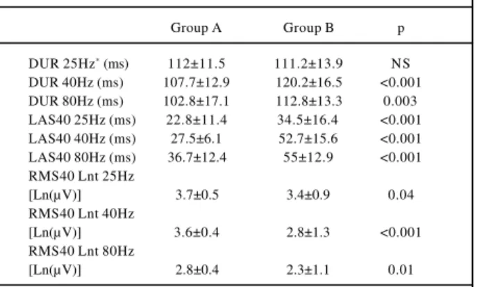
Table IV – Comparison of the magnitude vector variables in patients with sustained monomorphic ventricular tachycardia in
groups A and B according to the high-pass cut-off frequency of the filter used
Group A Group B p
DUR 25Hz* (ms) 112±11.5 111.2±13.9 NS DUR 40Hz (ms) 107.7±12.9 120.2±16.5 <0.001 DUR 80Hz (ms) 102.8±17.1 112.8±13.3 0.003 LAS40 25Hz (ms) 22.8±11.4 34.5±16.4 <0.001 LAS40 40Hz (ms) 27.5±6.1 52.7±15.6 <0.001 LAS40 80Hz (ms) 36.7±12.4 55±12.9 <0.001 RMS40 Lnt 25Hz
[Ln(µV)] 3.7±0.5 3.4±0.9 0.04
RMS40 Lnt 40Hz
[Ln(µV)] 3.6±0.4 2.8±1.3 <0.001 RMS40 Lnt 80Hz
[Ln(µV)] 2.8±0.4 2.3±1.1 0.01
ours. Rosas et al 10 analyzed patients with anterior and
in-ferior acute myocardial infarction aiming to assess the rela-tion between coronary artery patency and the presence of ventricular late potentials. These authors found differences in the results of the signal-averaged electrocardiogram between the 2 groups, both in the time and frequency doma-ins. However, Vatterott et al 26, using a high-pass cut-off
band of 40Hz, found no difference in the distribution of ven-tricular late potentials after acute myocardial infarction rela-ted to the damaged wall. Results similar to those observed in our study were found by Lander et al 27 and Savard et al 28.
The correlation between ventricular late potentials and sustained monomorphic ventricular tachycardia has been widely discussed in the literature 2-3,13,18. One week after
transmural myocardial infarction, approximately 30% to 60% of patients have ventricular late potentials on signal-avera-ged electrocardiography. However, within the first year after transmural myocardial infarction, both sensitivity and the positive predictive value of ventricular late potentials for sustained monomorphic ventricular tachycardia or sudden death are very low. This fact indicates that ventricular late potentials are better markers of structurally established myocardial lesions, which may eventually become a subs-trate for arrhythmias through a reentry mechanism, than proper clinical markers of repetitive ventricular arrhythmia. Considering the presence of ventricular late potentials after transmural myocardial infarction, the mechanisms triggering repetitive arrhythmia are not necessarily related to the pre-sence of these signals 11. We speculate that other factors,
such as unidirectional block 11 and an instantaneous heart
rate 24, associated with alterations in the pattern of
sympa-thovagal autonomic myocardial modulation may contribute to the transformation of the substrate. In group B patients with documented episodes of sustained monomorphic ven-tricular tachycardia, all variables of the vector magnitude analyzed had durations and voltages significantly lower and more prolonged than those in group A patients for all cut-off bands, except for the DUR variable at 25Hz. The pvalence of ventricular late potentials in group A did not re-gister significant values, but, in group B, this prevalence was significantly high at 40Hz and 80Hz cut-off bands using the conventional definitions and at 40Hz and 80Hz cut-off bands assessed on the isolated RMS40 variable. Conside-ring the reduced number of elements in each group, these findings support the electrophysiological principle that the ventricular activation of the anteroseptal regions occurs be-fore that occurring in the inferobasal regions. This streng-thens the hypothesis that fragmented potentials origina-ting in the first regions have their time course parallel to
ventricular activation, being covered during depolarization, while those originating later go beyond ordinary ven-tricular activation reaching the ST-segment.
A lower prevalence of ventricular late potentials is ex-pected in chronic coronary artery disease rather than after transmural myocardial infarction, because no anatomical substrate is supposed to exist in that disease. However, the high prevalence of ventricular late potentials in group C, si-milar to that observed in patients with anterior transmural myocardial infarction (group A), suggests that chronic is-chemia may cause structural myocardial lesions compatible with the concept of ischemic cardiomyopathy, which are not detected using some conventional methods 29.
All patients with episodes of sustained monomorphic ventricular tachycardia were using Vaughan-Williams gro-up III antiarrhythmic drugs. Even though these drugs pro-long ventricular late potentials 30,31, their expected effects
were considered not to influence the comparative results, because they were distributed in both groups in a similar manner. The use of thrombolytic drugs, the use of invasive procedures to identify the culprit coronary artery, and the prevalence of episodes of aborted sudden death and of sus-tained monomorphic ventricular tachycardia in the acute phase of transmural myocardial infarction were not investi-gated in this study. Even though the patients analyzed constituted a group of late survivors of transmural myocar-dial infarction, when the anatomical spectrum of myocarmyocar-dial lesions undergo significant modifications, we consider that the impact of these variables on the results observed requi-res further investigation.
The prevalence of ventricular late potentials observed in a late phase (>2 years) after the first transmural myocardial infarction is greater in the inferior-inferodorsal wall lesions than in the anterior-anteroseptal wall lesions. The identi-fication of these arrhythmogenic potentials is significantly influenced by the high-pass cut-off band used for the time-domain analysis of the signal-averaged electrocardiogram. In groups of patients after transmural myocardial infarction with documented clinical episodes of severe repetitive ventricular arrhythmias, ventricular late potentials are better identified at elevated cut-off frequencies for both anterior-anteroseptal wall lesions and inferior-inferodorsal wall lesions, confirming the high-frequency nature of these signals.
Acknowledgments
We thank Professor Justiniano Simões Lopes for re-viewing this manuscript and for important contributions.
References
1. Berbari EJ, Sherlag BJ, Hoper R, Lazzara R. Recordings from the body surface of arrhythmogenic ventricular activity during ST segment. Am J Cardiol 1978; 41: 697-702.
variables in signal-averaged ECG as a predictor of inducible sustained mono-morphic ventricular myocardial infarction. Circulation 1992; 86: 780-9. 4. Barbosa PRB. A signal-averaging system for surface ECG signal analysis.
Phys Med Biol 1994: 39: 412.
5. Barbosa PRB, Barbosa EC, Ginefra P. Avaliação do ruído do ecg-ar em pacientes com infarto agudo do miocárdio. Estudo preliminar. Rev SOCERJ 1997: 10(supl A): 28.
6. Barbosa PRB. Análise dos potenciais tardios da ativação ventricular baseada no histograma de intervalos RR modais. Tese de Mestrado, Programa de Engenharia Biomédica, COPPE/UFRJ, Rio de Janeiro, Brasil, 1997.
7. Dopico LR, Nadal J, Infantosi AFC. Análise dos potenciais tardios do eletrocar-diograma de alta resolução de pacientes chagásicos usando a média ponderada. Rev Bras Eng Biomed 2000; 16: 49-59.
8. Kuchar DL, Thorburn CW, Sammel NL. Late potentials detected after myocardial infarction: natural history and prognostic significance. Circulation 1986; 74: 1280-9.
9. Gomes JA, Mehra R, Barreca P, el-Sherif N, Hariman P, Holtzman R. Quantitative analysis of the high frequency components of the signal-averaged QRS complex in patients with acute myocardial infarction: a prospective study. Circulation 1985; 72: 105-11.
10. Rosas M, Hermosillo AG, Infante O, Kuri J, Cardenas M. Relationship between the site of a myocardial infarction and signal-averaged electrocardiogram indices. Int J Cardiol 1998; 63: 129-40.
11. El-Sherif N, Gough WB, Restivo M. Electrophysiologic correlates of ventricular late potentials. In: El-Sherif N, Turitto G (eds): High-Resolution Electrocardio-graphy. Mount Kisco, NY: Futura Publishing Co., Inc., 1992: 279-98. 12. Gomes JA, Horowitz SF, Millner M, Mochec J, Winters SL, Barreca P. Relation of
late potentials to ejection fraction and wall motion abnormalities in acute myocardial infarction. Am J Cardiol 1987; 59: 1071-4.
13. Gomes JA, Winters SL, Stewart D, Horowitz S, Millner M, Barreca P. A new noninvasive index to predict sustained ventricular tachycardia and sudden death in the first year after myocardial infarction: based on signal-averaged electrocardiogram, radionuclide ejection fraction and Holter monitoring. J Am Coll Cardiol 1987; 10: 349-57.
14. Sakai Y, Tsunoda K, Ishibashi I, et al. Time course of left ventricular remodeling after myocardial infarction: a two-dimensional echocardiographic study. Jpn Circ J 2000; 64: 421-9.
15. Zimmermann M, Sentici A, Adamec R, et al. Long-term prognostic significance of ventricular late potentials after a first acute myocardial infarction. Am Heart J 1997; 134: 1019-28.
16. De Sousa MO. O eletrocardiograma de alta resolução na fase crônica da cardiopa-tia isquêmica: importância de diferentes bandas-passantes na identificação de potenciais tardios da ativação ventricular. Tese de mestrado, FCM-UERJ, Rio de Janeiro, Brasil, 2000.
17. Tranchesi J. Eletrocardiograma Normal e Patológico: Noções de Vectorcardio-grafia. São Paulo: Atheneu Editora, 1967.
18. Barbosa EC, Barbosa PRB, Ginefra P, Albanesi-Fo FM. O eletrocardiograma de alta resolução no domínio da freqüência: utilização técnicas estatísticas de cor-relação espectral para a identificação de pacientes com taquicardia ventricular monomórfica sustentada. Arq Bras Cardiol 1998; 71: 595-9.
19. Gomes JA, Winters SL, Stewart D, Targonski A, Barreca P. Optimal bandpass fil-ters for time-domain analysis of the signal-averaged electrocardiogram. Am J Cardiol 1987; 60: 1290-8.
20. Caref EB, Turitto G, Ibrahim BB, et al. Role of bandpass filters in optimizing the value of the signal-averaged electrocardiogram as a predictor of the results of pro-grammed stimulation. Am J Cardiol 1989; 64: 16-26.
21. Scherlag BJ, Lazzara R. High-Resolution Electrocardiography: Historical Pers-pectives. In: El-Sherif N, Turitto G (eds): High-Resolution Electrocardiogra-phy. Mount Kisco, NY: Futura Publishing Co. Inc., 1992: 3-20.
22. Hochman JS, Buller CE, Sleeper LA, et al. Cardiogenic shock complicating acute myocardial infarction - etiologies, management and outcome: a report from the SHOCK Trial Registry. Should we emergently revascularize occluded coronaries for cardiogenic shock? J Am Coll Cardiol 2000; 36(3 suppl A): 1063-70. 23. Barbosa PRB, Barbosa EC, Ginefra P, Nadal J. Butterworth bi-directional and
BispecTM filters in the assessment of ventricular late potentials: a comparative study. Comput Cardiol 1999; 26: 579-82.
24. Barbosa PRB, Barbosa Filho J, Sá CAM, Nadal J. Assessment of ventricular late potentials in HIV positive patients based on the RR interval histogram. Comput Cardiol 1999; 26: 327-30.
25. Gomes JA, Winters SL, Martinson M, Mochec J, Stewart G, Jargoriski A. The prognostic significance of quantitative signal-averaged variables relative to clinical variables, site of myocardial infarction, ejection fraction and ventricular premature beats: a prospective study. J Am Coll Cardiol 1989; 13: 377-84. 26. Vatterott PJ, Bailey KR, Hammil CS. Improving the predictive ability of the
sig-nal-averaged electrocardiogram with a linear logistic model incorporating clini-cal variables. Circulation 1990; 81: 797-804.
27. Lander P, Gomis P, Goyal R, et al. Analysis of abnormal intra-QRS potentials: im-proved value for arrhythmogenic events with the signal-averaged electrocardio-gram. Circulation 1997; 95: 1386-93.
28. Savard P, Rouleau JL, Ferguson J. Risk stratification after myocardial infarction using signal-averaged electrocardiographic criteria adjusted for sex, age, and myocardial infarction location. Circulation 1997; 96: 202-13.
29. Tamura K, Tsuji H, Masui A, et al. Prevalence, resolution and determinants of late potentials in patients with unstable angina and left ventricular wall motions ab-normalities. Am Heart J 1996; 131: 731-5.
30. Telichowski A, Banasiak W, Bobak J, et al. The effect of long term use of amioda-rone hydrochloride on time and frequency domain parameters of signal averaged electrocardiogram in patients with ischemia heart disease. Pol Merku-riusz Lek 1997; 2: 378-81.