CLINICAL SCIENCE
Heart Institute (InCor HCFMUSP), Acute Coronary Care Unit, Hospital das Clínicas da Faculdade de Medicina da Universidade de São Paulo - São Paulo/SP, Brazil.
Email: [email protected] Tel.: 55 11 3069.5058
Received for publication on November 26, 2009 Accepted for publication on December 07, 2009
EFFECT OF
Β
-BLOCKERS ON THE RISK OF
ATRIAL FIBRILLATION IN PATIENTS WITH ACUTE
MYOCARDIAL INFARCTION
Antonio Eduardo Pesaro, Alexandre de Matos Soeiro, Carlos Vicente Serrano, Roberto Rocha Giraldez, Renata Teixeira Ladeira, José Carlos Nicolau doi: 10.1590/S1807-59322010000300005
Pesaro AE, Soeiro AM, Serrano CV, Giraldez RR, Ladeira RT, NicolauJC. Effect of β-blockers on the risk of atrial ibrillation in patients with acute myocardial infarction. Clinics. 2010;65(3):265-70.
INTRODUCTION: Oral β-blockers improve the prognosis of patients with acute myocardial infarction, while atrial ibrillation worsens the prognosis of this population. The reduction of atrial ibrillation incidence in patients treated with β-blockers could at least in part explain the beneits of this drug.
OBJECTIVE: To investigate the effect of β-blockers on the incidence of atrial ibrillation in patients with acute myocardial infarction. METHODS: We analyzed 1401 patients with acute myocardial infarction and evaluated the occurrence or absence of atrial ibril-lation, the use of oral β-blockers and mortality during the irst 24 hours.
RESULTS: a) The use of β-blockers was inversely correlated with the presence of atrial ibrillation (ρ = 0.004; OR = 0.54). b) Correlations with mortality were as follows: 31.5% in patients with atrial ibrillation, 9.2% in those without atrial ibrilla-tion (ρ < 0.001; Odds Ratio = 4.52), and 17.5% in patients not treated with β-blockers and 6.7% in those who received the drug (ρ < 0.001; OR = 0.34). c) Adjusted Models: The presence of atrial ibrillation was independently correlated with mortality (OR = 2.48, ρ = 0.002). The use of β-blockers was inversely and independently correlated with mortality (OR = 0.53; ρ = 0.002). The patients who used β-blockers showed a lower risk of atrial ibrillation (OR = 0.59; ρ = 0.029) in the adjusted model.
CONCLUSION: The presence of atrial ibrillation and the absence of oral β-blockers increased in-hospital mortality in patients with acute myocardial infarction. Oral β-blockers reduced the incidence of atrial ibrillation, which might be at least partially responsible for the drug’s beneit.
KEYWORDS: Acute myocardial infarction; β-blockers; Atrial ibrillation; Mortality; Arrhythmias.
contraindications.
It has classically been accepted that the main mechanisms responsible for the beneicial effects of β-blockers involve blocking myocardial sympathetic stimulation, a decrease in heart rate and blood pressure and a benefit for heart remodeling.1 However, some recent publications have
suggested that the reduction in the incidence of arrhythmias after AMI, seen after β-blocker treatment, could also have a leading role in explaining the beneits obtained with the use of these drugs.2,11-17 It is also well demonstrated that atrial
ibrillation (AF) is considered a factor of poor prognosis in myocardial infarction, even in adjusted models.14,18-25
In this context, we analyzed data from 1401 patients with AMI in a single institution in order to investigate the effect of β-blockers on the incidence of AF and to analyze the relationships between mortality in 24 hours and 1) the use of β-blockers and 2) the incidence of AF.
INTRODUCTION
In the United States, more than one million people suffer an acute myocardial infarction (AMI) each year. Even with recent advances in diagnosis and treatment, global mortality rates are still around 30%.1 Several studies have shown that
the early use of β-blockers in patients with AMI is able to limit the extent of myocardial injury and improve the short- and long-term prognosis.1-9 Thus, routine use of β-blockers
METHODS
This study was a retrospective unicentric study. All included patients with AMI (n = 1401; median age = 63 years) were hospitalized in a single coronary intensive care unit and were prospectively included in a specific database. The patients were analyzed during the irst 24 hours after hospitalization. The deinitions and medical procedures followed the institutional routines, in accordance with recent guidelines. During this period, AF was treated with synchronized electrical cardioversion and the use of amiodarone in all patients.
A diagnosis of AMI was established when patients had chest pain at rest with concomitant ischemic ST-T changes and positive serum troponin.26 The left ventricular
ejection fraction (LVEF) was calculated by Doppler echocardiography (Simpson). Only the period when patients were hospitalized was analyzed, taking into account the presence of AF, the use of oral β-blockers and all-cause mortality. Categorical variables were compared using Pearson’s chi-square test or Fisher’s exact test, as indicated. The Student’s t test was used to compare continuous variables.
In adjusted models, the analyses were performed by stepwise logistic regression. In the irst model, AF was included as a dependent variable. The adjusted R2 was
0.114. The following variables were considered independent: LVEF, age, gender, previous diabetes mellitus, previous myocardial infarction, current myocardial infarction location, ST elevation, admission creatinine, coronary
surgery and angioplasty during hospitalization, use of aspirin, angiotensin-converting enzyme inhibitor and use of
β-blockers. In the second model, death was the dependent variable. AF was added to the other independent variables included in the irst model. The adjusted R2 of this model
was 0.226.
In all models, statistical signiicance was set at 5% (ρ < 0.05).
RESULTS
a. Population studied
As stated above, 1401 patients were examined. The average age of the population was 63.19 + 12.7 years and 1021 patients (72.9%) were men. The left ventricular ejection fraction was, on average, 51.1% + 15.5. During the hospitalization, 150 patients (10.7%) died.
b. Univariate analysis
b.1. Occurrence of AF
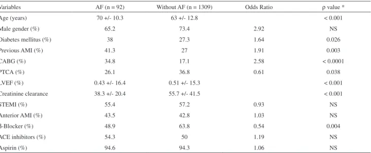
The baseline characters and univariate analysis of their association with AF is shown in Table 1. The use of β-blockers was inversely correlated with the presence of AF. As shown in Table 1, age, diabetes mellitus, previous AMI, coronary surgery, angioplasty, creatinine clearance and LVEF also had a signiicant correlation with the presence of AF. Excluding patients who used intravenous β-blockers followed by oral
β-blockers did not change the results; the ρ value was 0.009 for the correlation between oral β-blockers and AF.
Table 1 - Baseline characters in patients with AF and without AF
Variables AF (n = 92) Without AF (n = 1309) Odds Ratio ρ value *
Age (years) 70 +/- 10.3 63 +/- 12.8 < 0.001
Male gender (%) 65.2 73.4 2.92 NS
Diabetes mellitus (%) 38 27.3 1.64 0.026
Previous AMI (%) 41.3 27 1.91 0.003
CABG (%) 34.8 17.1 2.58 < 0.0001
PTCA (%) 26.1 36.8 0.61 0.038
LVEF (%) 0.43 +/- 16.4 0.51 +/- 15.3 < 0.001
Creatinine clearance 38.3 +/- 20.4 55.7 +/- 41.5 < 0.001
STEMI (%) 55.4 57.2 0.93 NS
Anterior AMI (%) 43.5 42.8 1.03 NS
β-Blocker (%) 48.9 63.8 0.54 0.004
ACE inhibitors (%) 54.3 50 1.19 NS
Aspirin (%) 94.6 94.3 1.06 NS
b.2. Mortality
As noted in Figure 1, 31.5% of the patients in the group that presented with AF died, compared to 9.2% in the group without arrhythmia (ρ < 0.001; Odds Ratio = 4.52). Figure 1 clearly shows an inverse correlation between the use of
β-blockers and mortality (17.5% mortality in patients who did not use β-blockers and 6.7% mortality in those that did; ρ < 0.001; OR = 0.34). LVEF and age also showed a correlation with mortality (Table 2). The incidence of death among women was 13.2% (50/380), while in males it was 9.8% (100/1021, OR = 0.72, ρ = 0.07). Two hundred and forty patients used both oral and intravenous β-blockers. The results did not change when these patients were excluded. Values of ρ <0.001 were found for the correlations between mortality and the use of oral β-blockers, AF, and also found between use of oral β-blockers or presence of AF and mortality.
c. Adjusted Models
c.1. Occurrence of AF
The patients who used β-blockers showed a lower risk of AF (OR = 0.59; ρ = 0.029) in the adjusted model. Age, coronary surgery and LVEF also had a signiicant correlation with the presence of AF, as shown in Table 3.
c.2. Mortality
The multivariate analysis of the association between
different clinical variables and mortality is shown in Table 4. The presence of AF was independently correlated with mortality (OR = 2.48, ρ = 0.002). The use of β-blockers was inversely and independently correlated with mortality (OR = 0.52; ρ = 0.002). Table 4 also shows the correlation between mortality and age, LVEF, creatinine clearance and use of angiotensin-converting enzyme inhibitors.
Limitations
This study was a retrospective study based on a databank. However, the fact that the data were included prospectively decreases the chance of any bias related to the results. Due to retrospective design of the study,
Figure 1 - The relationship between use of β-Blocker and AF with mortality. Legend: AF = atrial ibrilation. * = ρ < 0.001.
Table 2 - The relationship between age, ejection fraction and mortality LVEF = left ventricle ejection fraction
Age (years)* Ejection fraction of LVEF*
Deaths 72 +/- 11.9# 0.41 +/- 15.0#
Live 62 +/- 12.4# 0.52 +/- 15.1#
* = average; +/- standard deviation; # = ρ value <0.001.
Table 3 - Multivariate analysis of the association between different clinical variables and the occurrence of AF
Variables Odds Ratio CI 95% ρ value*
Age 1.045 1.024 – 1.067 < 0.0001
Diabetes mellitus 1.164 0.711 – 1.907 NS
Previous AMI 1.297 0.792 – 2.124 NS
CABG 3.085 1.886 – 5.057 < 0.0001
PTCA 0.925 0.531 – 1.611 NS
LVEF 0.968 0.952 – 0.983 < 0.001
Creatinine clearance 1.248 0.960 – 1.621 NS
STEMI 1.144 0.703 – 1.861 NS
Anterior AMI 1.008 0.621 – 1.634 NS
β-Blocker 0.59 0.367 – 0.948 0.029
ACE inhibitors 1.492 0.925 – 2.408 NS
Aspirin 0.9 0.328 – 2.471 NS
some important data, including echocardiography parameters (ejection fraction, left atrial size, left ventricular hypertrophy, diastolic function), electrolyte levels and oxygen status are missing.
DISCUSSION
In accordance with previous evidence, this sample showed a significant association between the use of β-blockers and reduction in mortality during the hospitalization of patients with AMI. In these results, the lack of β-blocker use was related to increased mortality and AF. This correlation remained signiicant even in adjusted models. Most studies that evaluated the effect of β-blockers after AMI reported a short-term reduction of up to 50% in the risk of death, similar to our results.1,3,5,6,8 However, there
are no studies that clearly associated β-blockers, AF and in-hospital mortality after an AMI.
Dargie et al. conducted a randomized, multicenter, placebo-controlled study (CAPRICORN) examining the use of carvedilol in 1959 infarcted patients with left ventricular dysfunction (EF < 40%) over a period of 1.3 years. As expected, the observed mortality was lower in the group that received carvedilol than in the control group (12% vs. 15%, respectively, ρ = 0.03).9 Recently, the COMMIT/
CCS-2 study10,27 assessed the in-hospital use of intravenous
metoprolol followed by oral treatment in 45,852 patients with ST-segment elevation AMI. Despite the reduction in
ventricular arrhythmias in the β-blocker group, the drug was related to an increased risk of cardiogenic shock, especially in patients in Killip 3 and 4. These results raised some issues: should β-blockers be used orally or intravenously? Which mechanisms are responsible for the drug’s beneit? In what group of patients should we use the drug after AMI? In fact, a previous study, based on the GUSTO-1 trial, showed a larger beneit in patients who used oral β-blockers than in those who used intravenous followed by oral treatment.12 In
our study, intravenous β-blockers was not better than oral treatment. In any case, all of the available evidence has led the most recent guidelines to suggest caution when using intravenous β-blockers after AMI.26
The analysis of AF after AMI showed that, as in previous publications, patients who had had an arrhythmia during hospitalization had an increased mortality rate (31% vs. 9.2%, ρ < 0.001).14,15,18-25 Laurent et al. conducted
a retrospective study in patients with non-ST elevation AMI. The study showed that the occurrence of AF in the irst 24 hours after AMI signiicantly increased in-hospital mortality compared to patients who did not have the arrhythmia (21% vs. 6%, respectively, ρ = 0.03).21 In
another retrospective study, Pedersen et al. observed that patients with left ventricular dysfunction who had AF after AMI also showed increased in-hospital mortality (OR = 1.8, ρ <0.05).20 In a sub-analysis of the GUSTO-III trial,
13,858 patients were assessed after AMI. Investigators correlated the occurrence of AF and prognosis. The mortality in patients with AF was greater than in the group without the arrhythmia [OR = 1.63 (1.31-2.02)].28
Additionally, Asanin et al. demonstrated that the recurrence of AF during hospitalization for AMI further increases the risk of death compared to those with a single episode of arrhythmia (36.1% vs. 12.9%, respectively).14
Regarding the anti-arrhythmic effects of β-blockers, previous studies have shown controversial results regarding the ability of the drug to reduce the risk of AF after AMI.13,14,18,28,29 A study conducted by McCullough et
al. prospectively examined the benefits of the use of
β-blockers in 1724 patients after AMI with chronic renal failure. The authors reported a significant reduction in the incidence of AF in patients using β-blockers compared to patients who had not used the drug (9.5% vs. 16.4%, respectively, p < 0.0001).13 On the other hand,
other major studies have found different results. The retrospective analysis of the AIRE study, which assessed the use of β-blockers in patients with AMI and ventricular dysfunction, showed reduced mortality but no differences in the incidence of arrhythmia in patients who used the drug.18 Similarly, Yilmaz et al. reported an AF incidence
of 23.8% after AMI, without any reduction in the risk of Table 4 - Multivariate analysis of the association between
different clinical variables and mortality
Variables Odds Ratio CI 95% ρvalue*
Age 1.061 1.042 – 1.081 < 0.0001
Diabetes mellitus 1.097 0.703 – 1.712 NS
AF 2.476 1.399 – 4.382 0.002
Previous AMI 0.781 0.495 – 1.233 NS
CABG 1.222 0.720 – 2.075 NS
PTCA 0.703 0.436 – 1.133 NS
LVEF 0.955 0.940 – 0.969 < 0.001
Creatinine clearance 1.694 1.297 – 2.214 < 0.001
STEMI 1.127 0.732 – 1.737 NS
Anterior AMI 1.089 0.709 – 1.671 NS
β-Blocker 0.521 0.342 – 0.794 0.002
ACE inhibitors 0.481 0.312 – 0.740 0.001
Aspirin 1.027 0.451 – 2.337 NS
arrhythmia in the group treated with β-blockers.29 On the
other hand, our study found a signiicant increase in the incidence of AF in patients who had not used β-blockers compared to those who used the drug. Even after multivariate analysis, the risk of AF in patients who had not used β-blockers remained high and signiicant.
CONCLUSION
The presence of AF and the absence of oral β-blocker use increased in-hospital mortality in patients with AMI. Oral
β-blockers reduced the incidence of AF, a mechanism that might be at least partially responsible for the drug’s beneit.
REFERENCES
1. Kopecky SL. Effect of beta blockers, particularly carvedilol, on reducing the risk of events after acute myocardial infarction. Am J Cardiol. 2006;98:1115-9.
2. Stenestrand U, Lindback J, Wallentin L, RIKS-HIA Registry. Anticoagulation therapy in atrial ibrillation in combination with acute myocardial infarction inluences long-term outcome: a prospective cohort study from the Register of Information and Knowledge About Swedish Heart Intensive Care Admissions (RIKS-HIA). Circulation. 2005;112:3225-31.
3. Herlitz J, Waagstein F, Lindqvist J, Swedberg K, Hjalmarson A. Effect of metoprolol on the prognosis for patients with suspected acute myocardial infarction and indirect signs of congestive heart failure (a subgroup analysis of the Goteborg Metoprolol Trial). Am J Cardiol. 1997;80(9B):40J-44J.
4. Pitt B, Fonarow GC, Gheorghiade M, Deedwania PC, Duprez DA. Improving outcomes in post-acute myocardial infarction heart failure: incorporation of aldosterone blockade into combination therapy to optimize neurohormonal blockade. Am J Cardiol.;97(10A):26F-33F. 5. Berger AK, Duval S, Krumholz HM. Aspirin, beta-blocker, and
angiotensin-converting enzyme inhibitor therapy in patients with end-stage renal disease and an acute myocardial infarction. J Am Coll Cardiol.;42:201-8.
6. Anzai T, Yoshikawa T, Takahashi T, Maekawa Y, Okabe T, Asakura Y, et al. Early use of beta-blockers is associated with attenuation of serum C-reactive protein elevation and favorable short-term prognosis after acute myocardial infarction. Cardiology. 2003;99:47-53.
7. Hognestad A, Dickstein K, Myhre E, Snapinn S, Kjekshus J; OPTIMAAL Investigators. Effect of combined statin and beta-blocker treatment on one-year morbidity and mortality after acute myocardial infarction associated with heart failure. Am J Cardiol. 2004;93:603-6. 8. Weir R, McMurray JJ. Treatments that improve outcome in the patient
with heart failure, left ventricular systolic dysfunction, or both after acute myocardial infarction. Heart. 2005;91 Suppl 2:ii17-20; discussion ii31, ii43-8.
9. Dargie HJ. Effect of carvedilol on outcome after myocardial infarction in patients with left-ventricular dysfunction: the CAPRICORN randomised trial. Lancet. 2001;357:1385-90.
10. III Diretriz sobre tratamento do infarto agudo do miocárdio. Arq Bras Cardiol. 2004 (83) Suplemento IV.
11. Garton M. COMMIT/CCS-2 studies. Lancet. 200619;368:642. 12. Goodman SG, Langer A, Ross AM, Wildermann NM, Barbagelata A,
Sgarbossa EB, et al. Non-Q-wave versus Q-wave myocardial infarction after thrombolytic therapy: angiographic and prognostic insights from the global utilization of streptokinase and tissue plasminogen activator
for occluded coronary arteries-I angiographic substudy. GUSTO-I Angiographic Investigators. 1998;97:444-50.
13. McCullough PA, Sandberg KR, Borzak S, Hudson MP, Garg M, Manley HJ. Beneits of aspirin and beta-blockade after myocardial infarction in patients with chronic kidney disease. Am Heart J. 2002;144:226-32. 14. Asanin M, Perunicic J, Mrdovic I, Matic M, Vujisic-Tesic B,
Arandjelovic A, et al. Signiicance of recurrences of new atrial ibrillation in acute myocardial infarction. Int J Cardiol. 200610;109:235-40. 15. Kober L, Swedberg K, McMurray JJ, Pfeffer MA, Velazquez EJ, Diaz
R, et al. Previously known and newly diagnosed atrial ibrillation: a major risk indicator after a myocardial infarction complicated by heart failure or left ventricular dysfunction. Eur J Heart Fail. 2006;8:591-8. 16. Wong CK, White HD, Wilcox RG, Criger DA, Califf RM, Topol EJ, et al. Management and outcome of patients with atrial ibrillation during acute myocardial infarction: the GUSTO-III experience. Global use of strategies to open occluded coronary arteries. Heart. 2002;88:357-62. 17. McMurray J, Køber L, Robertson M, Dargie H, Colucci W,
Lopez-Sendon J, et al. Antiarrhythmic effect of carvedilol after acute myocardial infarction: results of the Carvedilol Post-Infarct Survival Control in Left Ventricular Dysfunction (CAPRICORN) trial. J Am Coll Cardiol. 200515;45:525-30.
18. Spargias KS, Hall AS, Greenwood DC, Ball SG. Beta blocker treatment and other prognostic variables in patients with clinical evidence of heart failure after acute myocardial infarction: evidence from the AIRE study. Heart. 1999;81:25-32.
19. Sakata K, Kurihara H, Iwamori K, Maki A, Yoshino H, Yanagisawa A, Ishikawa K. Clinical and prognostic signiicance of atrial ibrillation in acute myocardial infarction. Am J Cardiol. 1997;80:1522-7.
20. Pedersen OD, Bagger H, Kober L, Torp-Pedersen C; FACC, on behalf of the TRACE Study Group. Impact of congestive heart failure and left ventricular systolic function on the prognostic signiicance of atrial ibrillation and atrial lutter following acute myocardial infarction. Int J Cardiol. 20058;100:65-71.
21. Laurent G, Zeller M, Dentan G, Moreau D, Laurent Y, Beer JC, et al. Prognostic impact of new onset atrial ibrillation in acute non-ST elevation myocardial infarction data from the RICO survey. Heart. 200;91:369-70.
24. Petrina M, Goodman SG, Eagle KA. The 12-lead electrocardiogram as a predictive tool of mortality after acute myocardial infarction: current status in an era of revascularization and reperfusion. Am Heart J. 2006;152:11-8.
25. Asanin M, Perunicic J, Mrdovic I, Matic M, Vujisic-Tesic B, Arandjelovic A, et al. Prognostic signiicance of new atrial ibrillation and its relation to heart failure following acute myocardial infarction. Eur J Heart Fail. 2005;7:671-6.
26. Canadian Cardiovascular Society, American Academy of Family Physicians, American College of Cardiology, American Heart Association, Antman EM, Hand M, et al. 2007 focused update of the ACC/AHA 2004 guidelines for the management of patients with ST-elevation myocardial infarction: a report of the American College of Cardiology/American Heart Association Task Force on Practice Guidelines. J Am Coll Cardiol. 2008;51:210-47.
27. Chen ZM, Pan HC, Chen YP, Peto R, Collins R, Jiang LX, et al. Early intravenous then oral metoprolol in 45,852 patients with acute myocardial infarction: randomised placebo-controlled trial. Lancet. 20055;366:1622-32.
28. Wong CK, White HD, Wilcox RG, Criger DA, Califf RM, Topol EJ, et al. New atrial ibrillation after acute myocardial infarction independently predicts death: the GUSTO-III experience. Am Heart J. 2000;140:878-85.