Case 3 / 2009 - A 75 - Year - Old Man with Heart Failure due to
Previous Extensive Infarction and Ventricular Aneurysm Formation
Gustavo Ken Hironaka, Rodrigo Barbosa Ésper, Luiz Antonio Machado Cesar, Cinthia Goulart Fernandes Dias, Wilson
Mathias Jr., Jussara Bianchi Castelli
Instituto do Coração (InCor – The Heart Institute) HC-FMUSP, São Paulo - Brazil
Mailing address: Vera D. Aiello •
InCor – Av. Dr. Enéas de Carvalho Aguiar, 44 – 05403-000 – São Paulo, SP - Brazil
E-mail: [email protected]
Key Words
Atherosclerosis, acute myocardial infarction, heart failure, cardiac aneurysm.
Section Editor: Alfredo José Mansur ([email protected])
Associated Editors: Desidério Favarato ([email protected]) Vera Demarchi Aiello ([email protected])
A 75-year-old man sought medical assistance due to hemoptysis and general malaise. The patient presented dyspnea triggered by moderate exertion at the end of April. A week after the symptom onset, the dyspnea increased and started to be triggered by mild exertion. He also suffered a syncopal episode, which prompted him to seek emergency medical attention.
Two weeks later he sought medical assistance at Instituto do Coracao (InCor – The Heart Institute) due to worsening of the dyspnea and a sensation of suffocation in the chest.
The physical examination (May 10, 2008) showed a heart rate (HR) of 88 bpm and blood pressure (BP) of 90/60 mm Hg. The lung examination disclosed pulmonary rales. The remainder of the examination did not reveal any other alterations.
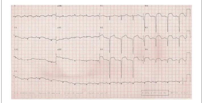
The electrocardiogram (May 10, 2008) showed sinus
rhythm; HR at 88 bpm; PR interval of 203 ms; QRS duration of 68 ms; low-voltage QRS complex in the frontal plane;
extensive anterior and inferior-wall areas that were electrically inactive; and ST-segment elevation from V1 to V6 and in I aVL, with positive T waves from V1 to V6 (Figure 1).
The laboratory assessment (May 10, 2008) showed glycemia of 94 mg/dl; urea of 43 mg/dl; creatinine of 1.07 mg/dl; sodium of 137 mEq/l; potassium of 4.5 mEq/l; hemoglobin of 12.6 g/dl; leukocytes at 6,500/mm³; platelets at 286,000/m³; INR of 1.1; APTT (patient/control ratio) of 1.12; CKMB of 6.46 ng/ml and troponin of 6.27 ng/ml.
The chest X-ray (05. 10. 2008) showed cardiomegaly at the expense of the left ventricle (LV).
The echocardiogram (05. 14. 2008) showed an enlarged left atrium (45 mm). The left ventricular ejection fraction (LVEF) was 20%. Akinesis of the LV septal and anterior segments and
signs suggestive of intracavitary thrombi in the apical region, one measuring 2.4 x 5 mm, fixed, and a mobile one in middle septal segment measuring 1.0 x 0.7 mm, were diagnosed. There was slight pericardial effusion. The pulmonary artery pressure was estimated at 30 mmHg.
The coronary angiography (May 14, 2008) identified occlusion of the anterior interventricular branch of the left coronary artery, a 90% lesion at the emergence of the first diagonal branch and a 70% in the left marginal branch; there were also irregularities in the right coronary artery. An angioplasty of the anterior interventricular branch was attempted; however, the guidewire did not surpass the lesion and an angioplasty in the first diagonal branch was performed with a balloon-catheter.
The patient was discharged from the hospital (May 20, 2008) with a daily prescription of 100 mg ASA, 12.5 mg carvedilol, 10 mg enalapril, 25 mg spironolactone, 20 mg simvastatin and 5 mg warfarin .
The patient developed dyspnea triggered by moderate exertion and three days later, stated to present dyspnea even at rest. On the morning of the following day, he presented an episode of precordial discomfort, followed by intense sudoresis and nausea that lasted for 30 minutes. He sought medical attention at InCor seven hours after the pain onset.
At physical examination (May 24, 2008), the patient presented regular general status, tachypnea (22 incursions per minute), heart rate (HR) of 84 bpm and blood pressure (BP) of 120/90 mmHg. There was no increase in the jugular venous pressure. The pulmonary semiology showed crackling rales in the lower two-thirds of both hemithoraxes. The cardiac semiology was normal. The abdominal assessment did not disclose any alterations. There was no edema of limbs and the pulses were palpable and symmetric.
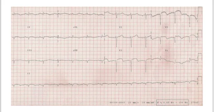
The electrocardiogram (May 24, 2008) showed sinus
rhythm; HR of 75 bpm; PR interval of 200 ms; QRS duration of 80 ms; low QRS voltage in the frontal plane; extensive
electrically inactive anterior area and ST-segment elevation from V1 to V6 and I and aVL, with positive T waves in the same leads (Figure 2).
The markers of myocardial necrosis were normal. There was improvement in the dyspnea and physical examination, with slight remaining pulmonary rales after the administration of diuretics. The patient was discharged on the following day, with an additional 40 mg of furosemide to the previously prescribed medication (May 25, 2008), for further outpatient treatment.
Figure 1 -ECG - low-voltage QRS complexes in the frontal plane, extensive anterior and inferior-wall areas that were electrically inactive and ST-segment elevation in I, aVL and from V1 to V6.
Figure 2 -ECG - extensive electrically inactive anterior and inferior wall areas, persistence of ST-segment elevation, suggesting aneurysm formation in the LV anterolateral and apical regions. The chest X-ray (May 24, 2008) showed pulmonary congestion.
coughing, hemoptysis and general malaise. He denied hematemesis or melena.
The physical examination (June 23, 2008) showed the patient in regular general status, with pale skin ++/4+, dehydrated +/4+, with a HR of 80 bpm, BP of 86 / 48
Figure 3 -ECG. No alterations in comparison to the previous ones, extensive electrically inactive anterior area and probable electrically inactive area in the lower wall, persistence of ST-segment elevation, suggesting aneurysm formation in the LV anterolateral and apical regions.
10 mg of phytomenadione and 250 ml of saline solution at 0.9% were administered.
The electrocardiogram (June 23, 2008) showed sinus
rhythm, HR of 75 bpm, PR interval of 200 ms, QRS interval of 80 ms, low-voltage QRS complex in the frontal plane,
extensive electrically inactive anterior area and probable electrically inactive area in the lower wall, in addition to the persistence of ST-segment elevation in V3 to V6 and I and aVL (Figure 3).
The chest X-ray (June 23, 2008) showed pulmonary congestion and pleural effusion in both hemithoraxes. The chest tomography (June 23, 2008) showed bilateral pleural effusion, atelectasis in the lower right lobe and alveolar infiltrate in the left lung.
Some hours after hospital admission, the patient started to present tachycardia, dyspnea, sudoresis and hypotension (BP of
80/50 mmHg). Initially, the tachycardia showed a narrow QRS.
During evolution, the patient started to present tachycardia with
wide QRS, which degenerated into ventricular fibrillation and was
then submitted to electrical cardioversion with 200 J, followed by 360 J twice, with sinus rhythm recovery.
The patient was submitted to orotracheal intubation and even with the administration of 300 mg of amiodarone, presented new cardiorespiratory arrests, which lasted approximately 10 minutes each.
In the afternoon of the same day, the patient presented cardiac arrest in asystole reversed with resuscitation maneuvers; however, he presented re-entry ventricular tachycardia, in spite of new intravenous administrations of amiodarone. The patient developed intense bradycardia and
received a subendocardial transvenous pacemaker implant in the right ventricle.
A new echocardiographic assessment (May 25, 2008) showed a 25-mm aorta, a 50-mm left atrium, 8-mm interventricular septum and LV posterior wall, 73-mm LV diastolic diameter and 67-mm LV systolic diameter, with a LV ejection fraction of 18%. A mid-apical aneurysm was identified in the left ventricle. The pulmonary artery pressure was estimated at 67 mmHg.
The patient received high-dose noradrenalin and dobutamine, in addition to ceftriaxone and clarithromycin. However, he developed refractory shock and presented irreversible asystolic cardiac arrest on the fourth day of hospital stay (June 26, 2008).
ECG Comments
The ECG is a poorly specific method to determine the size of the myocardial infarction. Several studies have shown how many errors are made when this type of assessment is used for this purpose. Among them, the study by Yusuf et al.1
showed that there was no correlation between the maximum
number of leads with ST-segment elevation, or of Q or QS
waves and CKMB levels.1
However, there is a situation in which the ECG can, with a good chance, identify the extensive myocardial infarction: when there are inactive areas in the I, aVL and V1 to V6 leads, followed by absence of R or a very small R, usually lower than 5mV. This is the situation of this clinical case, in which the ECG demonstrates how extensive the myocardial necrosis area is.
Echocardiographic comments
Given the high prevalence of coronary artery disease, the assessment of patients with suspected ischemic disease is one of the most common indications for echocardiography, as it allows a detailed assessment of the general systolic function and LV segmental function, both with a diagnostic and prognostic value.
Fundamental data on the correct treatment of the patient are supplied by the examination, and the role of echocardiography is well documented in the emergency department for the early diagnosis of acute myocardial infarction in patients with inconclusive ECG results.
The transthoracic echocardiogram, performed on May 14, 2008, showed an increase in the left chambers (ventricular volume of 173 ml), preserved thickness and decreased systolic function at the expense of an apical aneurysm and akinesis of the septal (middle segment) and anterior wall (middle and basal segment). The ejection fraction was estimated at 20%. The Doppler findings were compatible with the relaxation alteration. The right ventricle had normal systolic function. There was calcification of the mitral valvular ring and slight mitral insufficiency. The aortic valve presented signs of slight fibrocalcification, with preserved valve mobility. The tricuspid and pulmonary valves had normal aspect and presented normal cusp mobility and slight tricuspid insufficiency. The systolic pressure in the pulmonary artery was estimated at 30 mmHg. The aortic sinuses, the ascending aorta and the aortic arch had normal diameters and flow. There was pericardium thickening and slight pericardial effusion. There were signs of spontaneous contrast in the left ventricle and two thrombi, of 2.4 x 0.5 cm and 1.0 x 0.7 cm, respectively; the first was fixed in the apical segment of the septum, whereas the other was mobile, in the middle segment of the same wall.
During the evolution, a new transthoracic echocardiogram was performed on June 26, 2008, at bedside, after the recovery from the cardiorespiratory arrest, with the patient under mechanical ventilation and receiving vasoactive drugs. In comparison to the previous examination, there was significant left ventricular remodeling (281 ml) and the color flow Doppler examination showed worsening of the degree of valvular regurgitation (moderate mitral and tricuspid insufficiency), in addition to disclosing pulmonary artery hypertension (systolic pressure in the pulmonary artery estimated at 67 mmHg). There was no progression of the pericardial effusion. Regarding the segmental involvement of the left ventricle, marked systolic dysfunction was observed (with no new alterations in segmental mobility in comparison to the previous examination), with signs of significant spontaneous contrast. Therefore, in this clinical context, the echocardiography was essential to establish the patient’s diagnosis and conduct, disclosing ventricular dysfunction data with marked segmental involvement, the presence of thrombi in an aneurismal or akinetic area and absence of other possible complications post-acute myocardial infarction. It also ruled out the possibility of a new ischemic picture, associated to the clinical episode of hemodynamic instability.
The echocardiography has a crucial role in risk stratification and prognostic assessment of patients with coronary artery
disease, and, finally, in the assessment of acute myocardial infarction complications. The left ventricular thrombi are formed in regions of blood flow stasis. The evidence of intense decrease in global ventricular function, of an aneurysm, of an akinetic area and the onset of a spontaneous contrast effect increase the probability of thrombus formation in the LV. Rarely, as in the hypereosinophilic syndrome, ventricular thrombi can be detected in the absence of a segmental mobility alteration. The thrombus is identified as an area of increased echogenicity located inside the ventricular chamber, exhibiting a concave shape (laminar thrombus), following the curvature of the endocardium, or convex, projecting into the ventricular chamber. The diagnosis of apical thrombi is optimized with the use of a 5 MHz transducer, allowing a clear outline with the endocardium. The transesophageal echocardiography is rarely necessary for this purpose, as the apex is in the distal field of the image and is frequently not well visualized through this method.
Dr. Cinthia Goulart Fernandes Dias and Dr. Wilson Mathias Jr.
Clinical aspects
The present case is a 75-year-old male patient, with a picture of progressive dyspnea at exertion, of which onset was approximately two months before. He also presented, during the clinical evolution, some other symptoms such as hemoptysis, syncopal episodes, precordial discomfort, sudoresis and general malaise. Despite the fast evolution from symptom onset until death (about a month), several important clinical aspects will be discussed.
The first point to be discussed refers to the clinical picture at the initial presentation: dyspnea triggered by moderate exertion, which progressed in one week to dyspnea on minimum exertion. The patient presented, in the same period, a syncopal episode, which prompted him to seek emergency medical attention. These symptoms direct us to the initial syndromic diagnosis of acute heart failure (AHF). This clinical entity is characterized by the rapid or gradual onset of signs and symptoms of heart failure that result in the necessity of treatment in emergency medical services.2 Due to the high
rates of mortality and re-hospitalization related to acute heart failure, the first randomized and placebo-controlled studies on this syndrome were published in 2002.3,4 Until recently,
the epidemiological aspects of hospitalized patients with a diagnosis of AHF as the patients’ clinical characteristics, treatment strategies and clinical outcomes were unknown. In 2005, a large national registry, the ADHERE study, was published with more than 100,000 hospitalized patients with AHF, and these questions started to be clarified.5 The present
case shows that the patient’s initial treatment was directed at congestive heart failure therapy, with diuretics, digitalis and vasodilators. Although the patient presented signs of extreme clinical severity and poor prognosis, such as syncopal episode,6
there is no description of additional investigation for the causes of AHF at the moment of the first visit to the ER.
moment, the physical examination showed signs of pulmonary congestion and arterial hypotension, with both manifestations probably being associated to marked LV cardiac dysfunction. The ECG showed extensive electrically inactive anterior wall area as well as electrically inactive inferior wall area, associated to ST-segment elevation from V1 to V6, D1 and aVL. This extensive electrocardiographic involvement demonstrated that the etiology of heart failure in this patient had an ischemic origin. The coronary disease is responsible for the etiology of around 60% to 70% of cases with AHF, particularly in elderly patients.7 The laboratory assessment confirmed the myocardial
involvement with muscular necrosis, with a higher proportional elevation of troponin in relation to CKMB. The chest X-ray showed cardiomegaly at the expense of an increased LV. At this moment, the diagnosis of ongoing acute myocardial infarction (AMI) was made, associated with heart failure (HF).
An echocardiogram was requested and performed, and showed a LV ejection fraction (EF) of 20% at the expense of akinesis of the septal and anterior segments, in addition to signs suggestive of intracavitary thrombi in the apical region, with one of these thrombi being mobile. The examination also disclosed a pulmonary artery pressure (PAP) of 30 mmHg and a mild pericardial effusion.
The diagnosis of ongoing AMI was then confirmed, with extensive LV involvement, resulting in HF. An invasive approach was then decided, in order to achieve a better clinical stratification of this patient through a coronary angiography. The examination showed occlusion of the anterior descending artery (ADA), obstructive lesion of 90% in the diagonal artery (DA) and an obstructive lesion of 70% in the left marginal artery (LMA). An angioplasty was attempted in the anterior descending artery, but it was unsuccessful. Therefore, a balloon angioplasty was performed only in the diagonal artery lesion.
The opening of total coronary obstructions correspond to approximately 6% to 10% of angioplasties performed in hemodynamic centers.8 The opening of total coronary
occlusions by angioplasty, after the maximum recommended time for reperfusion in AMI with ST-segment elevation (currently 12 hours of evolution or up to 24 hours in selected cases) has been the object of discussion and literature studies. In December 2006, the OAT study was published; it randomized 2,166 patients to undergo angioplasty of chronic occlusions (artery responsible for the infarction) versus
optimized clinical treatment, between the 3rd and 28th day after
the AMI. There was no significant difference between the two groups regarding the decrease in mortality, reinfarction and myocardial dysfunction.9 Some recently published articles
suggest the improvement in regional myocardial contractility, mainly when the revascularized territory corresponds to that of the ADA.10,11
In this case, the choice of an attempt at revascularization of the ADA territory was probably justified by the large ischemic territory involved. After the unsuccessful ADA angioplasty, a balloon angioplasty was performed in the DA and the clinical treatment of the LMA was chosen, as the latter is a vessel with lesser myocardial irrigation extension.
During the follow-up, medications for coronary artery
disease (CAD) and congestive heart failure (CHF) were initiated, in addition to oral anticoagulation therapy. The patient was discharged on the 10th day after hospital admission.
Currently, there is no consensus in the literature regarding the use of double platelet anti-aggregation (ASA + clopidogrel) concomitantly with the administration of anticoagulants (coumarins) to patients after AMI. It is known that this association is related to a higher risk of bleeding.12 We did
not find a randomized prospective study that evaluated the association of these three medications. The indications for anticoagulation in patients after AMI (for at least three months) followed the recommendations of the North-American Societies of Cardiology (ACC/AHA): LV aneurysm
or thrombus; LVEF < 30%, associated or not to symptoms of
heart failure; history of thromboembolic event and chronic atrial fibrillation.13
This patient had a LVEF < 30%, in addition to the presence
of intracavitary thrombus; hence, the full anticoagulation was well indicated in this case.
After four days of hospital discharge, the patient returned to the hospital with a complaint of chest discomfort that lasted 30 minutes, associated with sudoresis and nausea. The clinical examination showed signs of pulmonary congestion. At this moment, some diagnostic hypotheses must be raised at the emergency room environment:
Post-infarction angina - suspected due to the chest discomfort that suggested pain, probably of the angina type. This could be justified, in this context, by a possible ischemia in the territory irrigated by the left marginal artery (LMA), which was known to present an obstructive lesion and had not been revascularized at another moment, or due to reocclusion of the diagonal artery (DA), which had previously undergone balloon angioplasty, which could also justify myocardial ischemia.
Cardiogenic shock - in this case, secondary to post-extensive myocardial infarction. The patient presented clinical and radiographic signs of congestion. Symptoms of low cardiac output were also present, such as sudoresis, chest discomfort and nausea. The patient developed an extensive anterior myocardial fibrosis area, and it is very likely that the persistence of the ST-segment elevation at the ECG corresponded to the anterior dyskinetic area (aneurysm). The ventricular aneurysm is a factor that contributes to myocardial dysfunction and can also be the focus of ventricular arrhythmias.
Mechanical complications - The main mechanical complications in the post-AMI period are: acute mitral insufficiency due to rupture of dysfunction of the papillary muscle (mainly the anterolateral papillary muscle, which is irrigated by the circumflex artery and its branches); rupture of the LV free wall and interventricular communication.14 They
normally occur from the third to the seventh day post-AMI, presenting a dramatic and rapidly progressive clinical picture, with high rates of morbimortality.
Post-infarction pericarditis - occurs in approximately 5% of the patients that were submitted to myocardial reperfusion therapy and 12% to 20% of the patients that were not reperfused at the AMI.15,16 The diagnosis can be suspected in
alterations, such as ST-segment elevation, which does not respect a single arterial territory.
In the present case, the markers of myocardial necrosis were negative in due time. The patient presented significant symptom improvement after clinical measures were taken for congestive heart failure (CHF), secondary to ventricular dysfunction after the AMI, being discharged after 24 hours of hospitalization.
The ventricular dysfunction observed after the AMI (LVEF
< 30%) is the main predictor factor of mortality in six months
and one year after the AMI.11,12 The GISSI-Prevential Trial,
TRACE Trial and MADIT II studies showed that sudden death was higher in patients with myocardial dysfunction.17-19
Therefore, at this moment, in spite of the clinical compensation observed in this case, we must bear in mind that this is a patient at high risk of death due to arrhythmogenic, causes, cardiogenic shock and thromboemboligenic causes.
The patient returned to the ER after approximately one month, with coughing, hemoptysis and worsening of the general status. He also presented pale mucosas, dehydration and arterial hypotension (BP= 86/48mmHg). He was initially treated for hypovolemic/hemorrhagic shock secondary to bleeding (hemoptysis). Volemic expansion was carried out and plasma and vitamin K were administered for the reversion of probable coumarin intoxication. The ECG did not disclose any new alterations. The chest X-ray was compatible with pulmonary congestion and the chest CT showed bilateral pleural effusion, in addition to alveolar infiltrate in the left lung.
Some considerations can be made at this moment regarding the case. The symptoms of coughing and hemoptysis presented by the patient upon his arrival at the hospital raised the possibility of some differential diagnoses.
Hemoptysis is a common cause for seeking medical attention at the ER. Initially, one must try to differentiate the true hemoptysis, which originates in the lower airways, from the “pseudo-hemoptysis”, which can be originated from the upper airways or gastrointestinal tract. After verifying that it was a case of true hemoptysis, some hypotheses could justify this finding in the present patient, such coumarin intoxication, pulmonary congestion, pneumonia and pulmonary thromboembolism. Other causes of hemoptysis, such as neoplasia, bronchiectasis, vasculitis, arteriovenous malformation and tuberculosis, among others, did not seem to be adequate differential diagnoses for this patient and therefore, they will not be discussed.
The intoxication by coumarins, in this case, can be an initial diagnostic hypothesis, as the presence of hemoptysis is not uncommon in individuals with increased INR. The hemoptysis was not abundant; however, the patient was hypotensive. Examinations to assess hemostasis and coagulation must always be requested when coumarin intoxication is suspected. Eventual alterations must be corrected, as it was done in the present case.
The pulmonary congestion can be the cause of the bloody sputum in this case, as it is known that the patient presented markedly decreased ventricular function. However, in this case, there is a dissociation between the initial pulmonary auscultation, which did not suggest an important congestion
and the initial hypothesis of pulmonary congestion. The congestion later identified at the chest X-ray and CT might be justified by the volemic and plasma load that the patient received during the initial therapy.
The possibility of a pneumonic focus in this patient was verified at the chest CT. This could justify a bloody sputum, associated to coughing and worsening in the general status. It must be remembered that this is an individual with decreased ventricular function that could have pneumonia as a clinical decompensation factor and low cardiac output.
The diagnostic hypothesis of pulmonary thromboembolism must always be considered in individuals with a picture of sudden or new-onset dyspnea, associated or not to a picture of bloody sputum and chest pain. This patient did not present major risk factors for pulmonary thromboembolic events – such as neoplasia, recent trauma or orthopedic surgery –, asymmetric edema of lower limbs and was supposedly undergoing systemic anticoagulation therapy, which would hinder, but not completely rule out, the possibility of pulmonary thromboembolism.
During the clinical evolution, the patient presented homodynamic instability, characterized by hypotension, tachycardia and worsening in the respiratory pattern. He developed cardiorespiratory arrest, with ventricular fibrillation rhythm, which was initially reversed with defibrillation at 200 J, followed by two 360 J shocks, with return to sinus rhythm. It is worth considering that the most recent guideline on advanced cardiac life support (ACLS) states that, in cases of cardiorespiratory arrest with a shockable rhythm (pulseless VT or VF), the electrical defibrillation must be carried out with an initial energy of 360 J (in a monophasic equipment) and 200 J (in a biphasic equipment), followed by two minutes of cardiopulmonary resuscitation with massage and ventilation.
A new defibrillation after a cycle of massage and ventilation is only indicated if the case is a pulseless VT or VF. This change occurred because one took too long to start the external cardiac massage, according to the old protocol, in which three sequential defibrillations were initially performed with 200, 300 and 360 J. This delay in start of the cardiac compressions impaired the perfusion of vital organs during the cardiorespiratory reanimation. Therefore, initial high-energy shocks and fast external cardiac massage (at least 100 incursions per minute) are currently recommended as effective.
The patient developed significant hemodynamic worsening and was submitted to orotracheal intubation, presenting four cardiorespiratory arrests that were reversed during the first day of hospitalization. It was also necessary to implant a transvenous pacemaker after an episode of sinus bradycardia. Vasoactive drugs (noradrenaline and dobutamine) were administered and a specific antibiotic therapy was started for the pulmonary infectious focus. A new assessment with echocardiogram confirmed the previous hypothesis of LV aneurysm, with marked LV dysfunction and increase in the PAP to 67 mmHg.
presented by the patient was less likely. The myocardial necrosis markers were not available at the description.
The patient developed refractory shock, with cardiorespiratory arrest in asystole, non-responsive to the attempted clinical maneuvers of resuscitation.
Therefore, we believe that the probable cause of the patient’s hemodynamic instability was the infectious focus in the lung, which led to the clinical decompensation of the heart failure and low cardiac output.
Dr. Gustavo Ken Hironaka and Dr. Rodrigo Barbosa Ésper
Diagnostic hypothesis
Thus, our hypotheses for the main cause and for other diagnoses that contributed to the patient’s death are: ischemic heart disease, with extensive acute myocardial infarction that caused heart failure and was complicated by bronchopneumonia and mixed shock (septic and cardiogenic).
Dr. Gustavo Ken Hironaka and Dr. Rodrigo Barbosa Ésper
Necropsy
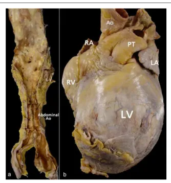
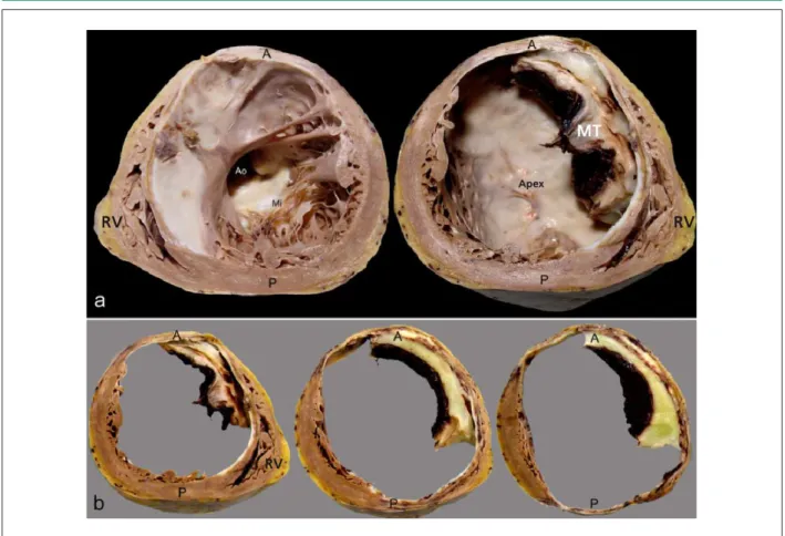
At the necropsy, the aorta presented moderate to intense atheromatosis, with marked calcification of its abdominal section. The heart presented a globus shape, at the expense of the LV anterior region (Figure 4). Cross-sections of the LV showed intense aneurysmatic dilatation in the LV cavity, the result of a previous extensive transmural myocardial infarction in the septal, anteroseptal, anterior and anterolateral walls, from the basis to the apex, with extensive mural thrombosis, as well as in the inferior wall, from the middle to the apex, with the involvement of approximately 60% of the ventricular muscle (Figure 5). In spite of the presence of cavitary thrombi, no systemic thromboembolism was detected. The histological analysis showed that extensive areas of myocardial tissue had been replaced by fibrosis. The pericardium presented fibrosis, with the parietal and visceral leaflets fastened firmly to each other in the anterior projection of ventricular aneurysm. There were signs of congestive HF represented by hydrothorax to the right and hydroperitoneum, with approximately 360 mL and 480 mL of citrine-yellow fluid, respectively. The dissection and histological analysis of the epicardial coronaries demonstrated severe obstructive atherosclerotic coronary lesions (Figure 6).
The lungs showed alterations that were suggestive of pulmonary infection, characterized by organized pleuritis, with firm and profuse adherences between the left lung visceral and parietal leaflets; agudization of chronic bronchitis, with intense edema and hyperemia of the respiratory mucosa, from both bronchial sources to the periphery of the respiratory tree, with marked increase in the left lung; slight cylindrical bronchiectasis in the lower left lobe and intense pulmonary parenchyma congestion (both lungs weighed, together, 1,700 g, with the normal weight being around 600 g). The histological analysis showed an interstitial-alveolar inflammatory process with epithelial abundant epithelial desquamation and foci of
intra-alveolar protein exudation. The screening for bacteria, fungi and alcohol-acid-resistant bacilli were negative. Thus, the histological analysis did not identify the infectious agent, but considering the inflammatory pattern, the viral etiology is a possibility, even though no viral cytopathic alterations were observed.
The patient presented acute splenitis, with a small spleen, but with a “raspberry jam” appearance, which is associated with infectious pictures, but also, less exuberantly, with myocardial infarction. There was no neoplasia.
Risk factors for cardiovascular disease present in the clinical history of this patient were previous smoking habit and systemic arterial hypertension. To the latter, we associated the findings of lacunar state in the nucleus basalis of the brain and renal arteriolosclerosis with nephrosclerosis, not mentioning the heart hypertrophy masked by the dilatation, but present when one considers the total weight of the heart, even when disregarding the weight of the thrombi (546 g).
The remainder of the organs showed mixed shock alterations (mainly cardiogenic ones, with a septic component) and this was the immediate morphological cause of death. Such alterations were represented by cerebral edema with slight herniation of the cerebellar amygdala; pulmonary congestion; submassive centrolobular liver necrosis; acute renal tubular necrosis; pancreatic steatonecrosis with interstitial hemorrhage and acute gastritis with multifocal mucosal hemorrhage. A less important finding for the moment was diffuse thyroid hyperplasia (38 g).
Dr. Jussara Bianchi Castelli
Figure 6 -Histological analysis of the coronaries in sections, showing severe obstructive atherosclerotic lesions in this patient. In (a) and (b), we observe cross-sections of the anterior interventricular branch of the left coronary artery (LCA), in its 4th and 7th cm. In (c), we see the 1st cm of the diagonal branch 1 (DI1) of the LCA. In (d), we see the posterior interventricular branch (DP2) of the right coronary artery (hematoxylin and eosin stain; magniication: 2.5 x for all images).
References
1. Yusuf S, Lopez R, Maddison A, Maw P, Ray N, McMillan S et al. Value of electrocardiogram in predicting and estimating infarct size in man. British Heart Journal 1979;42:286-93
2. Gheorghiade M, Zannad F, Sopko G, et al: Acute heart failure syndromes: current state and framework for future research. Circulation 2005;112:3958-68.
3. VMAC Investigators: Intravenous nesiritide vs. nitroglycerin for treatment of decompensated congestive heart failure: A randomized controlled trial. JAMA 2002;287:1531-40.
4. Cuffe MS, Califf RM, Adams Jr. KF, et al: Short-term intravenous milrinone for acute exacerbation of chronic heart failure: a randomized controlled trial. JAMA 2002; 287:1541-47.
5. Adams KF Jr, Fonarow GC, Emerman CL, LeJemtel TH, Costanzo MR, Abraham WT, Berkowitz RL, Galvao M, Horton DP, for the ADHERE Scientific Advisory Committee and Investigators. Characteristics and outcomes of patients hospitalized for heart failure in the United States: rationale, design, and preliminary observations from the first 100,000 cases in the Acute Decompensated Heart Failure National Registry (ADHERE). Am Heart J. 2005; 149: 209–16.
6. Olshansky B, Poole JE, Johnson G, Anderson J, Hellkamp AS, Packer D et al, for SCD-HeFT Investigators. Syncope predicts the outcome of cardiomyopathy patients: analysis of the SCD-HeFT study. J Am Coll Cardiol. 2008;51:1277-82.
7. Fox KF, Cowie MR, Wood DA, Coats AJS, Gibbs JSR, Underwood SR et al. Coronary artery disease as the cause of incident heart failure in the
population. Eur Heart J. 2001;22:228–36
8. Hochman JS, Lamas GA, Buller CE, Dzavik V, Reynolds HR, Abramsky SJ et al. Coronary Intervention for Persistent Occlusion after Myocardial Infarction. N Engl J Med 2006; 355:2395-407.
9. Cheng ASH, Selvanayagam JB, Jerosch-Herold M, van Gaal WJ, Karamitsos TD, Neubauer S et al. Percutaneous Treatment of Chronic Total Coronary Occlusions Improves Regional Hyperemic Myocardial Blood Flow
and Contractility: Insights From Quantitative Cardiovascular Magnetic
Resonance Imaging. J Am Coll Cardiol Intv. 2008; 1:44-53.
10. Safley DM, House JA, Marso SP, Grantham JA, Rutherford BD. Improvement in survival following successful percutaneous coronary intervention of coronary chronic total occlusions: Variability by target vessel. J Am Coll Cardiol Intv 2008;1:295-302
11. Mehran R and Dangas GD. Revascularization of a chronically occluded left anterior descending artery: Is it worth all the effort? J Am Coll Cardiol Intv 2008;1:303-4.
12. Rothberg, MB, Celestin, C, Fiore, LD, Lawler E, Cook JR. Warfarin plus aspirin after myocardial infarction or the acute coronary syndrome: meta-analysis with estimates of risk and benefit. Ann Intern Med 2005; 143:241-50
13. Antman EM, Hand M, Armstrong PW, Bates ER; et al. STEMI focused upadate. 2007 Focused update of the ACC/AHA 2004 Guidelines for the manegment of patients with ST-elevation myocardial infarction: a report of the American College of Cardiology/ American heart association Task Force on Practice Guideline. J Am Cardiol 2008;51:210-47.
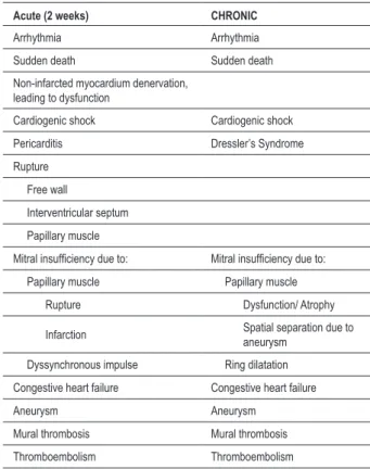
Table 1 - Myocardial infarction complications
Acute (2 weeks) CHRONIC
Arrhythmia Arrhythmia
Sudden death Sudden death
Non-infarcted myocardium denervation, leading to dysfunction
Cardiogenic shock Cardiogenic shock
Pericarditis Dressler’s Syndrome
Rupture
Free wall
Interventricular septum
Papillary muscle
Mitral insuficiency due to: Mitral insuficiency due to:
Papillary muscle Papillary muscle
Rupture Dysfunction/ Atrophy
Infarction Spatial separation due to
aneurysm
Dyssynchronous impulse Ring dilatation
Congestive heart failure Congestive heart failure
Aneurysm Aneurysm
Mural thrombosis Mural thrombosis
Thromboembolism Thromboembolism
(Reproduced from: Silver MD, Gotlieb A, Schoen FJ, eds. Cardiovascular Pathology. 3rd Ed. New York: Churchill Livingstone; 2001: p.228).
Anatomopathological diagnoses
Left ventricular aneurysm after extensive myocardial infarction; organized mural thrombosis; congestive heart failure; pulmonary bronchopneumonic process with undefined agent; mixed shock, but mainly cardiogenic one, with multiple-organ lesions
Dr. Jussara Bianchi Castelli
Comments
The patient presented a prior history of systemic arterial hypertension, previous smoking habit and myocardial infarction. Table 1 summarizes the post-infarction complications described in literature,20 of which three were
presented by the patient: ventricular aneurysm, cavitary thrombus and congestive heart failure. Large infarctions, affecting more than 40% of the left ventricle, generally course with cardiogenic shock, which is associated with a high mortality rate (of almost 70%) and it is responsible for more than 2/3 of in-hospital deaths. The extension of the infarction in the present case is remarkable, considering that the patient did not die sooner, but only after approximately a month, due to the association with the pulmonary involvement, thus complicating his borderline condition.
14. Reeder GS. Identification and treatment of complications of myocardial infarction. Mayo Clin Proc 1995; 70:880-84
15. Tofler GH, Muller JE, Stone PH, Willich SN, Davis VG, Poole WK et al. Pericarditis in acute myocardial infarction: Characterization and clinical significance. Am Heart J 1989;117:86-92.
16. Spodick, DH. Safety of ibuprofen for acute myocardial infarction pericarditis. Am J Cardiol. 1986;57:896.
17. Marchioli R, Barzi, F, Bomba E, Chieffo C, Di Gregorio D, Di Mascio R et al. Early protetion against sudden death by n-3 polyunsaturated fatty acids after myocardial infarction. Time-couse analysis of the results of the Gruppo Italiano per lo Studio della Sopravvivenza nell’Infarto Miocardio (GISSI)-Prevenzione. Circulation 2002;105:1987-903.
18. Moss AJ, Hall WJ, Cannom DS, Daubert JP, Higgins SL, Klein H, et al. for
the Multicenter Automatic Desfibrillator Implantation Trial Investigators. Improved survival with an implanted defribillator in patients with coronary desesa at high risk for ventricular arrhythmia. N Engl J Med 1996;335:1933-40
19. Torp-Pedersen, C, Kobler, for the TRACE Study GROUP. Effect of ACE inhibitor trandolapril on life expectancy of patients with reduced left-ventricular function after myocardial infarction. Lancet 1999;354:9-12.
20. Baroldi G. Myocardial cell death, including ischemic heart disease and its complications. In: Silver MD, Gotlieb A, Schoen FJ, eds. Cardiovascular Pathology. 3rd Ed. New York: Churchill Livingstone; 2001:226-35.