Research Article
Ecotin-Like ISP of
L. major
Promastigotes Fine-Tunes
Macrophage Phagocytosis by Limiting the Pericellular
Release of Bradykinin from Surface-Bound Kininogens:
A Survival Strategy Based on the Silencing of Proinflammatory
G-Protein Coupled Kinin B
2
and B
1
Receptors
Erik Svensjö,
1Larissa Nogueira de Almeida,
1Lucas Vellasco,
1Luiz Juliano,
2and Julio Scharfstein
11Instituto de Biof´ısica Carlos Chagas Filho, Universidade Federal do Rio de Janeiro, 21990-400 Rio de Janeiro, RJ, Brazil
2Escola Paulista de Medicina, Universidade Federal de S˜ao Paulo, 04044-020 S˜ao Paulo, SP, Brazil
Correspondence should be addressed to Julio Scharfstein; jscharf2@gmail.com
Received 13 June 2014; Accepted 17 August 2014; Published 10 September 2014
Academic Editor: Marcelo T. Bozza
Copyright © 2014 Erik Svensj¨o et al. his is an open access article distributed under the Creative Commons Attribution License, which permits unrestricted use, distribution, and reproduction in any medium, provided the original work is properly cited.
Inhibitors of serine peptidases (ISPs) expressed byLeishmania majorenhance intracellular parasitism in macrophages by targeting
neutrophil elastase (NE), a serine protease that couples phagocytosis to the prooxidative TLR4/PKR pathway. Here we investigated
the functional interplay between ISP-expressingL. majorand the kallikrein-kinin system (KKS). Enzymatic assays showed that NE
inhibitor or recombinant ISP-2 inhibited KKS activation in human plasma activated by dextran sulfate. Intravital microscopy in the
hamster cheek pouch showed that topically appliedL. majorpromastigotes (WT and���2/3mutants) potently induced plasma
leakage through the activation of bradykinin B2receptors (B2R). Next, using mAbs against kininogen domains, we showed that these
BK-precursor proteins are sequestered byL. majorpromastigotes, being expressed at higher % in the���2/3mutant population.
Strikingly, analysis of the role of kinin pathway in the phagocytic uptake ofL. majorrevealed that antagonists of B2R or B1R reversed
the upregulated uptake of���2/3mutants without inhibiting macrophage internalization of WTL. major. Collectively, our results
suggest thatL. majorISP-2 ine-tunes macrophage phagocytosis by inhibiting the pericellular release of proinlammatory kinins
from surface bound kininogens. Ongoing studies should clarify whetherL. majorISP-2 subverts TLR4/PKR-dependent prooxidative
responses of macrophages by preventing activation of G-protein coupled B2R/B1R.
1. Introduction
Integrated by 3 serine proteases, factor XII (FXII), factor XI (FXI), and plasma prekallikrein (PK) and by one nonen-zymatic cofactor, high molecular weight kininogen (HK), the kallikrein-kinin system (KKS), also referred to as the plasma contact pathway of coagulation, is assembled and activated when the blood comes in contact with negatively charged polymers of endogenous origin or microbial surfaces [1,2]. Upon binding to these negatively charged structures, the zymogen FXII undergoes a conformational change that
endows the unstable proenzyme with limited enzymatic activity. Activated FXII (FXIIa) then cleaves prekallikrein (complexed to the cofactor HK), generating PKa. Reciprocal cleavage reactions between FXIIa and PKa amplify the prote-olytic cascade, leading to downstream (i) generation of ibrin via the FXIIa/FXIa-dependent procoagulative pathway, (ii) release of the internal bradykinin (BK) moiety of HK by PKa. Once liberated, the short-lived BK induces vasodilation and increases microvascular permeability through the activation of bradykinin B2 receptors (B2R) expressed in the endothe-lium lining [1]. In addition, the multifunctional PKa generates Volume 2014, Article ID 143450, 12 pages
plasmin, an efector of ibrinolysis, and cleaves native C3 of the complement system C3 [3,4].
Although HK is classically regarded as the parental precursor of proinlammatory kinins, the cleaved form of HK (HKa), a disulide linked two-chain structure, has additional biological functions. For example, it has been reported that HKa reduces neutrophil adhesive functions upon binding to
�2-integrin Mac-1 (CR3, CD11b/CD18,�M�2) [1,5]. More recently, Yang et al. [6] appointed HK/HKa as the plasma-borne opsonins that drive eferocytosis of apoptotic cells via plasminogen activator receptor (uPAR)/RAC1-pathway. Ater binding to phosphatidyl serine (PS) exposed by apoptotic neutrophils, the surface-bound HK binds to uPAR before switching of proinlammatory responses of macrophages [6]. In contrast to this novel immunoregulatory function, HK (and low molecular weight kininogen, LK) are traditionally viewed as precursors of proinlammatory kinins. Once leaked into extravascular tissues, the plasma-borne HK/LK undergo proteolytic cleavage by tissue kallikrein, releasing the B2R agonist lysyl-BK (LBK) in inlammatory exudates [1]. It is noteworthy that oxidized forms of kininogens may release bioactive kinins as result of cooperation between neutrophil elastase (NE) and mast cell tryptase [7,8]. Acting as paracrine hormones, the short-lived kinins (BK or LBK) switly acti-vate G-protein coupled bradykinin B2 receptors (B2R), a subtype of receptor constitutively expressed by endothelial cells, nociceptive neurons, macrophages, and DCs [1, 9–
11]. he long-range signaling activity of intact kinins is controlled by kinin-degrading metallopeptidases, such as the angiotensin converting enzyme (ACE/kininase II) [1]. In addition, the liberated kinin peptides are metabolized by kininase I (carboxypeptidase N or M); removal of the C-terminal Arg residue generates des-Arg-kinins, the high-ainity ligands of B1R, a GPCR subtype whose expression is strongly upregulated in injured/inlamed tissues [12].
During the last decade, research conducted in our laboratory showed that kinins proteolytically released in peripheral sites ofT. cruzi orLeishmania chagasiinfection reversibly couple inlammation to antiparasite immunity [13–
17]. Another interesting twist came from studies showing that activation of the contact system/KKS promotes bacterial entrapment within ibrin meshes, thus providing a physical barrier against the systemic spread of microbial pathogens [18]. To this date, however, it is unclear whether the contact pathway modulates immunity at early stages of cutaneous leishmaniasis. Intravital microscopy studies conducted in the mouse ear model ofL. majorinfection [19–21] have shown that iniltrating neutrophils engulf the promastigotes before expressing the apoptotic markers required for eferocytosis by dermal DCs. Ater internalizing the parasitized/apoptotic neutrophils, the dermal DCs are no longer capable of steering protective TH1-responses in the draining lymph node [19–
21]. Although eferocytosis has strong impact on DC function and TH development in L. major infection, independent studies showed that macrophage clearance of apoptotic neutrophils may either induce pro- or anti-inlammatory responses in NE-dependent manner, the intracellular fate of the parasite being inluenced by the host genetic background [22,23].
In natural infection by blood-feeding arthropods, insect proboscis inevitably causes bleeding, which then causes the mixing of plasma and sandly saliva substances with parasites deposited in the injured dermis [24]. Interestingly,
Phlebotomy duboscq, a vector of Leishmania species,
con-tains high levels of a salivary protein (PdSP15) that inhibits the contact pathway [25] by binding to negatively charged polymers of endogenous origin—such as platelet-derived polyphosphates [2,18,26]. Considering that activation of the procoagulative contact system induces microvascular leakage through PKa-mediated release of BK, it is conceivable that sandly-transmittedLeishmaniapromastigotes have evolved the means to subvert the innate efector function of the kinin pathway at early stages of infection.
he current study was motivated by the recent discov-ery that Leishmania has three genes encoding ecotin-like inhibitors of serine peptidases (ISPs) [27]. Previous studies with the archetype of the familyEscherichia coliecotin [28] showed that this inhibitor targets neutrophil elastase (NE) [29]—a member of the trypsin-fold serine peptidases of clan PA/family S1A. Ater noting that theLeishmaniagenome [30] lacks these endogenous serine peptidase targets, Eschenlauer et al. [27] predicted that L. major ISPs might target S1A -family serine peptidases expressed by cells of the innate immune system, such as NE, tryptase, and cathepsin G [30]. In a series of elegant studies, Eschenlauer et al. [27] and Faria et al. [31,32] addressed this issue usingL. majorlines lacking ISP2 and ISP3 (Δ���2/3). Ater studying the outcome of interactions betweenL. majorpromastigotes and elicited macrophages, these authors found that these phagocytes internalized theΔ���2/3promastigotes far more eiciently than ISP-expressing wild-type (WT) parasites [27, 31] and linked the upregulated CR3-dependent phagocytosis of the
���2/3 L. major mutants to NE-dependent activation of innate immunity via the TLR4/PKR/TNF-�/IFN-�, a proox-idative pathway that limits intracellular parasite survival [31, 32]. Notably, the phenotype of���2/3 promastigotes was reversed by supplementing the macrophage cultures with puriied (recombinant) ISP-2 or with the synthetic NE inhibitor (MeOSuc-AAPV-CMK), at the onset of infec-tion [31]. Based on these collective indings, these authors suggested that ISP-2 expressing L. major promastigotes might downmodulate phagocytosis and limit microbicidal responses of macrophages by preventing NE-dependent acti-vation of TLR4[31,32]. More recently, we have documented that macrophages internalize and limit intracellularT. cruzi
growth in resident macrophages through activation pathways forged by the cross-talk between bradykinin B2receptors and C5a receptors [33]. Intrigued by the similarities that exist between the phenotype of theL. major���2/3mutant and
L. chagasi promastigotes [15] and T. cruzi trypomastigotes
(Dm28 strain) [33,34], in the current work we interrogated whether ISP-expressing L. major and the ISP-2 ���2/3 mutants difer in their ability to activate the KKSin vivoand
in vitro. Using intravital microscopy, we irst showed thatL.
majorpromastigotes topically applied to the hamster cheek
may subvert innate immunity by targeting kinin-releasing serine proteases (S1A family) exposed at the cell-surface of macrophages.
2. Materials and Methods
2.1. Parasites. L. majorPromastigotes of Friedlin (MHOM/
JL/80/Friedlin) were grown in modiied Eagle’s medium (HOMEM, Sigma) supplemented with 10% heat-inactivated fetal bovine serum (FBS, Gibco) at 25∘C, as previously described [31,32]. Suspensions of promastigotes were washed twice with PBS before being used either in vitro or in
vivo.Leishmania majordeicient in ISP2 and ISP3 (���2/3)
were generated as previously described by Eschenlauer et al. [27]. he following antibiotics were used at the indicated concentration for the selection of transfectants: 50 mg/mL hygromycin B (Roche), 25 mg/mL G418 (Invit-rogen), 10 mg/mL phleomycin (InvivoGen), and 50 mg/mL puromycin dihydrochloride (Calbiochem).
2.2. Intravital Digital Microscopy. Syrian hamsters,
3-month-old males, were maintained and anesthetized according to regulations given by the local ethical committee (IBCCF, protocol-014, 23/02/2008). Altogether 65 hamsters (114 ±
18g) (Anilab, S˜ao Paulo, Brazil) were used. Anesthesia was induced by intraperitoneal injection of sodium pentobarbital 3% that was supplemented with i.v.�-chloralose (2.5% W/V, solution in saline) through a femoral vein catheter. A tracheal cannula (PE 190) was inserted to facilitate spontaneous breathing and the body temperature was maintained at 37∘C by a heating pad monitored with a rectal thermistor. he hamster cheek pouch (HCP) was prepared and used for intravital microscopy as previously reported [34, 35]. he microcirculation of the HCP was observed using an Axioskop 40 microscope, objective 4x, and oculars 10x equipped with a LED light source Colibri (Carl Zeiss, Germany) and appropriate ilters (490/520 nm and 540/580 nm, rhodamine) for observations of luorescence in epiluminescence. A digital camera, AxioCam HRc, and a computer with the AxioVision 4.4 sotware program (Carl Zeiss, Germany) were used for image analysis of arteriolar diameter and total luorescence in a representative rectangular area (5 mm2) of the pre-pared HCP. Fluorescence was recorded for 30 min prior to experimental interventions to secure normal blood low and unaltered vascular permeability and the luorescence mea-sured at 30 min ater FITC-dextran (FITC-dextran 150 kDa, 100 mg/kg bodyweight, TdB Consultancy, Uppsala, Sweden) injection was adjusted to 2000 luorescent units (RFU = Relative Fluorescent Units) for statistical reasons. Leukocytes were labeledin vivoby injecting rhodamine 100�g/kg b.w i.v. (10 min prior to experimental interventions), reinforced by injection of the same tracer at 10�g/kg b.w. every 10 min until 60 min. he recorded luorescence at 10 min ater rhodamine injection in each experiment was adjusted to 3000 luorescent units (RFU) for statistical reasons. Two images of exactly the same area were recorded at every 5 min interval during the entire experiment. One was used to measure plasma leakage
and arteriolar diameter (490/520 nm) and the other to mea-sure total luorescence of rhodamine-labeled leukocytes in circulation, rolling, adherence, and migration (540/580 nm) in the observed area (5 mm2) here deined as leukocyte accumulation. Exposure time was limited to 15 s for each captured image in order to avoid phototoxicity. Following 30 min control period ater FITC-dextran injection HCPs were topically exposed to WT L. major promastigotes or
Δ���2/3(7.5×106/500�L) during interruption of the super-fusion for 10 min. Cromoglycate was injected i.p. (40 mg/kg b.w.) at time of pentobarbital anesthesia and dextran sulfate 500 kDa (TdB Consultancy, Uppsala, Sweden) was injected i.v. (2 mg/kg) prior to parasite application. HOE-140 tested at 0.5�M and the histamine receptor H1 mepyramine (10�M) were applied locally via a syringe pump into the superfusion during 10 min prior to application of promastigotes.
2.3. Isolation of Peritoneal Macrophages and Invasion Assays.
C57BL/6 mice received an intraperitoneal injection of 2 mL of 3% thioglycolate and macrophages were harvested from peritoneal lavage 3 days later. Macrophages were plated on 13 mm coverslips in 24-well plate and ater 20 h of incubation at 37∘C in complete medium (RPMI + 10% FBS, 100 U/mL penicillin, and 100�g/mL streptomycin) the nonadherent cells were removed by washing the monolayer of cells with PBS. Invasion assays were performed by adding stationary phase promastigotes to the monolayers at a ratio of 5 : 1 (parasite/macrophage) in medium containing 1 mg/mL albu-min from bovine serum (BSA, Sigma). he interaction was performed during 3 h in a humidiied chamber containing 5% CO2 at 37∘C. When indicated, the culture medium was supplemented with 100 nM of B2R antagonist (HOE-140, Sigma) or 1�M of B1R antagonist des-Arg9-[Leu8]-BK (DAL8-BK; Sigma), 5 minutes before addition of parasites. Ater interaction, extracellular promastigotes were removed by washing the monolayers twice with PBS, which were then ixed with Bouin overnight and stained with Giemsa (Merck). he number of intracellular amastigotes was determined by counting at least 100 cells per replicate under the light microscope. All assays were done in triplicates and results were expressed as mean values±SD.
2.4. Parasite Surface Staining. Promastigotes were
2.5. Contact Phase Activation of Human Plasma. he acti-vation of FXII/PK in human citrated (platelet free) plasma treated (or not) with dextran sulfate (DXS; 500 kDa, TdB Consultancy) was monitored by spectroluorimetry as pre-viously described [37], using internally quenched luo-rescent substrates whose sequences correspond to the C-terminal (Abz-GFSPFRSVTVQ-EDDnp) or N-terminal lanking region (Abz-MTEMARRPQ-EDDnp m) of BK of mouse kininogen. he hydrolysis of the cleaved substrate Abz-peptidyl-EDDnp (Abz = o-aminobenzoyl and EDDnp = ethylenediamine 2,4-dinitrophenyl) was monitored by mea-suring the luorescence at�ex. = 320nm and�em. = 420nm in a Spectramax M5 luorescence spectrophotometer. he reaction was carried out in PBS, pH 7.4, using citrated human plasma 1 : 20, 4�M of the Abz-peptidyl-EDDnp substrate and 20 nM of the contact system activator DXS (500 kDa). As internal controls, the plasma was pretreated with the synthetic PKa inhibitor (PKSI-527—5�M) [38]. Assays with recombinant ISP-2 (kindly supplied by A. P. C. A. Lima) were performed at inal concentrations of 142, 177, 240, and 355 nM; the neutrophil elastase (NE) inhibitor MeOSuc-AAPV-CMK (Calbiochem) was tested at 10, 20, and 30�M. PKSI or recombinant ISP2 and MeOSuc-AAPV-CMK were preincubated with human plasma for 15 min, at 37∘C, prior to the addition of DXS and the substrate. Plasma was prepared by centrifugation of blood samples at 2500 g for 20 min at 4∘C. Ater centrifugation, plasma samples were iltered using a 0.2�m membrane.
2.6. Statistical Analyses. Statistical analyses were done using
PRISM 5.0 (GraphPad Sotware). Comparisons of the means of the diferent groups were done by one-way analysis of variance (ANOVA). When the mean values of the groups showed a signiicant diference, pairwise comparison was performed with the Tukey test. A�value of 0.05 or less was considered to indicate a statistically signiicant diference. For intravital experiments, we used ANOVA or pairwiset-test, when appropriate.
3. Results
3.1. Analysis of the Dynamics of Inlammation in HCP Topically
Sensitized with L. major Promastigotes. Intravital microscopy
in HCP has provided a wealth of information about the inter-play between the KKS and the topically applied pathogens because this method dispenses the use of needles, thus ruling out the inluence of bleeding and collateral activation of the contact system in the analysis of microcirculatory parameters. As a starting point in this work, we asked whether the proinlammatory responses evoked by ISP-expressingL.
major promastigotes or ISP-deicient parasites were
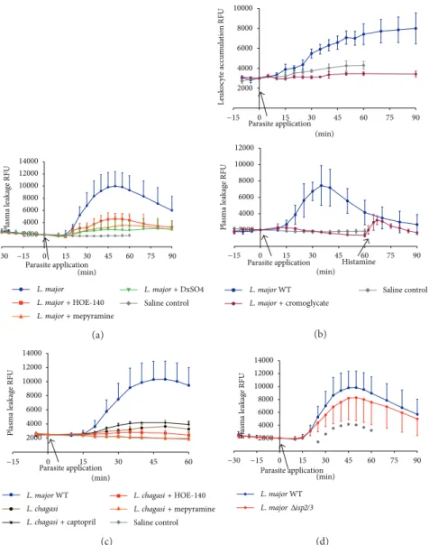
com-parable. Our results (Figures 1–3) revealed that L. major
(WT) promastigotes induced a very robust and reversible microvascular leakage that was detectable at 20 min and reached its maximal value 45 min ater pathogen application (Figures 1(a)–1(c)). In 4 out of 15 experiments we mea-sured leukocyte accumulation in and around postcapillary venules and noted that these circulating cells were promptly mobilized locally, the response being detectable up to 90 min
ater pathogen application (Figure 1(b)). he temporal course and dynamics of the plasma leakage response evoked by WT promastigotes were quite diferent from the classical responses elicited by BK, histamine, or leukotrienes, all of which cause a maximal increase within 10 min and reversed to steady-state conditions within 30 min [39,40]. Intriguingly, we found that the leakage responses evoked by L. major
(WT) promastigotes were generally more robust than those induced by the same inoculum ofL. donovanipromastigotes (Figure 1(c)) [16] orT. cruzi(tissue culture trypomastigotes, Dm28c strain) [35]. Akin to the indings made in the above-mentioned studies, we found that topically applied HOE-140 (B2R antagonist) markedly reducedL. major(WT-) induced plasma leakage (Figure 1(a)).
Considering that mast cells are innate sentinel cells strategically localized in the perivascular tissues, we next interrogated whether L. major promastigotes might evoke plasma leakage in mast cell-dependent manner. As shown in
Figure 1(a), the topical addition of mepyramine (histamine-1-receptor blocker) markedly inhibited the macromolecular leakage induced by L. major promastigotes. In a limited series of studies, we found that cromoglycate, a well-known mast cell stabilizer, abolished theL. major-induced leakage of plasma (Figure 1(b), lower panel). Of further interest, cro-moglycate prevented leukocyte accumulation in the parasite-laden microvascular beds, reducing this parameter to levels below controls (Figure 1(b), top panel). It is noteworthy that the doses of mepyramine and HOE-140 that were topically added to the HCP at the onset of infection were suicient to block the leakage induced by standard solutions of his-tamine (4�M) and BK (0.5�M) [35]. Further expanding this investigation, we next explored the possibility that the microvascular leakage elicited by L. major promastigotes requires the participation of circulating neutrophils. To this end, we injected separate group of hamsters intravenously with DXS, a negatively charged polymer (500 kDa) and found that it profoundly inhibited plasma leakage induced by L.
major promastigotes. Although DXS was initially thought
to inhibit neutrophil-dependent microvascular permeability by blocking endothelial interaction with neutrophil-derived cationic proteins [41–44], there is now awareness that these efects might result from DXS-mediated activation of the KKS, a systemic reaction that leads to hypotension as result of excessive BK formation [45].
Finally, we sought to compare the microvascular respons-es elicited by WTL. majorwith their counterparts genetically deicient in ISP-2/ISP-3. As shown inFigure 1(d), the dynam-ics of plasma leakage induced by topically appliedΔ���2/3 mutants was similar to that evoked by WT parasites, the peak response being observed at 45–50 min ater pathogen appli-cation. However, somewhat surprisingly, theΔ���2/3mutants were 20% less efective in eliciting transendothelial leakage of plasma as compared to WT promastigotes (Figure 1(d)); the diference in their proinlammatory phenotypes (� < 0.05) was already noticeable at 25 min ater parasite application.
3.2. Bradykinin Receptors Selectively Fuel Macrophage
2000 4000 6000 8000 10000 12000 14000
L. major
Saline control Parasite application
(min)
Plasma le
akag
e RFU
−30 −15 0 15 30 45 60 75 90
L. major+HOE-140
L. major+mepyramine
L. major+DxSO4
(a)
2000 4000 6000 8000 10000
Parasite application
L
eu
k
o
cyt
e acc
um
ula
tio
n RFU
2000 4000 6000 8000 10000 12000
Histamine
Plasma le
akag
e RFU
(min)
−15 0 15 30 45 60 75 90
Parasite application (min)
−15 0 15 30 45 60 75 90
L. major WT Saline control
L. major+cromoglycate
(b)
L. major WT L. chagasi
Saline control L. chagasi+captopril
L. chagasi+HOE-140
L. chagasi+mepyramine
2000 4000 6000 8000 10000 12000 14000
Plasma le
akag
e RFU
Parasite application (min)
−15 0 15 30 45 60
(c)
2000 4000 6000 8000 10000 12000 14000
Plasma le
akag
e RFU
Parasite application (min)
−30 −15 0 15 30 45 60 75 90
L. major WT
L. majorΔisp2/3
∗ ∗ ∗ ∗ ∗ ∗ ∗ ∗
(d)
Figure 1: Microvascular plasma leakage and leukocyte accumulation in the HCP sensitized withLeishmania. he data represent relative
luorescence units (RFU: mean±SD) induced by the topical application ofL. majorpromastigotes (MHOM/JL/80/Friedlin, 500�L of 1,5×
107/mL) on hamster cheek pouch preparations (HCPs) ater 30 min of stabilization period without signiicant increase in RFU. Applications of
promastigotes were made during 10 min of interrupted superfusion of the HCPs. (a) Pharmacological interventions. Four groups of HCP were
sensitized withL. majorWT promastigotes, whereas one group (saline control;� = 6) served as untreated control. he irst group (� = 15)
corresponds to the positive controls, that is, proile of HCP exposed toL. majoralone; the second group (� = 5) received HOE-140 (0.5�M)
5 min prior to promastigote application; the third group (� = 4) received the antagonist of histamine receptor (H1R) mepyramine (10�M)
5 min prior to challenge with promastigotes; and the fourth group (� = 7) was pretreated (i.v.) with dextran sulfate 500 (DXS-500; 2 mg/kg) at
time of FITC-dextran injection. Plasma leakage was signiicantly reduced (� < 0.05) in all experimental groups subjected to pharmacological
interventions. (b) Efect of the mast cell stabilizer cromoglycate. Data represent mean values±SD obtained in HCP sensitized byL. major
WT (� = 4) and a saline control group (� = 6). Two hamsters were given cromoglycate 40 mg/kg i.p. at time of anesthesia induction, and
this treatment resulted in a complete inhibition of plasma leakage and leukocyte accumulation elicited byL. majordespite the fact that the
HCPs responded to histamine stimuli (4�M) at the end of the experiment, that is, 60 min ater topical application ofL. majorpromastigotes.
As an internal control, one hamster from the DXS-treated group (� = 7,Figure 1(a)) received rhodamine i.v. prior to parasite challenge.
Measurements of leukocyte accumulation showed that DXS-500 reduced theLeishmaniaresponse to levels below the saline control group
while plasma leakage decreased to the level of controls depicted inFigure 1(a)(data not shown). (c) Comparative analysis of kinin/B2
R-driven microvascular plasma leakage induced by diferentLeishmaniaspecies. he graph depicts responses evoked byL. majorWT andL.
chagasipromastigotes (500�L de 1,5×10⁷/mL).L. majorWT (blue illed circles,� = 19);L. chagasi(brown illed circles,� = 6);L. chagasi+
captopril 1�M (black crosses,� = 4);L. chagasi+ o.5�M HOE-140 (red squares,� = 4);L. chagasi+ 10�M mepyramine (orange triangles,
� = 4); and saline control (grey diamonds,� = 3). he maximal microvascular response toL. majorwas 5-fold higher thanL. chagasiat
50 min ater parasite application. he tests involving pharmacological interventions in HCP sensitized withL. chagasigroups were diferent
(� < 0.05) from theL. chagasicontrol at 40 min. (d) Microvascular plasma leakage elicited byL. major���2/3. he data represent mean
values±SD. One group represents the microvascular responses evoked by WTL. major(MHOM/JL/80/Friedlin,� = 19) whereas the second
group represents responses induced byL. major���2/3(� = 16). he plasma leakage induced by WT versus���2/3promastigotes was
M
edi
um
H
O
E-140
0 10 20 30
L. major WT
Amas
tig
o
te
s/100 cells
DA
L
8-BK
M
edi
um
H
O
E-140
DA
L
8-BK L. major Δisp2/3
∗ ∗ ∗
∗
Figure 2: Diferential role for bradykinin receptors in the
phago-cytic response of macrophages infected by WT L. major
pro-mastigotes and���2/3mutants. hioglycolate elicited (peritoneal)
macrophages were incubated in medium containing 1 mg/mL BSA
in the presence or absence of 100 nM of HOE-140 or 1�m of
des-Arg9-[Leu8]-BK (DAL8-BK). Promastigotes were added
(para-site/cell ratio 5 : 1) and incubated for 3 h at 37∘C. White bars represent
L. major WT and black bars represent ���2/3. Data represent numbers of intracellular amastigotes per 100 macrophages (means
±SD) for triplicates and represent two diferent experiments (∗� <
0.05).
of acute Chagas disease [14] and visceral leishmaniasis [16] have recently showed that B2R-deicient mice exhib-ited impaired development of type-1 efector T cells, the immune dysfunction of the transgenic strain being ascribed to primary deiciency in the maturation of B2R−/− DCs in chagasic mice [14]. In a third study, we examined the role of the kinin pathway in the in vitro outcome of macrophage interactions with L. chagasi promastigotes [15]. Interestingly, these studies revealed that activation of the kinin/B2R pathway may either fuel intracellular par-asite outgrowth in splenic macrophages from hamsters, a species that is susceptible to visceral leishmaniasis, or limit parasite survival in thioglycolate-elicited mouse peritoneal macrophages [16]. Motivated by this groundwork, in the next series of experiments we examined the outcome of macrophage interaction (3 h in the absence of serum) withL.
majorpromastigotes or���2/3mutants. Consistent with the
phenotypic properties of ���2/3 promastigotes originally described by Eschenlauer et al. [27], we found that the phagocytic uptake of these mutants was strongly upregulated as compared to WT promastigotes (Figure 2). Next, we asked whether B2R (constitutively expressed) or B1R (NF� -B inducible; [35]) contributed to the phagocytic uptake ofL.
major. Infection assays performed in the presence of
HOE-140 or DAL8-BK (B1R antagonist) revealed that none of these GPCR antagonists inhibited macrophage uptake of ISP-expressing (WT)L. major. In striking contrast, however, both GPCR antagonists eiciently reduced the phagocytic uptake
of���2/3promastigotes by the phagocytes (resp., to 58% and 63%;Figure 2). For reasons that are not clear, the B1R antagonist had a mild but signiicant stimulatory efect (39% increase compared with medium) on the uptake of WT L. major.
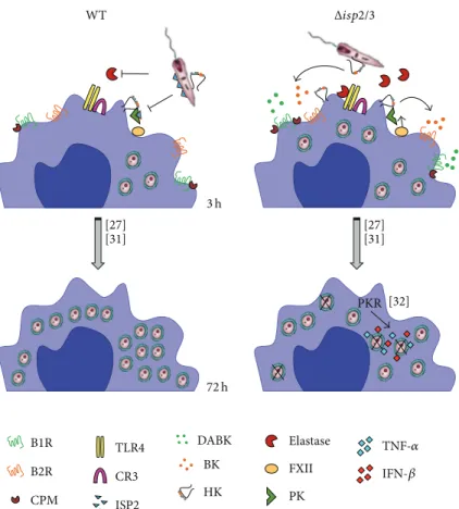
3.3. Surface Exposure of the BK Epitope Difers in WT and���2/3Promastigotes. Since the studies of macrophage infection by L. major promastigotes were routinely per-formed in the absence of serum, we reasoned that the kinin agonists should either originate from kininogens molecules bound to the surface of macrophages [46] or alterna-tively from kininogen molecules eventually sequestered from serum by promastigotes. To test the latter possibility, we washed stationary phaseL. majorpromastigotes extensively as described for infection assays and then stained the para-sites with two diferent domain-speciic mAbs: (i) MBK3, a monoclonal antibody that recognizes the BK epitope (domain D4) of kininogens (HK/LK), and (ii) HKH4, a mAb that recognizes domain D1of HK/LK [36]. FACS analysis showed that almost 70% of���2/3promastigotes are positive stained for HKH4, compared to less than 50% of the WT parasites (Figure 3(a)). Along similar lines, MBK3 antibody showed that the BK epitope of kininogens was present in a higher proportion of ���2/3 promastigotes as compared to WT promastigotes (Figure 3(b), 82,4%) as compared to WT par-asites (Figure 3(b), 73.1%). hese results suggest that both ISP-expressingL. majorpromastigotes and���2/3mutants are able to sequester kininogens (retaining the intact BK molecule) from FCS. According to our working hypothesis (Figure 5), the kininogen opsonins tethered on ISP-deicient parasites may be cleaved by pericellular serine proteases (S1a family) of macrophages, whereas the surface-bound kinino-gens associated to ISP-expressing (WT)L. majorshould be protected from proteolytic cleavage.
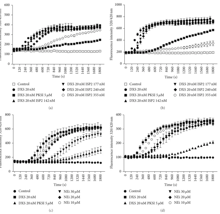
3.4. Targeting Activation of the Contact Phase/KKS in Human Plasma with Recombinant ISP-2 or Synthetic Inhibitor of
Neu-trophil Elastase. Considering that ISP-2 is hardly detected in
R2
0 165 330 495
660 L. major WT
49.3%
R2
0 165 330 495 660
68.3%
100 101 102 103 104 100 101 102 103 104
L. major Δisp2/3
HKH4 HKH4
(a)
R1
0 130 260 390 520
R1
0 130 260 390 520
82.4% 73.1%
100 101 102 103 104 100 101 102 103 104
MBK3 MBK3
(b)
Figure 3: Evidence of diferential display of kininogens and BK epitopes on surface of WTL. majorversus���2/3promastigotes. WT and
���2/3promastigotes were washed 3x before incubation with mAbs against D1 or D4 (BK epitope) of kininogens, HK/LK (HKH4(a) or
MBK3(b), resp.) for 1 h. Unrelated Ab (IgG2afor HKH4or IgG1 for MBK3staining) were used as speciicity controls. Binding of primary IgG
was assessed by incubating the cells with a secondary FITC-labeled anti-mouse IgG antibody for 1 h. he graphs represent the percentage of HK/LK adsorption and are representative of two independent experiments performed in duplicates.
the kininogen-like substrate by PKa [25]. Internal controls run in the presence of the synthetic PKa inhibitor (PKSI-257) show, as expected, pronounced inhibition of the contact phase enzyme by DXS. Assays performed with soluble ISP-2 revealed that the onset of hydrolysis was consistently delayed, in dose-dependent manner (range 142–355 nM; Figures4(a)
and 4(b)). A similar trend was observed when we added MeOSuc-AAPV-CMK (NE inhibitor) to the citrated plasma (range 10–30�M; Figures4(c)and4(d)). Collectively, these indings are consistent with the proposition that the activity of the contact phase enzyme complex (FXIIa/PKa) is at least partially inhibited by ISP-2 (soluble) or by the synthetic NE inhibitor.
4. Discussion
In the current study, we used genetically modiied���2/3 mutants of L. major to determine whether these ecotin-like inhibitors regulate the proinlammatory activity of the kinin/B2R pathway in vivo and in vitro. In the irst group of studies, we demonstrated that L. major promastigotes potently evoke plasma leakage and induce leukocyte accu-mulation in microvascular beds through mast cell-dependent activation of the kinin/B2R pathway, irrespective of the presence or absence of ISP-2. Extending this analysis to
in vitro infection models, we showed that antagonists of
B2R (HOE-140) or B1R (DAL8-BK) eiciently reversed the upregulated phagocytic uptake ofL. major ���2/3by TG-macrophages without interfering with the internalization of ISP-expressing (WT) promastigotes. As discussed further below, these indings suggested that, upon attachment to the macrophage surface, ISP-expressing promastigotes might suppress the activation of B2R/B1R-dependent proinlamma-tory responses by inhibiting the kinin-releasing activity of serine proteases (S1Afamily).
0 100 200 300 400 500 600
Time (s)
Fl
uo
re
scence in
te
n
si
ty
32
0
/
42
0
nm
0
120 240 360 480 600 720 840 960 1080 1200 1320 1440 1560 1680 1800
DXS20nM
DXS20nM PKSI5 �M
DXS20nM ISP2 142nM
DXS20nM ISP2 177nM
DXS20nM ISP2 240nM
DXS20nM ISP2 355nM
Control
(a)
0 200 400 600 800 1000
Time (s)
0
120 240 360 480 600 720 840 960 1080 1200 1320 1440 1560 1680 1800
Fl
uo
re
scence in
te
n
si
ty
32
0
/
42
0
nm
DXS20nM
DXS20nM PKSI5 �M
DXS20nM ISP2 142nM
DXS20nM ISP2 177nM
DXS20nM ISP2 240nM
DXS20nM ISP2 355nM
Control
(b)
0 200 400 600 800
Time (s)
0
120 240 360 480 600 720 840 960 1080 1200 1320 1440 1560 1680 1800
Fl
uo
re
scence in
te
n
si
ty
32
0
/
42
0
nm
Control
DXS20nM
DXS20nM PKSI5 �M
NEi30 �M
NEi20 �M
NEi10 �M
(c)
0 100 200 300 400
Fl
uo
re
scence in
te
n
si
ty
32
0
/
42
0
nm
Time (s)
0
120 240 360 480 600 720 840 960 1080 1200 1320 1440 1560 1680 1800
Control
DXS20nM
DXS20nM PKSI5 �M
NEi30 �M
NEi20 �M
NEi10 �M
(d)
Figure 4: Efect of ISP2 on DXS-induced contact phase activation of human plasma. Citrated human platelet free plasma diluted 1 : 20 in bufer
(described in methods) was supplemented with (i) 4�M Abz-MTEMARRPQ-EDDnp (a, c) or 4�M Abz-GFSPFRSVTVQ-EDDnp (b, d),
intramolecular quenched luorescent substrates whose sequences span the N-terminal or the C-terminal lanking sites (resp.) of BK in mHK (ii) dextran sulfate 500 kDa (DXS; 20 nM). he substrate was also tested in the absence of DXS (Control). Assays with the elastase inhibitor
(NEi—MeOSuc-AAPV-CMK-10, 20, and 30�M), the synthetic PKa inhibitor (PKSI-527—5�M), and the inhibitor of serine peptidase 2
(ISP2—142, 177, 240, and 355 nM) were performed using two diferent schemes. (a, b) PKSI or the elastase inhibitor was added to the plasma
together with DXS and the substrate. (c, d) PKSI or ISP2 was preincubated with plasma for 15 min, at 37∘C, prior to the addition of DXS and
the substrate. Hydrolysis was followed by measuring the luorescence at�ex. = 320nm and�em. = 420nm (up to 1800 seconds). he plot
shows the increase of luorescence with time, relecting substrate hydrolysis. he values in the igures represent the mean±SE of duplicate
determinations performed within 1 representative experiment of 2.
NE. Alternatively, our inding that soluble ISP-2 (or the NE inhibitor MeOSuc-AAPV-CMK) partially inhibit DXS-induced activation of the contact system in human citrated plasma (i.e., PKa-mediated hydrolysis of the lanking sites of BK in kininogen-like substrates) suggests that ISP-expressing promastigotes might rely on their surface-associated ISP-2 to target the contact phase enzymatic complex (FXIIa/PKa/HK)
WT
PKR [32]
B1R
B2R
TLR4
CR3
ISP2
DABK
BK
HK
Elastase
FXII
PK [27]
[31]
[27] [31]
CPM
72h
TNF-�
IFN-𝛽 3h
Δisp2/3
Figure 5: Scheme shows how ISP-2 limits the kinin-releasing activity of surface S1A-proteases of macrophages. As an extension of recently
published studies [27,31,32], here we propose that the proinlammatory phenotype of the���2/3mutant (right side of scheme) is due to
increased pericellular release of kinins mediated by NE and/or contact phase serine proteases (FXIIa/PKa). In the absence of ISP-2, the “eat me
signal” of kininogen tethered onL. majormutants might be inactivated by S1A-family proteases. In addition, the released kinin peptides fuel
phagocytosis and microbicidal function of macrophages via activation of B2R and B1R, a subtype of GPCR upregulated in inlamed tissues.
of TG-macrophages, preliminary results suggest that HOE-140 (tested at 100 nM) upregulates the outgrowth/survival of
���2/3mutants in TG-macrophages. If conirmed by genetic studies, our results may imply thatL. majorpromastigotes might limit ROS formation via the NE/TLR4
/PKR/TNF-�/IFN-� pathway originally described by Faria et al. [32] through ISP-2-dependent targeting of kinin-releasing pepti-dases assembled at the surface of macrophages.
A key event in many inlammatory processes is the adhesive interaction of circulating neutrophils and activated endothelial cells in postcapillary venules, a process that is oten coupled to increased microvascular permeability, which in turn leads to the progressive accumulation of protein-rich edema luid in interstitial tissues. Although conceding that the dynamics of the inlammatory responses that sand-ly-transmittedLeishmaniainduces in the injured dermis is far more complex than what is described in our intravital microscopy studies, the analysis of microvascular leakage and leukocyte accumulation in HCP topically sensitized with
L. major promastigotes (WT or ISP2/3-deicient parasites)
revealed that these parasites are far more potent inducers of plasma leakage and leukocyte accumulation than L.
dono-vani[15],L. chagasipromastigotes (Figure 1(c)), orT. cruzi
trypomastigotes [35]. Although the mechanisms underlying the discrepant phenotypes ofL. majorand L. chagasiorT.
cruziremain unknown, we were intrigued to ind out that a
3X-fold higher dose ofL. chagasipromastigotes did not evoke such a strong microvascular response, not even ater treating the HCP with captopril, an inhibitor of kinin degradation by angiotensin-converting enzyme (Figure 1(c), black curve). In contrast, T. cruzi and L. chagasi are potentially lethal pathogens that disseminate systemically and preferentially target tissue in organs irrigated by fenestrated capillaries. Under these circumstances, plasma-borne substrates, such as kininogens, difuse freely into the visceral tissues invaded by these visceralizing species of pathogenic trypanosomatids, both of which were empowered with kinin-releasing cysteine proteases [15, 47–49]. It is noteworthy that B2R-deicient mice acutely infected by T. cruzi [14] or L. chagasi [16] display heightened disease susceptibility, implying that the activation of the kinin/B2R pathway may preferentially shit the host/parasite balance towards protective immunity, at least during the acute phase.
with metacyclic parasites. Our studies in HCP topically sen-sitized withL. majorpromastigotes (which prevents bleeding and KKS activation due to pathogen inoculation through needles) suggest that these parasites potently evoke plasma leakage and leukocyte accumulation in microvascular beds via the kinin/B2R pathway. For reasons that are unclear, we found that���2/3promastigotes evoked a somewhat milder inlammatory response (20%). Incidentally, Eschenlauer et al. [27] have reported that mice subcutaneously infected with
L. major promastigotes transiently displayed higher tissue
burden of ISP2/ISP-3-deicient parasites as compared to WT parasites. Lasting 3 days, the parasite burden subsequently equalized, implying that the selective advantage conferred to ISP-deicient promastigotes has waned as the infection progressed.
Based on pharmacological approaches, we showed evi-dences that L. major activates the KKS via mechanisms that involve transcellular cross-talk between neutrophils (intravascularly) and mast cells, a subset of innate sentinel cells that are mostly localized in perivascular tissues. Beyond the vasoactive role of histamine, a potent inducer of vascular permeability, mast cells also release heparin and polyphos-phates, both of which were recently characterized as endoge-nous activators of the contact system [50,51]. Given the inter-dependent nature of inlammatory circuits, it is likely that mast cells and the KKS/complement cascades are reciprocally activated and fueled in the HCP sensitized with L. major
promastigotes. Although we have not studied the impact of the inlux of complement into peripheral sites of L. major
infection, it is well-documented thatLeishmania lipophos-phoglycan is opsonized by C3bi [52]. In the absence of other potent inlammatory cues, the engagement of macrophage CR3 by ISP-expressing L. major (WT) promastigotes may drive phagocytosis without necessarily stimulating the pro-duction of reactive oxygen intermediates, thereby creating a hospitable environment for the intracellular growth ofL.
major[31,32]. Although studies in CD11b-deicient BALB/c
mice have recently conirmed that activation of the C3bi/CR3 pathway increases host susceptibility toL. major infection [53], it will be interesting to know whether CR3-dependent suppression of IL-12 responses might depend on parasite-evoked extravasation of complement components to the extravascular compartment, as proposed here for plasma-borne kininogens.
Studies in the mouse ear model of sandly-transmitted infection showed that L. major metacyclic promastigotes deposited in the dermis are engulfed by the iniltrating neutrophils within approximately 3 h [19–21]. Based on the results described in HCP topically sensitized withL. major
promastigotes, it is conceivable that the proteolytic release of vasoactive kinins may further stimulate the transendothelial migration of neutrophils at very early stages of the infection. Furthermore, given evidence that parasitized neutrophils expose the apoptotic markers required for eferocytosis, it will be interesting to know whether plasma leakage may con-tribute to DC eferocytosis and to the ensuing suppression of h1-inductive functions of DCs in the draining lymph nodes [19–21]. Beyond the impact on adaptive immunity, it is well established that eferocytosis of apoptotic neutrophils has
profound efect on parasite survival in infected macrophages [22]. In this context, our inding that kininogen epitopes (N-terminal D1 and internal/BK/D4 domain) are tethered at the surface ofLeishmaniapromastigotes is an intriguing inding because it raises the possibility that ISP-expressing promastigotes might “protect” the integrity of surface bound kinin precursors from premature proteolytic degradation by host proteolytic enzymes. his hypothesis is worth exploring in light of recent studies showing that HK (an abundant protein in the bloodstream; 660 nM) binds to PS exposed on apoptotic neutrophils before stimulating uPAR-dependent eferocytosis by macrophages via the p130Cas-CrkII-Dock-180-Rac1 pathway [6]. In other words, ISPs may protect the integrity of HK opsonins “eat me signals” while at the same time preventing the liberation of proinlammatory kinins (see scheme, Figure 5) within sites of parasite attachment to phagocytes. Although we have not explored the poten-tial signiicance ofL. major opsonization by kininogens, it will be interesting to know whetherparasite subsets bearing the uPAR ligand HK/HKa “eat me signal” might render macrophage permissive to intracellular survival, perhaps reminiscent of the apoptotic mimicry paradigm originally described by Barcinski and coworkers [54].
5. Conclusions
Extending the breadth of our previous investigations about the role of the kallikrein-kinin system in the immunopatho-genesis of experimental Chagas disease and visceral leishma-niasis, the studies reported in this paper suggest that ecotin-like inhibitors expressed byL. majorpromastigotes ine-tune phagocytosis and may limit amastigote survival by inhibiting the pericellular activity of kinin-releasing serine proteases (S1afamily) of macrophages.
Conflict of Interests
he authors declare that there is no conlict of interests regarding the publication of this paper.
Acknowledgments
his study was supported by grants from CNPq/INBEB, Faperj, and PRONEX. he authors thank Professor Ana Paula Cabral de Araujo Lima (Instituto de Biof´ısica Carlos Chagas Filho, Universidade Federal do Rio de Janeiro) for kindly providing WT andΔ���2/3L. majorand inhibitor of serine peptidase 2 (ISP2) and Professor Werner M¨uller-Esterl (University of Frankfurt) for donation of kininogen anti-bodies. hey also wish to thank Dr. Marilia Faria (Instituto de Biof´ısica Carlos Chagas Filho, Universidade Federal do Rio de Janeiro) for independently testing the B2R and B1R antagonists in infection assays with macrophages (unpub-lished data).
References
inlammatory pathway,”Advances in Immunology, vol. 121, pp. 41–89, 2014.
[2] C. Maas and T. Renn´e, “Regulatory mechanisms of the plasma
contact system,”hrombosis Research, vol. 129, no. 2, pp. S73–
S76, 2012.
[3] U. Amara, M. A. Flierl, D. Rittirsch et al., “Molecular intercom-munication between the complement and coagulation systems,”
Journal of Immunology, vol. 185, no. 9, pp. 5628–5636, 2010. [4] D. Ricklin, G. Hajishengallis, K. Yang, and J. D. Lambris,
“Complement: a key system for immune surveillance and
homeostasis,”Nature Immunology, vol. 11, no. 9, pp. 785–797,
2010.
[5] Y. T. Wachtfogel, R. A. deLa Cadena, S. P. Kunapuli et al., “High molecular weight kininogen binds to Mac-1 on neutrophils by its heavy chain (domain 3) and its light chain (domain 5),”
Journal of Biological Chemistry, vol. 269, no. 30, pp. 19307–19312, 1994.
[6] A. Yang, J. Dai, Z. Xie et al., “High molecular weight kininogen binds phosphatidylserine and opsonizes urokinase
plasmino-gen activator receptor-mediated eferocytosis,”he Journal of
Immunology, vol. 192, no. 9, pp. 4398–4408, 2014.
[7] A. Kozik, R. B. Moore, J. Potempa, T. Imamura, M. Rapala-Kozik, and J. Travis, “A novel mechanism for bradykinin production at inlammatory sites: Diverse efects of a mixture of neutrophil elastase and mast cell tryptase versus tissue and
plasma kallikreins on native and oxidized kininogens,”Journal
of Biological Chemistry, vol. 273, no. 50, pp. 33224–33229, 1998. [8] K. M. Heutinck, I. J. M. ten Berge, C. E. Hack, J. Hamann, and A. T. Rowshani, “Serine proteases of the human immune system
in health and disease,”Molecular Immunology, vol. 47, no. 11-12,
pp. 1943–1955, 2010.
[9] J. Aliberti, J. P. B. Viola, A. Vieira-de-Abreu, P. T. Bozza, A. Sher, and J. Scharfstein, “Cutting edge: bradykinin induces IL-12 production by dendritic cells: a danger signal that drives h1
polarization,”Journal of Immunology, vol. 170, no. 11, pp. 5349–
5353, 2003.
[10] C. M. Bertram, S. Baltic, N. L. Misso et al., “Expression of kinin B1 and B2 receptors in immature, monocyte-derived dendritic
cells and bradykinin-mediated increase in intracellular Ca2+
and cell migration,”Journal of Leukocyte Biology, vol. 81, no. 6,
pp. 1445–1454, 2007.
[11] R. Gulliver, S. Baltic, N. L. Misso, C. M. Bertram, P. J. hompson, and M. Fogel-Petrovic, “Lys-des[Arg9]-bradykinin alters migration and production of interleukin-12 in
monocyte-derived dendritic cells,”American Journal of Respiratory Cell
and Molecular Biology, vol. 45, no. 3, pp. 542–549, 2011.
[12] F. Marceau and D. R. Bachvarov, “Kinin receptors,”Clinical
Reviews in Allergy and Immunology, vol. 16, no. 4, pp. 385–401, 1998.
[13] A. C. Monteiro, V. Schmitz, E. Svensj¨o et al., “Cooperative activation of TLR2 and bradykinin B2 receptor is required for induction of type 1 immunity in a mouse model of subcutaneous
infection byTrypanosoma cruzi,”Journal of Immunology, vol.
177, no. 9, pp. 6325–6335, 2006.
[14] A. C. Monteiro, V. Schmitz, A. Morrot et al., “Bradykinin B2 Receptors of dendritic cells, acting as sensors of kinins
pro-teolytically released byTrypanosoma cruzi, are critical for the
development of protective type-1 responses,”PLoS Pathogens,
vol. 3, no. 11, pp. 1730–1744, 2007.
[15] E. Svensj¨o, P. R. Batista, C. I. Brodskyn et al., “Interplay between parasite cysteine proteases and the host kinin system
modulates microvascular leakage and macrophage infection by
promastigotes of theLeishmania donovanicomplex,”Microbes
and Infection, vol. 8, no. 1, pp. 206–220, 2006.
[16] D. Nico, D. F. Feij´o, and N. Maran, “Resistance to visceral leishmaniasis is severely compromised in mice deicient of
bradykinin B2-receptors,”Parasites and Vectors, vol. 5, no. 1,
article 261, 2012.
[17] J. Scharfstein, D. Andrade, E. Svensj¨o, A. C. Oliveira, and C. R. Nascimento, “he kallikrein-kinin system in experimen-tal Chagas disease: a paradigm to investigate the impact of inlammatory edema on GPCR-mediated pathways of host cell
invasion byTrypanosoma cruzi,”Frontiers in Immunology, vol.
3, no. 396, pp. 1–20, 2007.
[18] B. Engelmann and S. Massberg, “hrombosis as an
intravascu-lar efector of innate immunity,”Nature Reviews Immunology,
vol. 13, no. 1, pp. 34–45, 2013.
[19] N. C. Peters, J. G. Egen, N. Secundino et al., “In vivo imaging reveals an essential role for neutrophils in leishmaniasis
trans-mitted by sand lies,”Science, vol. 321, no. 5891, pp. 970–974,
2008.
[20] N. C. Peters, “In vivo imaging reveals an essential role for
neutrophils in Leishmaniasis transmitted by sand lies,”Science,
vol. 322, no. 5908, p. 1634, 2008.
[21] F. L. Ribeiro-Gomes, N. C. Peters, A. Debrabant, and D. L. Sacks, “Eicient capture of infected neutrophils by dendritic cells in the skin inhibits the early anti-leishmania response,”
PLoS Pathogens, vol. 8, no. 2, Article ID e1002536, 2012. [22] F. L. Ribeiro-Gomes, M. C. A. Moniz-de-Souza, M. S.
Alexandre-Moreira et al., “Neutrophils activate macrophages
for intracellular killing ofLeishmania majorthrough
recruit-ment of TLR4 by neutrophil elastase,”he Journal of
Immunol-ogy, vol. 179, no. 6, pp. 3988–3994, 2007.
[23] A. A. Filardy, D. R. Pires, and G. A. Dosreis, “Macrophages and neutrophils cooperate in immune responses to Leishmania
infection,”Cellular and Molecular Life Sciences, vol. 68, no. 11,
pp. 1863–1870, 2011.
[24] R. G. Titus and J. M. C. Ribeiro, “he role of vector saliva in
transmission of arthropod-borne disease,”Parasitology Today,
vol. 6, no. 5, pp. 157–160, 1990.
[25] P. H. Alvarenga, X. Xu, F. Oliveira et al., “Novel family of insect salivary inhibitors blocks contact pathway activation by binding
to polyphosphate, heparin, and dextran sulfate,”Arteriosclerosis,
hrombosis, and Vascular Biology, vol. 33, no. 12, pp. 2759–2770, 2013.
[26] F. M¨uller, N. J. Mutch, W. A. Schenk et al., “Platelet polyphos-phates are proinlammatory and procoagulant mediators in
vivo,”Cell, vol. 139, no. 6, pp. 1143–1156, 2009.
[27] S. C. P. Eschenlauer, M. S. Faria, L. S. Morrison et al., “Inluence of parasite encoded inhibitors of serine peptidases in early
infection of macrophages with Leishmania major,” Cellular
Microbiology, vol. 11, no. 1, pp. 106–120, 2009.
[28] C. H. Chung, H. E. Ives, S. Almeda, and A. L. Goldberg,
“Puriication fromEscherichia coliof a periplasmic protein that
is a potent inhibitor of pancreatic proteases,”he Journal of
Biological Chemistry, vol. 258, no. 18, pp. 11032–11038, 1983. [29] C. T. Eggers, I. A. Murray, V. A. Delmar, A. G. Day, and
C. S. Craik, “he periplasmic serine protease inhibitor ecotin
protects bacteria against neutrophil elastase,”he Biochemical
Journal, vol. 379, no. 1, pp. 107–118, 2004.
[30] A. C. Ivens, C. S. Peacock, and E. A. Worthey, “he genome of
the kinetoplastid parasite,Leishmania major,”Science, vol. 309,
[31] M. S. Faria, F. C. G. Reis, R. L. Azevedo-Pereira, L. S. Morrison, J. C. Mottram, and A. P. C. A. Lima, “Leishmania inhibitor of serine peptidase 2 prevents TLR4 activation by neutrophil elastase promoting parasite survival in murine macrophages,”
he Journal of Immunology, vol. 186, no. 1, pp. 411–422, 2011. [32] M. S. Faria, T. C. Calegari-Silva, A. de Carvalho, J. C. Vivarini, U.
G. Lopes, and A. P. Lima, “Role of protein kinase R in the killing of Leishmania major by macrophages in response to neutrophil
elastase and TLR4 via TNF�and IFN�,”he FASEB Journal, vol.
28, no. 7, pp. 3050–3063, 2014.
[33] V. Schmitz, L. N. Almeida, E. Svensj¨o, A. C. Monteiro, J. K¨ohl, and J. Scharfstein, “C5a and bradykinin receptor cross-talk
regulates innate and adaptive immunity inTrypanosoma cruzi
infection,”he Journal of Immunology. In press.
[34] V. Schmitz, E. Svensj¨o, R. R. Serra, M. M. Teixeira, and J. Scharfstein, “Proteolytic generation of kinins in tissues infected byTrypanosoma cruzidepends on CXC chemokine secretion
by macrophages activated via Toll-like 2 receptors,”Journal of
Leukocyte Biology, vol. 85, no. 6, pp. 1005–1014, 2009.
[35] D. Andrade, R. Serra, E. Svensj¨o et al., “Trypanosoma cruzi
invades host cells through the activation of endothelin and bradykinin receptors: a converging pathway leading to chagasic
vasculopathy,”British Journal of Pharmacology, vol. 165, no. 5,
pp. 1333–1347, 2012.
[36] J. Kaufmann, M. Haasemann, S. Modrow, and W. M¨uller-Esterl, “Structural dissection of the multidomain kininogens. Fine mapping of the target epitopes of antibodies interfering with
their functional properties,”he Journal of Biological Chemistry,
vol. 268, no. 12, pp. 9079–9091, 1993.
[37] B. Korkmaz, S. Attucci, M. A. Juliano et al., “Measuring elastase, proteinase 3 and cathepsin G activities at the surface of human neutrophils with luorescence resonance energy transfer
substrates,”Nature Protocols, vol. 3, no. 6, pp. 991–1000, 2008.
[38] A. Fukumizu and Y. Tsuda, “Amino acids and peptides. LIII. Synthesis and biological activities of some pseudo-peptide analogs of PKSI-527, a plasma kallikrein selective inhibitor: the
importance of the peptide backbone,”Chemical and
Pharma-ceutical Bulletin, vol. 47, no. 8, pp. 1141–1144, 1999.
[39] E. Svensj¨o, “Bradykinin and prostaglandin E1, E2 and F2�
-induced macromolecular leakage in the hamster cheek pouch,”
Prostaglandines and Medicine, vol. 1, no. 5, pp. 397–410, 1978. [40] E. Svensj¨o, K. E. Andersson, E. Bouskela, F. Z. G. A. Cyrino,
and S. Lindgren, “Efects of two vasodilatory phosphodiesterase inhibitors on bradykinin-induced permeability increase in the
hamster cheek pouch,”Agents and Actions, vol. 39, no. 1-2, pp.
35–41, 1993.
[41] S. Rosengren, K. Ley, and K.-E. Arfors, “Dextran sulfate prevents LTB4-induced permeability increase, but not
neu-trophil emigration, in the hamster cheek pouch,”Microvascular
Research, vol. 38, no. 3, pp. 243–254, 1989.
[42] D. C. H¨ocking, T. J. Ferro, and A. Johnson, “Dextran sul-fate inhibits PMN-dependent hydrostatic pulmonary edema
induced by tumor necrosis factor,”Journal of Applied Physiology,
vol. 70, no. 3, pp. 1121–1128, 1991.
[43] R. A. Brown, R. Lever, N. A. Jones, and C. P. Page, “Efects of heparin and related molecules upon neutrophil aggregation and
elastase release in vitro,”British Journal of Pharmacology, vol.
139, no. 4, pp. 845–853, 2003.
[44] K. Ley, M. Cerrito, and K.-E. Arfors, “Sulfated polysaccharides
inhibit leukocyte rolling in rabbit mesentery venules,”American
Journal of Physiology. Heart and Circulatory Physiology, vol. 260, no. 5, pp. H1667–H1673, 1991.
[45] M. Siebeck, J. C. Cheronis, E. Fink et al., “Dextran sulfate activates contact system and mediates arterial hypotension via
B2 kinin receptors,”Journal of Applied Physiology, vol. 77, no. 6,
pp. 2675–2680, 1994.
[46] A. Barbasz and A. Kozik, “he assembly and activation of kinin-forming systems on the surface of human U-937
macrophage-like cells,”Biological Chemistry, vol. 390, no. 3, pp. 269–275,
2009.
[47] E. del Nery, M. A. Juliano, A. P. C. A. Lima, J. Scharfstein, and L. Juliano, “ Kininogenase activity by the major cysteinyl
proteinase (cruzipain) from Trypanosoma cruzi,” Journal of
Biological Chemistry, vol. 272, no. 41, pp. 25713–25718, 1997. [48] A. P. C. A. Lima, P. C. Almeida, I. L. S. Tersariol et al., “Heparan
sulfate modulates kinin release byTrypanosoma cruzithrough
the activity of cruzipain,”he Journal of Biological Chemistry,
vol. 277, no. 8, pp. 5875–5881, 2002.
[49] J. Scharfstein, V. Schmitz, V. Morandi et al., “Host cell invasion byTrypanosoma cruziis potentiated by activation of bradykinin
B2 receptors,”he Journal of Experimental Medicine, vol. 192, no.
9, pp. 1289–1299, 2000.
[50] C. Oschatz, C. Maas, B. Lecher et al., “Mast cells increase
vascu-lar permeability by heparin-initiated bradykinin formationin
vivo,”Immunity, vol. 34, no. 2, pp. 258–268, 2011.
[51] D. Moreno-Sanchez, L. Hernandez-Ruiz, F. A. Ruiz, and R. Docampo, “Polyphosphate is a novel pro-inlammatory
regu-lator of mast cells and is located in acidocalcisomes,”Journal of
Biological Chemistry, vol. 287, no. 34, pp. 28435–28444, 2012. [52] G. F. Sp¨ath, L. A. Garraway, S. J. Turco, and S. M. Beverley, “he
role(s) of lipophosphoglycan (LPG) in the establishment of
Leishmania majorinfections in mammalian hosts,”Proceedings of the National Academy of Sciences of the United States of America, vol. 100, no. 16, pp. 9536–9541, 2003.
[53] C. R. Carter, J. P. Whitcomb, J. A. Campbell, R. M. Mukbel, and M. A. McDowell, “Complement receptor 3 deiciency
inlu-ences lesion progression duringLeishmania majorinfection in
BALB/c Mice,”Infection and Immunity, vol. 77, no. 12, pp. 5668–
5675, 2009.
[54] J. L. M. Wanderley and M. A. Barcinski, “Apoptosis and
apoptotic mimicry: theLeishmaniaconnection,”Cellular and
Submit your manuscripts at
http://www.hindawi.com
Stem Cells
International
Hindawi Publishing Corporation
http://www.hindawi.com Volume 2014
Hindawi Publishing Corporation
http://www.hindawi.com Volume 2014
INFLAMMATION
Hindawi Publishing Corporation
http://www.hindawi.com Volume 2014
Behavioural
Neurology
Endocrinology
International Journal ofHindawi Publishing Corporation
http://www.hindawi.com Volume 2014
Hindawi Publishing Corporation
http://www.hindawi.com Volume 2014
Disease Markers
Hindawi Publishing Corporation
http://www.hindawi.com Volume 2014 BioMed
Research International
Oncology
Journal of Hindawi Publishing Corporationhttp://www.hindawi.com Volume 2014
Hindawi Publishing Corporation
http://www.hindawi.com Volume 2014 Oxidative Medicine and Cellular Longevity Hindawi Publishing Corporation
http://www.hindawi.com Volume 2014
PPAR Research
The Scientiic
World Journal
Hindawi Publishing Corporation
http://www.hindawi.com Volume 2014
Immunology Research Hindawi Publishing Corporation
http://www.hindawi.com Volume 2014
Journal of
Obesity
Journal ofHindawi Publishing Corporation
http://www.hindawi.com Volume 2014
Hindawi Publishing Corporation
http://www.hindawi.com Volume 2014
Computational and Mathematical Methods in Medicine
Ophthalmology
Journal ofHindawi Publishing Corporation
http://www.hindawi.com Volume 2014
Diabetes Research
Journal ofHindawi Publishing Corporation
http://www.hindawi.com Volume 2014
Hindawi Publishing Corporation
http://www.hindawi.com Volume 2014
Research and Treatment
AIDS
Hindawi Publishing Corporation
http://www.hindawi.com Volume 2014
Gastroenterology Research and Practice
Hindawi Publishing Corporation
http://www.hindawi.com Volume 2014
Parkinson’s
Disease
Evidence-Based Complementary and Alternative Medicine
Volume 2014