ORIGINAL ARTICLE
Nutritional status in patients with cutaneous leishmaniasis and
a study of the effects of zinc supplementation together with
antimony treatment
Miguel Guzman-Rivero
1,2,3*, Ernesto Rojas
1,2, Aleida Verduguez-Orellana
1,2,
Henry Pardo
1,2, Mary Cruz Torrico
1, Lieselotte Cloetens
3, Bjo¨rn A
˚ kesson
3,4and
Edgar Sejas
11
Instituto de Investigaciones Bio-Me´dicas (IIBISMED), Universidad Mayor de San Simo´n, Cochabamba, Bolivia;2Centro Universitario de Medicina Tropical (CUMETROP), Universidad Mayor de San Simo´n, Cochabamba, Bolivia;3Biomedical Nutrition, Pure and Applied Biochemistry, Lund University, Lund, Sweden;4Department of Clinical Nutrition, Ska˚ne University Hospital, Lund, Sweden
Abstract
Background: The role of micronutrient status for the incidence and clinical course of cutaneous leishmaniasis is not much studied. Still zinc supplementation in leishmaniasis has shown some effect on the clinical recovery, but the evidence in humans is limited.
Objective: To compare biochemical nutritional status in cutaneous leishmaniasis patients with that in controls and to study the effects of zinc supplementation for 60 days.
Design: Twenty-nine patients with cutaneous leishmaniasis were treated with antimony for 20 days. Fourteen of them got 45 mg zinc daily and 15 of them got placebo. Biomarkers of nutritional and inflammatory status and changes in size and characteristics of skin lesions were measured.
Results: The level of transferrin receptor was higher in patients than in controls but otherwise no differences in nutritional status were found between patients and controls. No significant effects of zinc supplementa-tion on the clinical recovery were observed as assessed by lesion area reducsupplementa-tion and characteristics or on biochemical parameters.
Conclusions: It is concluded that nutritional status was essentially unaffected in cutaneous leishmaniasis and that oral zinc supplementation administered together with intramuscular injection of antimony had no additional clinical benefit.
Keywords: nutritional biomarkers;zinc supplementation;cutaneous leishmaniasis;antimony treatment;clinical chemistry
Responsible Editor: Asim Duttaroy, University of Oslo, Norway.
To access the supplementary material to this article, please see Supplementary files under Article Tools online.
Received: 14 November 2013; Revised: 10 September 2014; Accepted: 30 September 2014; Published: 6 November 2014
C
utaneous leishmaniasis is caused by different species of the genus Leishmania, and the esti-mated number of new cases annually is around 1.1 million (1, 2). In Latin America with the exception of Chile and Antilles, 638,702 cases were reported in the period 20012011 (3). Cutaneous leishmaniasis resultsfrom multiplication of Leishmania in the phagocytes of the skin (4). In Bolivia, most cases are caused by
Leishmania (Viannia) braziliensis (5, 6). The cases are characterized by one or more lesions with raised borders
often associated with secondary bacterial infection and frequently localized in the exposed areas of the skin (79).
Among malnutrition conditions, micronutrient defi-ciencies can contribute to the exacerbation and delayed recovery of infectious and chronic non-infectious diseases (10, 11). In human cutaneous leishmaniasis, a significant decrease of zinc concentration and increase of copper concentration in plasma have been demonstrated which may be associated with the inability of the host to kill
the parasites or be an indication of the inflammatory pro-cess (12, 13). Other aspects of the biochemical nutritional status in leishmaniasis have not been studied previously. In Bolivia, no data regarding the prevalence of micro-nutrient deficiencies in the general population is available. Regarding therapy for cutaneous leishmaniasis, the sys-temic use of antimony compounds gives a cure rate of 7790% (1). The results of oral zinc treatment studies
in cutaneous leishmaniasis have been contradictory. A decrease of erythemas and size of the induration was reported (14, 15) but others found no effect of zinc on cutaneous leishmaniasis (16). Still the available data on the effects of zinc as nutritional supplementation in this disease are scant.
In the present study, we compared biochemical nu-tritional status in cutaneous leishmaniasis patients and matched controls and conducted a placebo-controlled study on the effect of zinc supplementation in patients with cutaneous leishmaniasis.
Methods
Patients
The patients were residents of the IsiboroSe´cure national
park, a tropical forest of Cochabamba province. The patients were selected on the basis of the following criteria: age 1550 years, diagnosis of cutaneous leishmaniasis
by any of the two laboratory tests described below, and no history of previous leishmaniasis episodes. Exclusion criteria were mucosal or mucocutaneous leishmaniasis, presence of more than two cutaneous lesions, pregnancy, lactation, use of nutritional supplements, presence of dia-betes mellitus, chronic renal failure, or liver disease. We contacted 87 patients with cutaneous leishmaniasis visit-ing Villa Tunari Hospital and the 34 patients meetvisit-ing the inclusion criteria were selected. All patients completed a health questionnaire prior to entering the study and signed a consent form for inclusion into the study.
Control subjects
The controls were age- and gender-matched subjects living in the same area as the corresponding patients. All con-trols completed a health questionnaire prior to entering the study and signed a consent form for inclusion into the study. Subjects were excluded if they had diabetes mellitus, had cardiovascular disease, were in pregnancy or lactation, or received regular medication or nutritional supplements.
Zinc supplementation and antimony treatment of cutaneous leishmaniasis
Patients were randomly allocated to receive zinc or placebo coded capsules for 60 days. Each zinc capsule contained 315 mg of zinc gluconate (45 mg zinc) and each placebo capsule contained 315 mg of corn starch
(Farmacia artesanal, Cochabamba, Bolivia). One capsule per day (zinc or placebo) was taken after a meal coinciding with the time of antimony injection during the therapy period and continued at the same time thereafter. All patients received for 20 days daily intramuscular injections of pentavalent antimony (Glucantime†, Sanofi Aventis
Farmaceˆutica Ltda, Sa˜o Paulo, Brazil), 20 mg Sb/kg/day. The physicians in the health care centers of Isiboro-Se´cure park administered the injection. The compliance was assessed by daily reporting of given capsule by the physicians. Control subjects were not given any drugs or capsules.
Materials and measurements
Venous blood collection tubes of the Vacutainer†system
(cat no 367874 and 368380) were obtained from Becton Dickinson AB (Stockholm, Sweden). Blood agar No2 (cat no DF 0027-17-0) was obtained from Difco Labora-tories Inc (Detroit, Michigan, USA). The Panotic fast staining system (cat no 620529) was obtained from LB (Laborclin, Sa˜o Paulo, Brazil) and disposable plastic calipers for the measurement of lesion dimensions were obtained in local commerce.
Collection of blood samples
For patients, blood was sampled three times, before the start of the treatment (T0), after 20 days at the end of antimony treatment (T1) and after 60 days of supplemen-tation with zinc or placebo (T2). For controls, blood was sampled at time zero only. Blood was collected by venipuncture after 12 h of fasting and 30 min of relaxation between 7 and 8 in the morning into polystyrene test tubes. They were centrifuged for 10 min at ]2,000 g and plasma samples were aliquoted and stored at 808C
until analysis.
Microbiological and biochemical analysis
Parasite identification was performed by microscopy of stained smears of scrapings of lesion borders and by isolation in culture (17). Briefly, smears of scrapings of lesion borders in triplicate were stained with the Panotic fast system and microscopically assessed for amastigote forms of the parasite. The isolation by culture was performed by inoculation of aspirates into tubes contain-ing blood agar base No2 and 10% rabbit defibrinated blood (18). The tubes were incubated at 268C for 10 days and the promastigote form of parasite was microscopically assessed. Total number of white blood cells, neutrophils, eosinophils and lymphocytes (as fraction of white blood cells), red blood cells, hemoglobin, and hematocrit were measured within 30 min after blood sampling using the Auto hematology analyzer BC-3000 Plus, MindrayTM
IIBISMED laboratories. The other clinical chemistry measurements were performed at the Clinical Chemistry Laboratory of Ska˚ne University Hospital, Lund, Sweden, using certified methods. These data were compared with the reference ranges for healthy subjects established at that laboratory.
Assessment of lesion healing
The cutaneous lesions were assessed in two time phases, the first one at 3, 9, 15, and 20 days, concomitant with antimony treatment and then every 10 days during the last 40 days. Area of lesion (mm2) and presence of raised edge of lesion, inflammatory halo, satellite lesions, and purulent material were measured. The area was calcu-lated using the formula for a circle or ellipse based on measurements with a caliper. The healing of lesions was expressed as percent reduction of the initial area.
Ethics permission
Ethics permission for procedures involving human vo-lunteers was obtained from the Bolivian Ethics Com-mittee of the Medical Faculty, Universidad Mayor de San Simo´n and the Regional Ethics Committee, Lund, Sweden (no. 2009/171).
Statistical analysis
The SPSS software was used. The MannWhitney test was
used for testing the significance of differences between two non-normally distributed continuous variables for comparisons between patients and controls and also for two non-normally distributed quantitative discrete vari-ables between zinc-supplemented and placebo groups. Chi-square analysis was used for comparison of individual characteristics of the lesions between the groups. The Wilcoxon signed rank test was used to compare the same variables of patients at T0, T2 and in controls for the two groups (zinc-supplemented or placebo). No corrections for multiple testing were made.
Results
Comparison of nutritional status in patients and controls
For patients and 29 matched controls there were no statistically significant differences in weight, height and body mass index (BMI) (Supplementary Table 1). The plasma concentrations of nutrient-related compounds (Table 1) showed in both patient groups increased values of transferrin receptor at T0 compared with the corre-sponding controls (p0.002 and 0.033, respectively). Still
a concentration above the reference range was observed in only two female patients at T2. This can reflect depleted iron storage and the two patients had also subnormal concentrations of iron and ferritin in plasma and high values of the total iron-binding capacity and also low blood hemoglobin values both at T0 and T2. A slightly lower concentration of sodium at T0 in the placebo group
compared to control was observed (p0.05). For plasma
vitamin B12, subnormal values were observed in five patients at T0 and also at T2 and in three controls but no significant differences between the group mean values were observed. For other nutrient-related compounds, there were no significant differences between patients at T0 and controls (Table 1).
Clinical observations after zinc supplementation
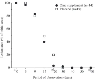
Thirty-four patients entered in the study and 29 completed it. The reasons for dropping out in the placebo group were low adherence to the supplementation and clinical follow-up in three cases and low adherence to the anti-mony treatment in one case. In the zinc-supplemented group one case dropped out due to low adherence to the clinical follow up. The time course for reduction of lesion area did not differ significantly between placebo and zinc-supplemented groups (Fig. 1). The differences in lesion area (%) were significant for the intervals day (915),
(1520), and (2030) in both groups (Table 2). Regarding the four lesion characteristics, a higher frequency of purulent material (an indicator of superinfection) and of inflammatory halo was found in the zinc-supplemented group at day 20 only (Chi-square test p50.05) (Fig. 2).
Biochemical markers after zinc supplementation
The number of red cells, hematocrit, and the hemoglobin concentration were decreased at T1 in both zinc-supple-mented and placebo groups compared to data at T0 (Supplementary Table 2). The total number of leucocytes was lower in both groups at T0 and the fraction of lymphocytes was lower at T1 and T2 in the placebo group compared with the data in controls (Supplementary Table 2). The enzymes ASAT and ALAT tended to be higher at T1 than at T0 and T2 (Supplementary Table 3). These findings may partly indicate side effects of antimony treatment.
Regarding inflammatory markers (Supplementary Table 4), an increase of CRP at T2 compared to at T0 was found in the zinc-supplemented group together with a small decrease in the placebo group although without statistical significance in both cases. The concentration of haptoglobin was decreased at T2 compared to T0 in both zinc-supplemented and placebo groups. Orosomu-coid was decreased in the placebo group at T2 compared to T0.
Regarding nutrient-related compounds (Table 1), the concentration of folate was decreased at T2 compared to T0 in the zinc-supplemented group. At T2 the cal-cium concentration in the placebo group was decreased (p0.012). The comparison of the differences T2T0
Regarding other clinical chemistry biomarkers (Supple-mentary Table 5), decreased concentration of creatinine was found at T2 in the zinc-supplemented group com-pared with T0 (p0.019). Regarding
gammaglutamyl-transferase (GT) only one patient had high values at T0 and T2 and also one control. Alkaline phosphatase (ALP) was slightly elevated in four patients at T0, nine patients at T2 and in seven controls. No differences were found between zinc-supplemented and placebo groups in these respects.
Discussion
Nutritional status
Regarding general nutritional status in leishmaniasis, a low BMI (B18.5 kg/m2) was found in 10% and hypoalbuminemia (B35 g/L) in 12% of the patients (19). In our patient group only one patient (of 29) has such a low BMI and three patients had a BMI below 20 kg/m2.
Only one of our patients had a plasma albumin below 35 g/L. In another study, children with visceral leishma-niasis were found to have a lower BMI and plasma albumin than the other groups studied (20). The loss of weight may be attributable in cases of American tegu-mentary leishmaniasis to difficulties to eat (19) and in visceral leishmaniasis to the cachexia caused by the infection (20).
Concerning the nutrient-related compounds of plasma, the concentration of transferrin receptor was higher in patients than in controls (Table 1). Also low levels of plasma iron and high levels of iron-binding capacity were ob-served in several patients. The subnormal iron status can at least partly be explained by a sequestration of iron which can limit the leishmania infection (2123). Other
authors also reported decreased levels of iron plasma concentration in cutaneous and visceral leishmaniasis which was interpreted partly as a response to the effect of immunoregulatory cytokines (24, 25).
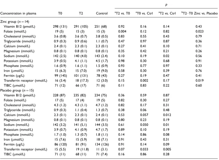
Table 1. Plasma concentration of nutrient-related compounds in cutaneous leishmaniasis patients before and after zinc supplementation and in controls
p
Concentration in plasma T0 T2 Control aT2 vs. T0 aT0 vs. Ctrl aT2 vs. Ctrl bT2T0 Zinc vs. Placebo
Zinc group (n14)
Vitamin B12 (pmol/L) 298 (131) 291 (105) 251 (68) 0.92 0.16 0.14 0.43 Folate (nmol/L) 19 (5) 15 (3) 15 (3) 0.004 0.12 0.82 0.023 Cholesterol (mmol/L) 3.6 (0.8) 3.6 (0.7) 3.8 (0.5) 0.83 0.55 0.43 0.79 Triglyceride (mmol/L) 0.9 (0.3) 0.9 (0.6) 1.1 (0.7) 0.47 0.97 0.87 0.36 Calcium (mmol/L) 2.4 (0.1) 2.3 (0.1) 2.3 (0.1) 0.27 0.41 0.10 0.71 Magnesium (mmol/L) 0.8 (0.1) 0.8 (0.1) 0.8 (0.1) 0.35 0.42 0.21 0.26 Sodium (mmol/L) 142 (2.5) 140 (4.0) 143 (2.4) 0.10 0.19 0.02 0.56 Potassium (mmol/L) 3.9 (0.5) 4.1 (1.1) 4.5 (1.7) 0.98 0.30 0.68 0.91 Phosphate (mmol/L) 1.6 (0.9) 1.6 (1.1) 1.5 (0.9) 0.93 0.77 0.97 0.33 Iron (mmol/L) 15 (6.3) 15 (7.0) 19 (9.0) 0.58 0.30 0.39 0.76 Ferritin (mg/L) 99 (145) 101 (131) 78 (43) 0.27 0.19 0.47 0.41 Transferrin receptor (nmol/L) 16 (3.4) 18 (17.3) 12 (3.0) 0.15 0.002 0.17 0.019 TIBC (mmol/L) 71 (12) 66 (17) 71 (6) 0.11 0.83 0.22 0.60 Placebo group (n15)
Vitamin B12 (pmol/L) 228 (87) 235 (82) 234 (75) 0.36 0.59 0.87 Folate (nmol/L) 17 (5) 17 (4) 19 (5) 0.82 0.30 0.27 Cholesterol (mmol/L) 4.3 (1.2) 4.3 (1.1) 4.7 (1.2) 0.82 0.17 0.31 Triglyceride (mmol/L) 0.9 (0.3) 1.1 (0.4) 1.3 (0.7) 0.38 0.06 0.48 Calcium (mmol/L) 2.3 (0.1) 2.3 (0.1) 2.4 (0.1) 0.53 0.057 0.012 Magnesium (mmol/L) 0.8 (0.1) 0.8 (0.1) 0.8 (0.1) 0.80 0.23 0.19 Sodium (mmol/L) 142 (3.2) 141 (5,1) 144 (3.5) 0.61 0.050 0.01 Potassium (mmol/L) 3.9 (0.7) 4.1 (0.9) 4.7 (1.7) 0.89 0.10 0.19 Phosphate (mmol/L) 1.7 (1.0) 1.3 (0.7) 1.8 (1.1) 0.14 0.86 0.08 Iron (mmol/L) 15 (7.6) 15 (5.9) 18 (7.1) 0.91 0.43 0.31 Ferritin (mg/L) 86 (120) 81 (91) 134 (126) 0.91 0.14 0.09 Transferrin receptor (nmol/L) 15 (5.5) 19 (11.8) 11 (3.1) 0.07 0.033 0.005 TIBC (mmol/L) 71 (11) 68 (11) 71 (7.4) 0.16 0.86 0.28
The data are expressed as mean (SD). Ctrl, Control. TIBC, total iron-binding capacity. aWilcoxon’s signed-rank test, level of significance
Zinc supplementation in leishmaniasis
This study explored the effects of zinc provided as a nutritional supplement in addition to antimony treatment on changes in lesions and biochemical markers in patients with cutaneous leishmaniasis. No additional effect of zinc on lesion healing was found which can be explained by the high efficacy of antimony alone. Previously oral zinc sulfate was studied as an anti-leishmania drug and the cure rates were 83.9, 93.1 and 96.9% with the doses of 2.5, 5 and 10 mg/kg/day, respectively (14). A comparison of treatment by zinc sulfate (10 mg/kg/day) and the intra-muscular antimony treatment (20 mg/kg/day) showed no difference in cure rates (30.2 and 35.5%, respectively) (16). The dose of zinc used by us (45 mg/day) is close to 2.5 mg/kg/day of zinc sulfate (assuming that the latter was given as the heptahydrate). The present study seems
to be the only one in which oral zinc as nutritional supplementation has been combined with intramuscular antimony injections. Zinc has also been used as intrale-sional treatment. Injection of 2% zinc sulfate, 7% sodium chloride or pentavalent antimony all gave cure rates of 8595% (26). In a similar study of intralesional injections
zinc sulfate gave a cure rate of 83.8% and pentavalent antimony of 60% (27). Other intralesional treatments have been proposed (28) and therapy options in general have been summarized in a Cochrane review (9). There is a need to further study the effects of intralesional treatments and of oral supplementation with zinc and other nutrients.
The many effects of zinc on wound healing in general have been reviewed (2931). An important effect is
ex-erted by zinc metalloenzymes involved in membrane stability and in the maturation of collagen during the proliferative and remodeling phases of wound healing. Many factors also affect the defense against parasites and how parasites can escape control. This may be due to a complex interaction between molecules released by the parasites to assure survival (32) and the host immune response through release of oxidant molecules and increase of the concentration of pro-inflammatory cytokines (33).
Biochemical markers
In visceral leishmaniasis, other authors found a signi-ficantly increased level of CRP both before and after treatment compared to controls but then CRP decreased to similar levels as in controls at 90 days after treatment (3436). In our study CRP was above the reference
value 3.0 mg/L in nine patients at T0, nine patients at T2 and in six controls but the data did not show signi-ficant changes with time. The indicators of liver func-tion before and after therapy plus supplementafunc-tion in our study did not show significant changes between zinc-supplemented and placebo groups and also compared
(a)
0 3 9 15 (b)20 30 40 50 (c)60 0
20 40 60 80 100
Zinc supplement (n=14) Placebo (n=15)
Period of observation (days)
Lesion area (% of initial area)
Fig. 1. Changes of the lesion area in patients with cutaneous leishmaniasis during the supplementation period with zinc or placebo. aBefore starting treatment, bend of antimony
therapy, cend of supplementation period. Mann-Whitney test,p0.05 at all time points.
Table 2. The changes in lesion area in patients with cutaneous leishmaniasis as measured as differences of area between occasions of clinical observation in zinc-supplemented and placebo groups
Differences in lesion area (mm2) p
Differences in days Zinc suppl. (n14) Placebo (n15) aZinc suppl. aPlacebo bZinc vs. Placebo
03 22.5 (143) 0 (49) 0.72 0.50 0.59
39 34 (155) 35 (130) 0.65 0.023 0.62 915 81 (192) 22 (181) 0.017 0.009 0.72
1520 29.5 (93) 9 (106) 0.003 0.012 0.38 2030 6.5 (73) 3 (52) 0.012 0.043 0.51
3040 0 (12.5) 0 0.71 0.18 0.65
4050 0 (44) 0 0.06 0.31 0.56
5060 0 0 1.00 0.18 0.71
Data were expressed as median (IQR).
aWilcoxon’s signed-rank test, level of significancepB0.05,bMann
to their controls. Some authors reported increased levels of ASAT during treatment with antimony (37, 38), but other trials evaluating the tolerability of antimony and miltefosine reported no changes in hepatic parameters (39, 40).
Strengths and weaknesses
This pilot study is the first one on the effects of oral zinc supplementation combined with intramuscular antimony therapy. A careful characterization of nutritional status in leishmaniasis patients was performed and compared with that in matched controls. Extensive documentation was made using clinical chemistry measurements and the study provides a possible model for performing interven-tion studies in Bolivia. In future studies, a larger number of patients should be included and additional biomarkers be used.
Conclusions
Nutritional status in cutaneous leishmaniasis was es-sentially normal. No additional clinical benefit of zinc supplementation could be documented probably because antimony treatment alone had a high efficiency. Several changes were observed in different biochemical markers which could be attributed to other factors than a direct effect of zinc. There is a need to further study different treatments in leishmaniasis and the possible additive effects of zinc and other nutrients.
Acknowledgements
The study was part of a collaborative program between Universidad Mayor de San Simo´n and Lund University on Health and Nutrition supported by SIDA (Swedish International Development Agency). Further support was obtained from the EU project ECNIS2. We thank the patients for their participation. We also thank the personnel at the Villa Tunari Hospital, Dr. Daniel Illanes, Dr. Claudia Lazarte, Dr. Yvonne Granfeldt, and Prof Leif Bu¨low for helpful support.
Conflict of interest and funding
The authors declare no conflict of interests.
References
1. World Health Organization (2010). Control of leishmaniasis: report of a meeting of the WHO expert committee on the con-trol of leishmaniasis. Geneva. http://www.who.int/iris/handle/ 10665/44412 [cited 19 January 2013].
2. World Health Organization (2010). Working to overcome the global impact of neglected tropical diseases. First WHO report on neglected tropical diseases. http://www.who.int/iris/handle/ 10665/44440 [cited 19 January 2013].
3. Pan American Health Organization (2013). Leishmaniasis: 2007 update. http_www.paho.org_leishmaniasis [cited 6 June 2013]. 4. Salehizadeh E, Nahrevanian H, Farahmand M, Hajihosseini R,
Saghiri R, Khalili G. In vivo application of killed leishmania vaccine and imiquimod as adjuvant in Balb/c mice infected with Leishmania major MHRO/IR/75/ER as Iranian strain. Res J Parasitol 2011; 8: 11626.
5. Rojas E, Parrado R, Delgado R, Reithinger R, Garcia AL. Leishmaniasis in Chapare´, Bolivia. Emerg Infect Dis 2009; 15: 67880.
1 2 3 4 1 2 3 4 1 2 3 4 1 2 3 4 1 2 3 4 1 2 3 4 1 2 3 4 1 2 3 4 1 2 3 4 0
20 40 60 80 100
Zinc supplement (n =14) Placebo (n =15)
Day 0a Day 3 Day 9 Day 15 Day 20b Day 30 Day 40 Day 50
Days of clinical observation
Day 60c 1. lesion raised edge 2. Inflammatory halo 3. Satellite lesion 4. Purulent material
Frequency of lesion characteristics in percentage
Fig. 2. Clinical characteristics of cutaneous leishmaniasis lesions in zinc or placebo supplemented groups (expressed as percent of occurrence).
a
6. Bermudez H, Rojas E, Garcia L, Desjeux P, Dujardin J-C, Boelater M, et al. Generic sodium stibogluconate is as safe and effective as branded meglumine antimoniate, for the treatment of tegumentary leishmaniasis in Isiboro Secure Park, Bolivia.
Ann Trop Med Parasitol 2006; 100: 591600.
7. Jones TC, Johnson WDJ, Barretto AC, Lago E, Marsden PD. Epidemiology of American cutaneous leishmaniasis due to Leishmania braziliensis. J Infect Dis 1987; 156: 7383.
8. Vieira-Gonc¸alves R, Pirmez C, Jorge ME, Souza WJ, Oliveira MP, Rutowitsch MS, et al. Clinical features of cutaneous and disseminated cutaneous leishmaniasis caused by Leishmania (Viannia) braziliensis in Paraty, Rio de Janeiro. Int J Dermatol 2008; 47: 92632.
9. Gonza´lez U, Pinart M, Rengifo-Pardo M, Macaya A, Alvar J, Tweed JA. Interventions for American cutaneous and mucocu-taneous leishmaniasis (Review). Cochrane Database Syst Rev
2009; (2): CD004834. doi: 10.1002/14651858.CD004834.pub2.
10. Tulchinsky TH. Micronutrient deficiency conditions: Global
health issues. Public Health Rev 2010; 32: 24355.
11. Taylor CE, Higgs ES. Micronutrients and infectious diseases: thoughts on integration of mechanistic approaches into
micro-nutrient research. J Infect Dis 2000; 182(Suppl 1): S14.
12. Amini M, Nahrevanian H, Khatami S, Farahmand M, Mirkhani F, Javadian S. Biochemical association between essential trace elements and susceptibility to Leishmania major in BALB/c and
C57BL/6 mice. Braz J Infect Dis 2009; 13: 835.
13. Van Weyenbergh J, Santana G, D’Oliveira A, Jr, Santos AF, Jr, Costa CH, Carvalho EM, et al. Zinc/copper imbalance reflects immune dysfunction in human leishmaniasis: an ex vivo and in vitro study. BMC Infect Dis 2004; 17: 50.
14. Sharquie KE, Najim RA, Farjou IB, Al-Timimi DJ. Oral zinc sulphate in the treatment of acute cutaneous leishmaniasis. Clin
Exp Dermatol 2001; 26: 216.
15. Prasad AS. Impact of the discovery of human zinc deficiency on
health. J Am Coll Nutr 2009; 28: 25765.
16. Yazdanpanah MJ, Banihashemi M, Pezeshkpoor F, Khajedaluee M, Famili S, Tavakoli Rodi I, et al. Comparison of oral zinc sulfate with systemic meglumine antimoniate in the treatment of cutaneous leishmaniasis. Dermatol Res Pract 2011; 2011: 269515. 17. Torrico-Rojas MC. Manual de normas y procedimientos te´cnicos de laboratorio para Leishmaniasis. Ministerio de Salud y Deportes Bolivia; 2010. La Paz Bolivia. ISBN:978-99954-50-13-7.
18. Bermu´dez H, Solano M, Torrico-Rojas MC, Carballo-Montero M, Lafuente-Covarrubias O, Lara-Arias MP, et al. Diagnostico de leishmaniasis utilizando medio de cultivo TSTB en pacientes
del tropico de Cochabamba. Gac Med Bol 2005; 28: 315.
19. Oliveira AGL, Brito PD, Schubach AO, Oliveira RV, Saheki MN, Lyra MR, et al. Influence of the nutritional status in the clinical and therapeutical evolution in adults and elderly with American Tegumentary Leishmaniasis. Acta Trop 2013; 128: 3640.
20. Maciel BL, Lacerda HG, Queiroz JW, Galva˜o J, Pontes NN, Dimenstein R, et al. Association of nutritional status with the response to infection with Leishmania chagasi. Am J Trop Med
Hyg 2008; 79: 5918.
21. Baynes RD. Assessment of iron status. Clin Biochem 1996; 29: 20915.
22. Shaw JG, Friedman JF. Iron deficiency anemia: focus in infectious diseases in lesser developed countries. Anemia 2011; 2011: 260380.
23. Kumar V, Choudhry VP. Iron deficiency and infections. Indian J Pediatr 2010; 77: 78993.
24. Machado-Coelho GL, Caiaffa WT, Genaro O, Magalha˜es PA, Mayrink W. Risk factors for mucosal manifestation of
American cutaneous leishmaniasis. Trans R Soc Trop Med Hyg 2005; 99: 5561.
25. Lal CS, Kumar S, Ranjan A, Rabidas VN, Verma N, Pandey K, et al. Comparative analysis of serum zinc, copper, magnesium calcium and iron level in acute and chronic patients of visceral
leishmaniasis. J Trace Elem Med Biol 2013; 27: 98102.
26. Sharquie KE, Najim RA, Farjou IB. A comparative controlled trial of intralesionally-administered zinc sulphate, hypertonic sodium chloride and pentavalent antimony compound against acute cutaneous leishmaniasis. Clin Exp Dermatol 1997; 22: 16973.
27. Iraji F, Vali A, Shahtalebi AM, Momeni AZ. Comparison of intralesionally injected zinc sulphate with meglumine antimoni-ate in treatment of acute cutaneous leishmaniasis. Dermatology 2004; 209: 469.
28. Soto J, Rojas E, Guzman M, Verduguez A, Nena W, Maldonado M, et al. Intralesional antimony for single lesions of Bolivian
cutaneous leishmaniasis. Clin Infect Dis 2013; 56: 125560.
29. Burns JL, Mancoll JS, Phillips LG. Impairments to wound
healing. Clin Plast Surg 2003; 30: 4756.
30. Harris CL, Fraser C. Malnutrition in the institutionalized elderly: the effect on wound healing. Ostomy Wound Manage 2004; 50: 5463.
31. Guo S, Dipietro LA. Factors affecting wound healing. J Dent
Res 2010; 89: 21929.
32. Zambrano-Villa S, Rosales-Borjas D, Carrero JC, Ortiz-Ortiz L. How protozoan parasites evade the immune response. Trends Parasitol 2002; 18: 2728.
33. Bogdan C, Ro¨llinghoff M. The immune response to Leishma-nia: mechanisms of parasite control and evasion. Int J Parasitol 1998; 28: 12134.
34. Ansari NA, Sharma P, Salotra P. Circulating nitric oxide and C-reactive protein levels in Indian Kala azar patients:
correla-tion with clinical outcome. Clin Immunol 2007; 122: 3438.
35. Paltrineri S, Ravicini S, Rossi G, Roura X. Serum concentration of the derivatives of reactive oxygen metabolites (d-ROMs) in
dogs with leishmaniosis. Vet J 2010; 186: 3935.
36. Wasunna KM, Raynes JG, Were JB, Muigai R, Sherwood J, Gachihi G, et al. Acute phase protein concentrations predict parasite clearance rate during therapy for visceral leishmaniasis.
Trans R Soc Trop Med Hyg 1995; 89: 67881.
37. Franke ED, Wignall FS, Cruz ME, Rosales E, Tovar AA, Lucas CM, et al. Efficacy and toxicity of sodium stibogluconate for
mucosal leishmaniasis. Ann Intern Med 1990; 113: 93440.
38. Oliveira LF, Schubach AO, Martins MM, Passos SL, Oliveira RV, Marzochi MC, et al. Systematic review of the adverse effects of cutaneous leishmaniasis treatment in the New World. Acta
Trop 2011; 118: 8796.
39. Soto J, Valda-Rodriguez L, Toledo J, Vera-Navarro L, Luz M, Monasterios-Torrico H, et al. Comparison of generic to branded pentavalent antimony for treatment of new world cutaneous
leishmaniasis. Am J Trop Med Hyg 2004; 71: 57781.
40. Mohebali M, Fotouhi A, Hooshmand B, Zarei Z, Akhoundi B, Rahnema A, et al. Comparison of miltefosine and meglumine antimoniate for the treatment of zoonotic cutaneous leishma-niasis (ZCL) by a randomized clinical trial in Iran. Acta Trop 2007; 103: 3340.
*Miguel Guzman-Rivero
Biomedical Nutrition, Pure and Applied Biochemistry Lund University
PO Box 124
SE-22100 Lund, Sweden