CLINICAL SCIENCE
I Department of Diagnostic Imaging, Hospital A C Camargo - São Paulo / SP, Brazil.
II Thoracic Surgery, Hospital A C Camargo - São Paulo/SP, Brazil. Email: [email protected]
Tel: 55 11 2189.5000
Received for publication on July 22, 2009 Accepted for publication on August 11, 2009
PREDICTIVE SUCCESS FACTORS FOR CT-GUIDED
FINE NEEDLE ASPIRATION BIOPSY OF PULMONARY
LESIONS
Marcos Duarte Guimarães,I Rubens Chojniak,I Jefferson L Gross,II Almir G.V. BitencourtI
doi: 10.1590/S1807-59322009001200002
Guimarães MD, Chojniak R, Gross JL, Bitencourt AGV. Predictive success factors for ct-guided ine needle aspiration biopsy of pulmonary lesions. Clinics. 2009;64(12):1139-44.
OBJECTIVE: Computed tomography-guided percutaneous ine needle aspiration biopsy of lung lesions is a simple, safe and reproducible procedure. Currently, it is widely used to diagnose lung lesions. However, different factors can inluence the success rates of this procedure. The purpose of this study was to determine the inluence of radiological and procedural characteristics in predicting the success rates of computed tomography-guided ine needle aspiration biopsy of lung lesions.
SUBJECTS AND METHODS: A retrospective study was developed and involved 340 patients who were submitted to a consecu-tive series of 362 computed tomography-guided ine needle aspiration biopsies of lung lesions, between July 1996 and June 2004, using 22-gauge needles (Chiba). Variables such as the radiological characteristics of the lesions, secondary pulmonary radiological indings, and procedural techniques were studied.
RESULTS: For this study, 304 (84%) ine needle aspiration biopsies of lung lesions provided suficient material for cytological evaluation. The variables that predicted suficient material for cytological evaluation were lesions larger than 40 mm (p=0.02), lesions on the superior lung lobes (p=0.02), and suspicion of primary lung malignancy (p=0.03). From the multivariate analysis, the only predictive variable for success of the biopsies was localization on the superior lobes (p=0.01).
CONCLUSIONS: Computed tomography-guided percutaneous ine needle aspiration biopsy of lung lesions showed greater rates of success in biopsies performed in patients with suspicion of primary lung malignancy, with lesions located in the superior lobes, and that have diameters equal to and larger than 40 mm.
KEYWORDS: Lung neoplasms. Computed tomography. Fine needle aspiration biopsy.
INTRODUCTION
Computed tomography (CT)-guided percutaneous ine needle aspiration biopsy (FNAB) of lung lesions is currently one of the most commonly used techniques to determine the nature of lung lesions, such as nodules and tumors.1 Because
it is a simple, safe and reproducible technique with lower costs than a surgical biopsy, it has been widely accepted.2-4
Signiicant variations in the accuracy of the procedure
have been reported in the literature. Some studies had over 90% accuracy. However, very few studies have examined the inluence of the radiological characteristics of pulmonary lesions on the results of CT-guided FNAB.5-7
The purpose of this study was to evaluate the inluence of radiological and procedural characteristics in predicting the success rates of CT-guided FNAB of lung lesions.
SUBJECTS AND METHODS
This is a retrospective analysis of all consecutive patients who were received CT-guided FNAB of pulmonary lesions at an oncological center in Brazil between 1996 and 2004.
were deined as being on the chest wall or pleura after cytological evaluation or on follow-up; 26 (5.3%) were excluded for having divergences between the admission and chart registrations; and 97 (19.8%) were excluded because the procedure was performed with cutting needles, which was not the subject of this study. A total of 362 procedures performed on 340 patients are reported. Of these 362 procedures, 319 were of lesions submitted to biopsy once; 20 were of lesions submitted to biopsy twice; and one was of a lesion submitted to biopsy three times. Written informed consent was obtained from all patients.
Information was collected from the charts available at the hospital’s Medical Archive Service (MAS) and the percutaneous biopsy forms. These forms contained data regarding the location, size of the lesions, distance between the lesion and the needle entrance site, the number of lesions, contour aspects of the lesions, and the relationship of the contour aspects to the adjacent structures such as the mediastinum or chest wall. Secondary radiological indings such as atelectasis, pleural effusion, cavitations, necrosis, iniltrates, adenopathy and the presence of other tumors were also included. Radiologic indings were based on the CT report prior to the procedure.
The FNABs were conducted by an oncologic radiologist with over ten years experience in CT-guided FNAB or by a resident in radiology under his guidance, following the standard procedure deined by the Imaging Department.8
At the Imaging Department, coagulation tests were routinely checked. When these tests were within normal parameters, the biopsy was planned and performed using a chest CT acquired with thin slices (3-5 mm). The computer’s cursor was used to measure the size of the lesions and the distances of the lesions from the biopsy needle entrance site. The skin was prepped with an antiseptic solution, and local anesthesia with 1% lidocaine was applied. The punction needle was introduced, and new tomographic cuts were obtained in order to conirm or modify the needle position. The biopsies were performed within a breath-hold.
The FNABs were performed using 22-gauge “CHIBA” type needles; subsequently, the obtained material was prepared in smears and immersed in 90% alcohol. Due to the retrospective nature of our study, it was not possible to determine the number of thoracic transixations done by the FNAB procedure. However, the number of thoracic transixations was indirectly obtained using the number of smears made by the puncture. In the experience of the Department of Imaging at the Hospital of Cancer, each puncture made around four smears.
The collected material was sent to the Department of Pathological Anatomy for analysis, where it was stained with hematoxylin-eosin (HE) and classiied as adequate or
inadequate for analysis, following department policy. When a sample was considered adequate by the pathologist, it was further categorized as positive, negative or suspicious for malignancy, and a speciic diagnosis was provided when possible. For this study, we deined the FNAB as a success when the samples obtained were adequate for analysis by the pathologist.
The possible results included the following: malignant, when a specific type of cancer could be recognized; suspicion of malignancy, when atypical cells were present but were insuficient for a deinitive diagnosis; negative for malignancy, when the sample was representative and without atypical cells; and possibly benign, when a benign tumor, inlammatory process or infection could be recognized.
By correlating the patient radiological data with the sample analysis results and complications, we were able to determine the predictive success rates of CT-guided FNAB of lung lesions. Descriptive statistics were used when applied. The chi-square test or Fisher’s exact test was applied when appropriate. A P value was considered statistically signiicant when equal to or less than 0.05. The multivariable model of logistic regression analysis was applied using the variables that had p < 0.05 in univariate analysis; this model was used to identify possible predictive factors for the success of CT-guided FNAB.
RESULTS
From the 362 FNABs performed, 212 (58.6%) were performed on male patients, and 150 (41.4%) were performed on female patients. The group mean age was 61 ± 16 years, and the median age was 63 years old. The reason for the FNAB referral was to obtain a primary diagnosis of a focal lesion suspected to be malignant in 215 (59.4%) procedures and to document possible secondary malignancy in 147 (40.6%) procedures.
The lung lesions were right-sided in 163 (45%) procedures and left-sided in 154 (42.5%) procedures. The lesion distribution by pulmonary lobes was as follows: 76 (21.0%) on the left superior lobe, 60 (16.6%) on the right superior lobe, 58 (16.0%) on the right inferior lobe, 48 (13.3%) on the left inferior lobe, and 27 (7.5%) on the middle lobe. In addition, this information was not available in 93 cases (25.7%).
contour type was irregular in 96 patients (26.5%), spiculated in 38 patients (10.5%), smooth in 37 patients (10.2%), and lobulated in 22 patients (6.0%). This information was not available in 169 cases (46.8%). In 148 (40.9%) of the procedures, the patient had one lung lesion, and in 25 (6.9%) of the procedures, the patient had two lesions. In 22 (6.1%) of the procedures, the patients had three lesions, and in 43 (11.9%) of the procedures, four or more lung lesions were present.
The most common secondary radiologic indings were adenopathy (34 patients; 9.4%), additional tumor (32 patients; 8.8%), cavitation (16 patients; 4.4%), necrosis (14 patients; 3.9%), iniltration (13 patients; 3.3%), pleural effusion (10 patients; 2.8%), and opaciication on the present lesion (four patients; 1.1%).
From a total of 362 needle biopsies, a cytological result was obtained from the patients’ charts for 357 (98.5%). The success rate for obtaining adequate material for cytological analysis is shown in table 1.
Of the 357 punctions evaluated by the pathologist, the material was considered adequate for analysis in 304 biopsies (84%) and inadequate for analysis in 53 biopsies (14.6 %) .
The number of smears obtained per procedure varied from one to thirty-six (mean ± SD: 8.4 ± 5.7 median: 7.0). Procedures that provided more than four smears had a proportionately larger amount of adequate material for analysis (88.1% vs. 78.5%; p = 0.04).
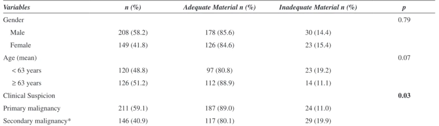
The proportion of biopsies with material judged adequate for analysis was higher in the patients referred for lesions suspected to be primary malignancy, as compared to the patients referred for lung lesions suspected to be secondary malignancy (Table 2). There was no difference in the material adequacy rate in patients of different gender or age groups (Table 2).
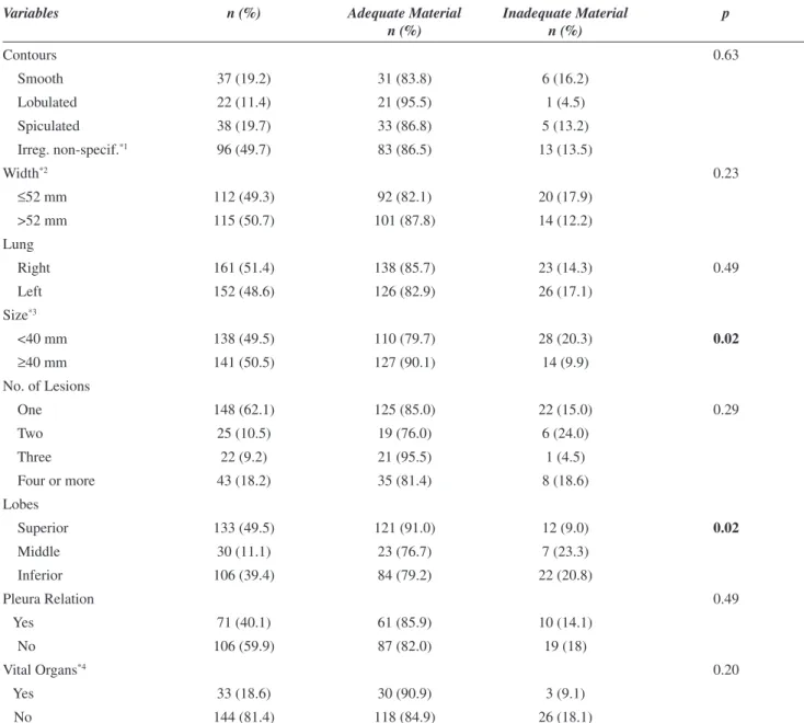
Table 3 shows the distribution of the radiological characteristics of the lesions and the corresponding rates of cytological material adequacy. The frequency of adequate material was higher in the lesions located in the superior lobes, as compared with the middle and inferior lobes. The larger lesion diameters (≥ 40 mm) also resulted in higher rates of success in obtaining adequate cytological material (Table 3). There was no statistical difference in material adequacy rates for series of biopsies performed in groups of patients with different secondary radiological lung indings. The total number of smears was not predictive of biopsy success.
In the multivariate analysis model, the only predictive variable for success of the biopsies was localization on the superior lobes (Table 4).
DISCUSSION
The overall success rate in our study was 84%, which is similar to other studies of FNAB of lung lesions without
Table 1 - Cytological analysis results of 357 FNABs
Material n (%)
Adequate for Analysis
Malignant 204 56.4
Suspicion of malignancy 38 10.5
Negative for malignancy 45 12.4
Possibly Benign 17 4.7
Total 304 84.0
Inadequate for Analysis
Insuficient 44 12.1
Inadequate 9 2.5
Total 53 14.6
No information 5 1.4
Total 362 100
Table 2 - Material adequacy according to patient gender and age
Variables n (%) Adequate Material n (%) Inadequate Material n (%) p
Gender 0.79
Male 208 (58.2) 178 (85.6) 30 (14.4)
Female 149 (41.8) 126 (84.6) 23 (15.4)
Age (mean) 0.07
< 63 years 120 (48.8) 97 (80.8) 23 (19.2)
≥ 63 years 126 (51.2) 112 (88.9) 14 (11.1)
Clinical Suspicion 0.03
Primary malignancy 211 (59.1) 187 (89.0) 24 (11.0)
Secondary malignancy* 146 (40.9) 117 (80.1) 29 (19.9)
Table 3 - Analysis of the radiological characteristics of the lung lesions in relation to the adequate and inadequate material
Variables n (%) Adequate Material
n (%)
Inadequate Material n (%)
p
Contours 0.63
Smooth 37 (19.2) 31 (83.8) 6 (16.2)
Lobulated 22 (11.4) 21 (95.5) 1 (4.5)
Spiculated 38 (19.7) 33 (86.8) 5 (13.2)
Irreg. non-specif.*1 96 (49.7) 83 (86.5) 13 (13.5)
Width*2 0.23
≤52 mm 112 (49.3) 92 (82.1) 20 (17.9)
>52 mm 115 (50.7) 101 (87.8) 14 (12.2)
Lung
Right 161 (51.4) 138 (85.7) 23 (14.3) 0.49
Left 152 (48.6) 126 (82.9) 26 (17.1)
Size*3
<40 mm 138 (49.5) 110 (79.7) 28 (20.3) 0.02
≥40 mm 141 (50.5) 127 (90.1) 14 (9.9)
No. of Lesions
One 148 (62.1) 125 (85.0) 22 (15.0) 0.29
Two 25 (10.5) 19 (76.0) 6 (24.0)
Three 22 (9.2) 21 (95.5) 1 (4.5)
Four or more 43 (18.2) 35 (81.4) 8 (18.6)
Lobes
Superior 133 (49.5) 121 (91.0) 12 (9.0) 0.02
Middle 30 (11.1) 23 (76.7) 7 (23.3)
Inferior 106 (39.4) 84 (79.2) 22 (20.8)
Pleura Relation 0.49
Yes 71 (40.1) 61 (85.9) 10 (14.1)
No 106 (59.9) 87 (82.0) 19 (18)
Vital Organs*4 0.20
Yes 33 (18.6) 30 (90.9) 3 (9.1)
No 144 (81.4) 118 (84.9) 26 (18.1)
*1 Irreg. non-specif., when the contour of the pulmonary injury was classiied as irregular by the radiologist without other speciica-tions.
*2 Supericial, when less than or equal to medium size; deep, when larger than medium size. *3 Smaller Size, when less than or equal to medium size; larger size, when larger than medium size.
*4 Vital Organs, when there is a relation to structures considered as vital: aorta, main bronchi, heart, pulmonary hilum, trachea, peri-cardium, inferior vena cava, and pulmonary arteries and veins.
Table 4 - Multivariate model analysis for success in patients undergoing CT-guided FNAB of lung lesions
Variables 95% Conidence Interval p
Odds Ratio Inferior Limit Superior Limit
Primary Diagnosis 1.99 0.87 4.55 0.10
Localization 3.37 1.36 8.35 <0.01
Size 0.44 0.19 1.01 0.05
immediate cytological examination.9-13 On univariate
analysis, procedures that were able to provide more than four smears obtained a larger amount of adequate material for analysis when compared to the procedures that provided less than or equal to four smears. These results tend to support the recommendation of more than one chest transixation by the FNAB procedure for thoracic lesions. Regarding the radiological characteristics, the lesions larger than 40 mm, those located in the superior lobes and suspicion of primary malignancy were predictive of the FNAB success.
Layield et al. (1996) demonstrated that the localization and size of thoracic injuries affect the diagnostic accuracy of FNAB of the thorax, with the best results in peripheral and larger lesions.5 Yankelevitz et al. (1997), when studying
small lesions (less or equal to 30 mm), demonstrated that the distance between the nodule and the pleural surface did not inluence the biopsy accuracy.6 In our study, there was
also no difference in obtaining adequate material for analysis between the supericial and deep lesions.
Miller et al. (1998) demonstrated that spiculated contours and deep seated lesions were predictive characteristics of success in obtaining adequate material for analysis and diagnosis.7 In this study, we did not observe this relationship.
Many authors have found larger amounts of adequate material for analysis and greater accuracy in the lesions that were considered large (greater than 1.5 cm) as compared with the lesions that were considered small.3,11,13-16 In this
study, lesions with diameters equal to or larger than 40 mm supplied larger amounts of adequate material for analysis than lesions with diameters of less than 40 mm, and this difference was statistically signiicant.
In our study, we also observed that the location of the superior lobe lesions supplied a proportionally larger amount
of adequate material for analysis when compared with other locations, and this difference was statistically signiicant. A possible explanation for this result is that it is often easier to reach the superior lobe injuries. Regardless of the position of the patient on the examination table, the punction needle generally reaches the injury at a straight angle. These lesions frequently have a pleural base and, consequently, a close-itting surface or even an iniltration of the chest wall, and this position favors the introduction of the needle to collect material for analysis without having to transpose the pleural space.
The patients who were referred due to suspicion of lung primary malignancy presented proportionally higher rates of adequate material for analysis in our study. One explanation for this inding could be the greater concern of the radiologist during the procedure for establishing a new cancer diagnosis rather than documenting a metastatic disease; however, this explanation is only an assumption.
Some lesion characteristics described in this study, such as the location on superior lobes and the suspicion of primary lung malignancy, were not described in previous studies. The only independent predictive factor of needle biopsy success was location in the superior lobes.
CONCLUSION
CT-guided percutaneous FNAB of lung lesions had a great rate of success in our study. Some radiologic characteristics of lung lesions may predict higher rates of success, such as a diameter equal to or larger than 40 mm, suspicion of primary lung malignancy, and location in the superior lobes. These characteristics should be considered when ordering this procedure.
REFERENCES
1. Murphy JM, Gleeson FV, Flower CD. Percutaneous needle biopsy of the lung and its impact on patient management. World J Surg. 2001;25:373-9.
2. Conces DJ Jr, Schwenk GR Jr, Doering PR, Glant MD. Thoracic needle biopsy. Improved results utilizing a team approach. Chest. 1987;91:813-6.
3. Perlmutt LM, Johnston WW, Dunnick NR. Percutaneous transthoracic needle aspiration: A Review. Am J Roentgenol. 1989;152:451-5. 4. Westcott JL. Percutaneous transthoracic needle biopsy. Radiology.
1988;169:593-601.
5. Layield LJ, Coogan A, Johnston WW, Patz EF. Transthoracic ine needle aspiration biopsy. Sensitivity in relation to guidance technique and lesion size and location. Acta Cytol. 1996;40:687-90.
6. Yankelevitz DF, Henschke CI, Koizumi JH, Altorki NK, Libby D. CT-guided transthoracic needle biopsy of small solitary pulmonary nodules. Clin Imaging. 1997;21:107-10.
7. Miller JA, Pramanik BK, Lavenhar MA. Predicting the rates of success and complications of computed tomography-guided percutaneous core-needle biopsies of the thorax from the indings of the preprocedure chest computed tomography scan. J Thorac Imaging. 1998;13:7-13. 8. Chojniak R, Isberner RK, Viana LM, Yu LS, Aita AA, Soares FA.
Computed tomography guided needle biopsy: experience from 1,300 procedures. Sao Paulo Med J. 2006;124:10-4.
10. Austin JH, Cohen MB. Value of having a cytopathologist present during percutaneous ine-needle aspiration biopsy of lung: report of 55 cancer patients and metaanalysis of the literature. Am J Roentgenol. 1993;160:175-7.
11. Milman N. Percutaneous lung biopsy with a fine bore cutting needle (Vacu-Cut): improved results using drill technique. Thorax. 1995;50:560-2.
12. Arakawa H, Nakajima Y, Kurihara Y, Niimi H, Ishikawa T. CT-guided transthoracic needle biopsy: a comparison between automated biopsy gun and ine needle aspiration. Clin Radiol. 1996;51:503-6.
13. Li H, Boiselle PM, Shepard JO, Trotman-Dickenson B, McLoud TC. Diagnostic accuracy and safety of CT-guided percutaneous needle aspiration biopsy of the lung: comparison of small and large pulmonary nodules. Am J Roentgenol. 1996;167:105-9.
14. Westcott JL. Direct percutaneous needle aspiration of localized pulmonary lesions: result in 422 patients. Radiology. 1980;137:31-5. 15. Stanley JH, Fish GD, Andriole JG, Gobien RP, Betsill WL, Laden SA, et
al. Lung lesions: cytologic diagnosis by ine-needle biopsy. Radiology. 1987;162:389-91.