CLINICAL SCIENCE
Predictive complication factors for ct-guided fine
needle aspiration biopsy of pulmonary lesions
Marcos Duarte Guimara˜es,IMarcony Queiroz de Andrade,IAlexandre Calabria da Fonte,IGustavo Benevides,IRubens Chojniak,IJefferson Luiz GrossII
IDepartment of Radiology, Hospital A. C. Camargo, Sa˜o Paulo, SP, Brazil.IIDepartment of Thoracic Surgery, Hospital A. C. Camargo, Sa˜o Paulo, SP, Brazil.
OBJECTIVE:Distinct aspects can influence the complication rates of computed tomography-guided percutaneous fine needle aspiration biopsy of lung lesions. The purpose of the current study is to determine the influence of radiological techniques and clinical characteristics in predicting complications from this procedure.
SUBJECTS AND METHODS:A retrospective study was developed involving 340 patients who were submitted to a consecutive series of 362 computed tomography-guided fine needle aspiration biopsies of lung lesions between July 1996 and June 2004, using 22-gauge needles (CHIBA). Variables such as the radiological characteristics of the lesions, secondary pulmonary radiological findings, co-morbidities, and aspects concerning the procedure were studied.
RESULTS:The diameters of the lung lesions varied from 9 to 140 mm, with a mean of 51.5¡24.3 mm and median of 40 mm. The depth of the lesions varied from 10 mm to 130 mm, with a mean of 44¡20.9 mm, and median median of 52 mm. Complications occurred in 52 (14.4%) cases, pneumothorax being the most frequent, with 40 (11.1%) cases, followed by hemoptisis with 7 (1.9%) cases, and hematoma with 4 (1.1%) cases. Lesions that did not contact the pleura, with normal pulmonary tissue interposition between lesion and pleura, had higher complication rates, with 22 (22%) cases, than lesions that contact the pleura, with 6 (9%) cases, with a statistically significant difference (p = 0.03).
CONCLUSIONS: CT-guided percutaneous fine needle aspiration biopsy of lung lesions had a lower rate of complications in our study and presented more rates of complications on lesions that lack pleural contact.
KEYWORDS: Fine; Needle; Biopsy; Lung; Complication.
Guimara˜es MD, Andrade MQ, Fonte AC, Benevides G, Chojniak R, Gross JL. Predictive complication factors for ct-guided fine needle aspiration biopsy of pulmonary lesions. Clinics. 2010;65(9):847-850.
Received for publication onMay 4, 2010;Review completed onJune 5, 2010;Accepted for publication onJune 9, 2010 E-mail: [email protected]
Tel.: 55 11 2189-5000 (1050)
INTRODUCTION
There is a significant variation in literature regarding the potential for complications with computed tomography (CT)-guided percutaneous fine needle aspiration biopsy (FNAB) of thoracic lesions. The incidence of complications ranges between 7% and 62%; pneumothorax is the most frequent complication, and thoracic draining ranges between 1% and 31%.1-5
Identifying characteristics such as the radiological aspects of the lesions, secondary pulmonary radiological findings, procedural aspects, and clinical characteristics such as co-morbidities can help uncover the reason for complications in patients submitted to the CT-guided FNAB of lung lesions.6-7 The purpose of this study is to evaluate the influence of these characteristics in predicting complications from CT-guided FNAB of lung lesions.
SUBJECTS AND METHODS
This is a retrospective analysis of all consecutive patients who received CT-guided FNAB of pulmonary lesions at an oncological center in Brazil between 1996 and 2004. From a total of 491 procedures evaluated, 362 (73.7%) were considered eligible for this study. Of the 129 excluded procedures, 6 (1.2%) were excluded because after cytologi-cal evaluation or upon follow-up, the lesions were defined as being on the chest wall or pleura; 26 (5.3%) were excluded for having divergences between the admission and chart registrations; and 97 (19.8%) were excluded because the procedure was performed with cutting needles, which are not the subject of this study. A total of 362 procedures performed on 340 patients are reported here; of these 362 procedures, 319 were of lesions that were submitted to biopsy once; 20 were of lesions submitted to biopsy twice; and one was of a lesion submitted to biopsy three times. Written informed consent was obtained from all patients. Information was collected from the charts available at the hospital’s Medical Archive Service (MAS) and the percutaneous biopsy forms. These forms contain data regarding the location, size, and number of the lesions, the
Copyrightß2010CLINICS– This is an Open Access article distributed under the terms of the Creative Commons Attribution Non-Commercial License (http:// creativecommons.org/licenses/by-nc/3.0/) which permits unrestricted non-commercial use, distribution, and reproduction in any medium, provided the original work is properly cited.
CLINICS 2010;65(9):847-850 DOI:10.1590/S1807-59322010000900006
distance between the lesion and the needle entrance site, contour aspects of the lesions, and the relationship of the contour aspects to adjacent structures such as the mediastinum, or chest wall. Secondary radiological find-ings such as atelectasis, pleural effusion, cavitations, necrosis, infiltrates, adenopathy, and the presence of other tumors and comorbidities were also included. Radiologic findings were based on the CT report prior to the procedure; the FNABs were conducted by an oncologic radiologist with over 10 years experience in CT-guided FNAB, or by a resident in radiology under his guidance, following the standard procedure defined by the Department of Radiology.8
At the Department of Radiology, coagulation tests were checked routinely; when these tests were within normal parameters, the biopsy was planned and performed using a chest CT with thin slices (3–5 mm). The computer’s cursor was used to measure the size of the lesions and the distances of the lesions from the biopsy needle entrance site. The skin was prepped with an antiseptic solution, and local anesthesia, with 1% lidocaine, was applied. The puncture needle was introduced, and new tomographic cuts were obtained in order to confirm or modify the needle position. The biopsies were performed within a breath-hold.
The FNABs were performed using 22-gauge CHIBA-type needles; subsequently, the obtained material was prepared in smears and immersed in 90% alcohol. Due to the retrospective nature of our study, it was not possible to determine the number of thoracic transfixations done via the FNAB procedure. However, the number of thoracic transfixations was indirectly obtained using the number of smears made by the puncture. In the experience of the Department of Radiology at the Hospital of Cancer, each puncture will supply approximately 4 smears.
Complications were considered when any new radiolo-gical occurrence was presented at the control exams with thoracic tomography being performed after biopsy, as well as new clinical symptoms and signs such as shortness of breath, intense dyspnea, thoracic pain, and hemoptisis appearing after biopsy.
Control exams with 10 mm axial tomographic cuts involving the entire thorax were performed immediately and 2 to 4 hours after the procedure to confirm the presence of complications such as pneumothorax and hematoma.
Pneumothorax was considered present when any material with aerated density accumulated in the pleural space. The tomographic control exam was anticipated if the patient demonstrated any signs or symptoms of acute respiratory discomfort. Hematoma was considered for each new opacification that was related to the lesion and to the conti-guous pulmonary normal tissue that appeared following the puncture.
The presence of hemoptisis during and in the first 24 hours following the puncture was considered a compli-cation as well.
By correlating the patient radiological data and co-morbidities with complication occurrence, we were able to determine the predictive factors for complication rates of CT-guided FNAB of lung lesions. Descriptive statistics were used when applied; the chi-square test or Fisher’s exact test was applied when appropriate. AP value was considered statistically significant when equal to or less than 0.05.
RESULTS
Of the 362 FNABs performed, 212 (58.6%) were per-formed on male patients, and 150 (41.4%) were perper-formed on female patients. The mean age was 61¡16 years, and
the median age was 63 years old. The purpose of the FNAB referral was to obtain a primary diagnosis of a suspected malignant focal lesion in 215 (59.4%) procedures and to document possible secondary malignancy in 147 (40.6%) procedures.
The lung lesions were right-sided in 163 (45%) proce-dures, and left-sided in 154 (42.5%) procedures. The lesion distribution by pulmonary lobes was as follows: 76 (21.0%) on the left superior lobe, 60 (16.6%) on the right superior lobe, 58 (16.0%) on the right inferior lobe, 48 (13.3%) on the left inferior lobe, and 27 (7.5%) on the middle lobe. In addition, this information was not available in 93 cases (25.7%).
The diameters of the lung lesions varied from 9 to 140 mm (mean: 51.5 ¡ 24.3 mm; median: 40 mm); the distance
between the lesion and the biopsy entry point on the skin varied from 10 to 130 mm (mean: 44¡ 20.9 mm; median: 52 mm). Additionally, 71 (40.1%) lesions were in contact with the pleura, and 106 (59.9%) had pulmonary tissue between the lesions and the pleura. The lesion contour type was irregular in 96 patients (26.5%), spiculated in 38 patients (10.5%), smooth in 37 patients (10.2%), and lobulated in 22 patients (6.0%); this information was not available in 169 cases (46.8%). In 148 (40.9%) procedures, the patient had one lung lesion, and in 25 (6.9%) of the procedures, the patient had two lesions. In 22 (6.1%) of the procedures, the patients had three lesions, and in 43 (11.9%) of the procedures, four or more lung lesions were present.
The most common secondary radiologic findings were adenopathy (34 patients; 9.4%), additional tumors (32 patients; 8.8%), cavitation (16 patients; 4.4%), necrosis (14 patients; 3.9%), infiltration (13 patients; 3.3%), pleural effusion (10 patients; 2.8%), and opacification on the present lesion (4 patients; 1.1%).
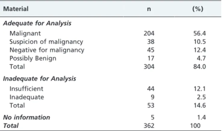
From a total of 362 needle biopsies, a cytological result was obtained from the patients’ charts for 357 (98.5%) of those biopsies. Of the 357 punctures evaluated by the pathologist, the material was considered adequate for analysis in 304 biopsies (84%) and inadequate for analysis in 53 biopsies (14.6 %). The frequency of material adequacy for cytological analysis is shown in Table 1.
From a total of 362 biopsies, complications occurred in 51 (14.1%), pneumothorax being the most frequent. No
Table 1 -Cytological analysis results of 357 FNABs.
Material n (%)
Adequate for Analysis
Malignant 204 56.4
Suspicion of malignancy 38 10.5
Negative for malignancy 45 12.4
Possibly Benign 17 4.7
Total 304 84.0
Inadequate for Analysis
Insufficient 44 12.1
Inadequate 9 2.5
Total 53 14.6
No information 5 1.4
Total 362 100
Ct-guided fine needle aspiration biopsy of pulmonary lesions
Guimara˜es MD et al. CLINICS 2010;65(9):847-850
complications were reported in 281 (77.6%) cases, and there was no available information in 30 (8.3%) cases, according to Table 2.
There was no difference in the complication rates in patients of different gender or age groups. There was also no statistical difference in complication rates for biopsies performed in groups of patients with different secondary radiological lung findings.
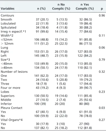
Table 3 shows the distribution of the radiologic char-acteristics of the lesions and the corresponding rates of complications. The frequency of complications was higher
in lesions that lacked contact with the pleura and with normal pulmonary tissue interposition between lesion and pleura, than in the lesions that maintained contact with the pleura (p = 0,03). There was no statistical difference in complication rates with the others radiological character-istics of lesions.
There was no difference in the complication rates of patients belonging different co-morbidity groups. The information of co-morbidity was presented in 96 patients, as shown in Table 4.
Of the 51 (14.1%) patients who had complications, only 11 (21.1%) needed thoracic draining. Chronic obstructive pulmonary disease (COPD) was the most common co morbidity, with 5 (17.2%) cases of thoracic drainage. In contrast, of the patients without COPD, 4 (5.8%) cases demonstrated a statistically significant difference (p,0.01). The total number of smears was not predictive of compli-cation rates.
DISCUSSION
Pneumothorax, pulmonary hematoma, and hemoptisis are the most frequent complications of pulmonary biopsy guided by imaging methods.4,6,7,10,11,14,16
The incidence of complications in CT-guided FNAB of lung lesions depends on several factors. Radiological and clinical characteristics such as size and depth of lesions, advanced age, abnormal tests of pulmonary function, and co-morbidities can contribute to the occurrence of complica-tions.11-13
Austin and Cohen (1993) demonstrated that lesions that contact the pleura do not develop into pneumothorax.16 Yankelevitz et al. (1997) demonstrated that the rate of pneumothorax was 20%, and that only 5.3% needed draining. They also observed that an increase in the frequency of pneumothorax occurred in lesions that were more than 30 mm away from the pleura, or nodules smaller than 10 mm.5Wallace et al. (2002) demonstrated the highest rate of complications among the revised works, with CT-guided FNAB of lung lesions smaller than 10 mm present-ing pneumothorax in 62% of the cases and thoracic drainage in 31% of the cases.5One reason for this is that the multiple needles tranfixations of the pleural space could be needed to reach small and deep lesions when compared with super-ficial and large lesions.
Li et al. (1996) had not demonstrated significant differ-ences in the rates of complications of FNAB of lung lesions Table 2 -Distribution of complications rates of CT-guided
FNAB of lung lesions.
Complications n (%)
Pneumothorax 40 11.1
Hemoptisis 07 1.9
Hematoma 04 1.1
No Complications 281 77.6
No Informations 30 8.3
Total 362 100
Table 3 -Analysis of lung lesions radiological
characteristics and occurrence of complications in CT-guided FNAB of lung lesions.
Variables n (%)
n No Complic (%)
n Yes Complic (%) p
Contours 0.96
Smooth 37 (20.1) 5 (13.5) 32 (86.5)
Lobulated 22 (11.9) 3 (13.6) 19 (86.4)
Spikulated 34 (18.4) 4 (11.8) 30 (88.2)
Irreg n especif.*1 91 (99.6) 14 (15.4) 77 (84.6)
Width*2 0.11
#52mm 106 (48.8) 15 (14.2) 91 (85.8)
.52mm 111 (51.2) 25 (22.5) 86 (77.5)
Lung 0.06
Right 153 (51.3) 26 (17.0) 127 (83.0)
Left 145 (48.7) 23 (15.9) 122 (84.2)
Size*3 0.79
,40mm 133 (49.9) 20 (15.0) 113 (85.0)
$40mm 134 (50.1) 24 (17.9) 110 (82.1)
Number of lesions 0.32
One 141 (62.3) 24 (17.0) 117 (83.0)
Two 24 (10.6) 5 (20.8) 19 (79.2)
Three 18 (7.9) 1 (5.6) 17 (94.4)
Four or more 43 (19.2) 4 (9.3) 39 (90.7)
Lobes 0.23
Superiors 130 (50.5) 19 (14.6) 111 (85.4)
Middle 27 (10.5) 2 (7.4) 25 (92.6)
Inferior 100 (39) 20 (20) 80 (80)
Pleura Contact 0.03
Yes 67 (40.1) 6 (9.0) 61 (91.0)
No 100 (59.9) 22 (22.0) 78 (78.0)
Vital Organs*4 0.27
Yes 30 (17.9) 3 (10) 27 (90)
No 137 (82.1) 25 (18.2) 112 (81.8)
*1 Irreg. non-specif., when contour of pulmonary injury was classified as
irregular by radiologist without other specifications.
*2 Superficial, when less or equal to the medium size; deep, when larger
than the medium size.
*3 Smaller Size, when less or equal to the medium size; larger size, when
larger than the medium size.
*4 Vital Organs,when there is a relation to structures considered as vital:
heart, main bronchi, heart, pulmonary hilum, trachea, pericardium, inferior vena cava, aorta and pulmonary arteries and veins.
Table 4 -Complications rates according to co-morbidity frequency.
Comorbity n (%)
COPD*1 30 8.3
Hypertension 29 8.0
Diabettes 18 5.0
IHD*2 05 1.4
CHF*3 04 1.2
Obesity 04 1.1
Others*4 06 1.6
*1 COPD - Chonic Obstructive Pulmonary Disease.
*2 IHD- Ischemic Heart Disease.
*3 CHF- Congestive Heart Failure.
*4 Others– Rheumatoid Arthritis, Lupus.
CLINICS 2010;65(9):847-850 Ct-guided fine needle aspiration biopsy of pulmonary lesions Guimara˜es MD et al.
with equal and smaller diameter than 15 mm versus FNAB of lung lesions larger than 15 mm.[22] In the present work there was no statistically significant difference in the ratio of complications (p = 0,11) or the FNAB of superficial or deep lesions, taking as reference the median (52 mm). However, our study demonstrated more rates of complications on lesions that lack contact with pleura, which could be considered a landmark in differentiate that is superficial from deep lesions.
Chojniak et al. (2006) demonstrated a rate of complication in 16%, and thoracic draining in 4.9% of patients submitted to CT-guided FNAB of lung lesions.8In our work, the rate of pneumohorax was 11.1% and thoracic draining, 3.0%.
The Department of Radiology of Hospital AC Camargo is a reference service and routinely performs CT-guided FNAB.4,8,19 Perhaps the acquired experience, with proce-dures over the past years, can minimize the occurrence of complications in small and deep lesions of FNAB. However, according the retrospective nature, this study could have underestimated the rates of complications.
Yu’s et al. (2002) study on CT-guided percutaneous biopsies with cutting needles presented a rate of 17.3% complications, including: small pneumothorax with no draining required (11.5%), hemoptisis (1.9%); pulmonary hematoma (1.9%) and hematoma in the chest wall upon needle trajectory (1.9%).4Sulhatin et al. (2002) demonstrated rates of 1.7% for hemoptisis and 1.4% for pulmonary hematoma, also without significant clinical repercussions.11 Conces et al. (1987) reveal rates of pulmonary hematoma and hemoptisis as 11% and 2%, respectively, with sponta-neous resolution.16In our study, the occurrence of compli-cations such as hemoptisis and pulmonary hematoma was 1.9% and 1.1% respectively, with the same frequency found in the literature. No cases of artery embolism and tumor implantation were observed.
Pneumothorax occurrence increased following FNAB in patients with COPD.13 Romano et al. (2004) found a pneumothorax rate of 11.8% (27) in his study on CT-guided FNAB lung lesions. Of these, only 6 (22.2%) required thoracic draining, but they all suffered from COPD. 15 Vitulo et al. (1996) demonstrated that no parameter of functional results were predictive for pneumothorax. The only variables that correlated significantly with pneu-mothorax in this study were the interposition of normal tissue (p,0.01) and the depth of needle penetration in pareˆnquima (p,0.05).12 On its own, COPD was not predictive for pneumothorax in our study. This result could have been influenced by the reduced amount of information collected in relation to the expected amount for the co-morbidities.
Kazerooni et al. (1996) demonstrated that there was a higher rate of thoracic drainage in the population with severe COPD.13 In our study, thoracic draining also occurred with more frequency in patients with COPD, with a statistically significant difference (p,0.01) in relation to those without this co-morbidity. Although pulmonary function has not been evaluated in the present study, these patients usually have weak lung function and are suscep-tible to more precocious and evident clinical manifestations, even with a small pneumothorax, which makes them require thoracic draining more frequently than those with-out COPD.
CONCLUSION
CT-guided percutaneous FNAB of lung lesions had a lower rate of complications in our study and presented higher rates of complications on lesions that lack pleural contact. COPD was the most common co-morbidity finding, with higher rates of thoracic drainage. These radiological and clinical characteristics should therefore be considered when ordering this procedure.
REFERENCES
1. Ku¨c¸u¨k CU, Yilmaz A, Yilmaz A, Akkaya E. Computed tomography-guided transthoracic fine-needle aspiration in diagnosis of lung cancer: a comparison of single-pass needle and multiple-pass coaxial needle systems and the value of immediate cytological assessment. Respirology. 2004;9:392-6, doi: 10.1111/j.1440-1843.2004.00607.x.
2. Wallace MJ, Krishnamurthy S, Broemeling LD, Gupta S, Ahrar K, Morello FA Jr, et al. CT-guided percutaneous fine-needle aspiration biopsy of small (,or = 1-cm) pulmonary lesions. Radiology. 2002; 225:823-8, doi: 10.1148/radiol.2253011465.
3. Perlmutt LM, Johnston WW, Dunnick NR. Percutaneous transthoracic needle aspiration: A Review. Am J Roentgenol. 1989;152:451-5. 4. - Yu LS, Deheinzelin D, Younes RN, Chojniak R. Computed
tomography-guided cutting needle biopsy of pulmonary lesions. Rev Hosp Clin Fac Med Sao Paulo. 2002;57:15-8.
5. Yankelevitz DF, Henschke CI, Koizumi JH, Altorki NK, Libby D. CT-guided transthoracic needle biopsy of small solitary pulmonary nodules. Clin Imaging. 1997;21:107-10, doi: 10.1016/S0899-7071(96)00011-3. 6. Greif J, Marmur S, Schwarz Y, Man A, Staroselsky AN. Percutaneous
core cutting needle biopsy compared with fine-needle aspiration in the diagnosis of peripheral lung malignant lesions: results in 156 patients. Cancer.1998;25;84:144-7, doi: 10.1002/(SICI)1097-0142(19980625)84: 3,144::AID-CNCR4.3.0.CO;2-O.
7. Ng YL, Patsios D, Roberts H, Walsham A, Paul NS, Chung T, Herman S, Weisbrod G. CT-guided percutaneous fine-needle aspiration biopsy of pulmonary nodules measuring 10 mm or less. Clin Radiol. 2008;63:272-7, doi: 10.1016/j.crad.2007.09.003.
8. Chojniak R, Isberner RK, Viana LM, Yu LS, Aita AA, Soares FA. Computed tomography guided needle biopsy: experience from 1,300 procedures. Sao Paulo Med J. 2006;124:10-4.
9. Lourenc¸o R, Camacho R, Barata MJ, Cana´rio D, Gaspar A, Cyrne C. CT-guided percutaneous transthoracic biopsy in the evaluation of unde-termined pulmonary lesions. Rev Port Pneumol. 2006;12:503-24. 10. Miller JA, Pramanik BK, Lavenhar MA. Predicting the rates of success
and complications of computed tomography-guided percutaneous core-needle biopsies of the thorax from the findings of the preprocedure chest computed tomography scan. J Thorac Imaging. 1998;13:7-13, doi: 10. 1097/00005382-199801000-00003.
11. Sulhattin A, Yilmaz A, Bayramgurler B, Uzman O, Unver E, Akkaya E. Ct-guided transthoracic fine needle aspiration of pulmonary lesions: accuracy and complications in 294 patients. Med Sci Monit 2002;8:493-7. 12. Vitulo P, Dore R, Cerveri I, Tinelli C, Cremaschi P. The role of functional respiratory tests in predicting pneumothorax during lung needle biopsy. Chest. 1996; 109:612-5, doi: 10.1378/chest.109.3.612.
13. Kazerooni EA, Lim FT, Mikhail A, Martinez FJ. Risk of pneumothorax in CT-guided transthoracic needle aspiration biopsy of the lung. Radiology. 1996; 198:371-5.
14. van Sonnenberg E, Casola G, D’Agostino HB, Goodacre B, Sanchez R. Interventional radiology in the chest. Chest. 1992; 102:608-12, doi: 10. 1378/chest.102.2.608.
15. Romano M, Griffo S, Gentile M, Mainenti PP, Tamburrini O, Iaccarino V, et al. CT-guided percutaneous fine needle biopsy of small lung lesions in outpatients. Safety and efficacy of the procedure compared to inpatients. Radiol Med (Torino). 2004;108:275-82.
16. Austin JH, Cohen MB. Value of having a cytopathologist present during percutaneous fine-needle aspiration biopsy of lung: report of 55 cancer patients and metaanalysis of the literature. AJR Am J Roentgenol. 1993;160:175-7.
17. Conces DJ Jr, Schwenk GR Jr, Doering PR, Glant MD. Thoracic needle biopsy. Improved results utilizing a team approach. Chest. 1987;91:813-6, doi: 10.1378/chest.91.6.813.
18. Li H, Boiselle PM, Shepard JO, Trotman-Dickenson B, McLoud TC. Diagnostic accuracy and safety of CT-guided percutaneous needle aspiration biopsy of the lung: comparison of small and large pulmonary nodules. Am J Roentgenol. 1996;167:105-9.
19. Guimara˜es MD, Chojniak R, Gross JL, Bitencourt AG. Predictive success factors for CT-guided fine needle aspiration biopsy of pulmonary lesions. Clinics. 2009;64:1139-44, doi: 10.1590/S1807-59322009001200002.
Ct-guided fine needle aspiration biopsy of pulmonary lesions
Guimara˜es MD et al. CLINICS 2010;65(9):847-850