Intracellular Bacterium
Rickettsia prowazekii
Andrew Woodard, David O. Wood*
Department of Microbiology and Immunology, University of South Alabama College of Medicine, Mobile, Alabama, United States of America
Abstract
Termination of transcription is an important component of bacterial gene expression. However, little is known concerning this process in the obligate intracellular pathogen and model for reductive evolution, Rickettsia prowazekii. To assess transcriptional termination in this bacterium, transcripts of convergent gene pairs, some containing predicted intrinsic terminators, were analyzed. These analyses revealed that, rather than terminating at a specific site within the intervening region between the convergent genes, most of the transcripts demonstrated either a lack of termination within this region, which generated antisense RNA, or a putative non-site-specific termination that occurred throughout the intervening sequence. Transcripts terminating at predicted intrinsic terminators, as well as at a putative Rho-dependant terminator, were also examined and found to vary based on the rickettsial host environment. These results suggest that transcriptional termination, or lack thereof, plays a role in rickettsial gene regulation.
Citation:Woodard A, Wood DO (2011) Analysis of Convergent Gene Transcripts in the Obligate Intracellular BacteriumRickettsia prowazekii. PLoS ONE 6(1): e16537. doi:10.1371/journal.pone.0016537
Editor:Samithamby Jeyaseelan, Louisiana State University, United States of America
ReceivedNovember 2, 2010;AcceptedDecember 21, 2010;PublishedJanuary 26, 2011
Copyright:ß2011 Woodard, Wood. This is an open-access article distributed under the terms of the Creative Commons Attribution License, which permits unrestricted use, distribution, and reproduction in any medium, provided the original author and source are credited.
Funding:This study was supported by National Institute of Allergy and Infectious Disease grant AI020384 to DOW. http://www.niaid.nih.gov/Pages/default.aspx. The funders had no role in study design, data collection and analysis, decision to publish, or preparation of the manuscript.
Competing Interests:The authors have declared that no competing interests exist.
* E-mail: [email protected]
Introduction
The obligate intracellular bacterium,Rickettsia prowazekii,is the causative agent of epidemic typhus, a louse-borne disease usually associated with non-hygienic conditions arising in crowded human populations during war, famine, and as a result of extreme poverty. Due to its potential as an instrument of bioterrorism,R. prowazekiiis classified as a category B Select Agent. The unique life style of this pathogen involves growth in the widely different environments of an arthropod louse vector and a human host. In addition, a zoonotic reservoir, the flying squirrel, has been identified in the United States [1,2]. R. prowazekii pathogenicity results from the intracytoplamsic growth of the rickettsiae, leading to cell lysis and the subsequent infection of additional host cells. In
R. prowazekii, this lifestyle is dependent on a relatively small genome that contains a high proportion of pseudogenes and non-coding sequences [3].
Rickettsial gene expression studies have focused primarily on transcription initiation. R. prowazekii has been shown to utilize regulated promoters and to organize genes into operons [4,5,6,7,8,9], and rickettsial RNA polymerase was shown to exhibit properties that distinguish it from the Escherichia coli
polymerase, such as the requirement for a supercoiled template for promoter binding, [10,11]. However, an area of gene regulation that has not been evaluated in rickettsiae is transcriptional termination. In fact, very few functional terminators have been experimentally confirmed outside of model organisms such asE. coliandBacillus subtilis[12]. The conservation of genes associated with the termination process (nusA, nusB, nusG, mfd, and rho) suggests that transcription termination has a role in rickettsial gene regulation [3]. Preliminary studies in our laboratory indicated that
rickettsial termination of thegreAgene transcript exhibited unusual properties. Transcripts of this gene were shown to extend through an intervening region and into the coding sequence of the convergingpntgene [13].
To gain insight into this aspect of rickettsial gene regulation, we elected to focus on areas of the rickettsial genome where gene organization suggests the most probable location for termination events; namely, the intervening regions between convergent genes [12]. Analysis of these regions revealed functional intrinsic terminators and a putative Rho-dependent terminator. Also identified were convergent genes that exhibit a lack of termination or a non-site-specific termination and the generation of rickettsial antisense RNAs resulting from the extension of transcripts into convergent gene sequences. Most importantly, we discovered that
R. prowazekii may regulate termination efficiency of specific terminators depending on the rickettsial host.
Materials and Methods
Bacterial strains, host cell lines, and culture conditions
(Mediatech, Inc.). For L929 cell rickettsial infections, rickettsiae propagated in hen egg yolk sacs were used to infect L929 cells at a multiplicity of infection of 5–10 rickettsiae per cell. Infected cells were planted into tissue culture flasks and incubated for 48 hours ($90% infected and 100–400 rickettsiae per cell) before harvesting. Rickettsiae were released from the cells using ballistic shearing (mini-Beadbeater-1, BioSpec Products Inc., Bartlesville, OK). Cells were subjected to 3 cycles at the maximum agitation setting for 10 seconds with at least 10 seconds of cooling on ice between each agitation. Sheared samples were placed in a 50 ml conical vial, diluted to 30 ml with SPG-Mg, and centrifuged at 9006g for 10 minutes at 4uC. The supernatant was collected and centrifuged in a Beckman J2-21 centrifuge (Beckman Coulter, Inc., Brea, CA) at 12,0006g, 4uC for 15 minutes to pellet rickettsiae for
RNA isolation.
RNA Isolation
For RNA isolation, rickettsial pellets were solubilized using 1 ml of Tri-Reagent (Applied Biosystems, Austin, TX) per-flask equivalent for L929-grown rickettsiae or per 0.5 ml of hen egg yolk sac rickettsial preparations. Total RNA was extracted following the manufacturer’s guidelines and suspended in THE RNA Storage Solution (Applied Biosystems). RNA concentration was determined using a NanoDrop ND-1000 spectrophotometer (NanoDrop Products, Wilmington, DE). Approximately 80mg of
total RNA was further purified using RNeasy-Mini columns (Qiagen Inc., Valencia, CA). Columns were treated with RNase-free DNase I (Qiagen), eluted in 100ml (2650ml) nuclease-free
water and treated with 1ml of the RNase inhibitor SUPERase-In
(Applied Biosystems). The quality and concentration of the RNA was determined utilizing the micro-fluidic Agilent 2100 Bioana-lyzer (Agilent Technologies, Inc., Santa Clara, CA). Aliquots were immediately precipitated, re-hydrated in THE RNA Storage Solution and stored at280uC.
Probe Design and Production
PCR templates for probe synthesis were generated using sequence-specific primers containing T7 and T3 promoters (Integrated DNA Technologies, Inc., Coralville, IA) with R. prowazekii genomic DNA as template (Table S1). Amplified products were purified with Geneclean Turbo for PCR kit (MP Biomedicals, Solon, OH) and size-verified by gel electrophoresis. Radiolabled RNA probes and markers (RNA Century or Century-Plus, Applied Biosystems) were synthesized utilizing the Riboprobe T7-T3 transcription system (Promega Corp., Madison, WI). Approximately 300 ng of the purified template was added to a reaction mixture, as directed by the manufacturer, containing: Transcription Buffer (1X), 100 mM DTT, 40 U RNasin, 2.5 mM of unlabled rATP, rGTP, rCTP, 0.2 mM rUTP, 20 U T7 or T3 polymerase, and 50mCi of [a-32P] rUTP (MP Biomedicals). The
reaction mixture was incubated in a dry heat-block for one hour at 37uC, and immediately treated with 1 U of RQ1 DNase (Promega Corp.) for 15 minutes. Unincorporated nucleotides were removed with an RNase-free G-50 column (GE Healthcare Biosciences, Pittsburg, PA). Radiolabeled probes was then diluted to a final activity of 5.06103cpm. Verification of a single, labeled product
of the predicted size was accomplished by analyzing each labeled probe on a 5% acrylamide/8 M urea gel. Visualization was achieved using a phosphor imaging screen and detection on a Cyclone Plus phosphoimager (Perkin Elmer, Waltham, MA). Unlabeled probes were synthesized in the same manner, analyzed on a 5% acrylamide/8 M urea gel, and visualized by ethidium bromide staining. Concentrations of unlabeled probes were determined using a NanoDrop spectrophotometer.
Ribonuclease Protection Assay
RPA analyses were conducted using the RPA III Kit (Applied Biosystems). Briefly, purified rickettsial RNA samples were pooled, separated into 50mg aliquots, and precipitated with ethanol.
Precipitated RNA was washed with 70% ethanol and dried for 4 minutes. RNA samples were then hydrated in a mixture of 3ml of
RNase-free water, 1ml of the radiolabeled probe (5.06103cpm/
ml) and 10ml of Hybridization Buffer III. Hybridization samples
were brought to 94uC for 4 minutes and then incubated in a dry heat block overnight at 42uC. RNase digestion buffer was added to the sample, and the sample incubated for 30 minutes at 37uC. Inactivation Solution III was then added and samples were placed at 220uC for a minimum of 15 minutes. The samples were centrifuged at 16,0006g for 15 minutes, and the resulting pellet suspended in 10ml of Gel Loading Buffer II. Experimental, and control samples as well as 5.06103cpm of the labeled RNA
Century-Plus Marker (Applied Biosystems) were incubated at 94uC for 3 minutes, analyzed by electrophoresis using a 5% acrylamide/8 M urea gel, and detected using a phosphopimager. Controls were included for each RPA analysis to confirm the validity of the assay (data not shown). Controls for each RPA analysis included: a positive control for the hybridization consisting of the labeled probe and 300 pg of its unlabeled complement. A negative control of labeled probe only, at the amount used in the experimental assay, was included to ensure that all unprotected probe would be digested. DNA contamination would be reflected by full length protection of every probe under all conditions. Therefore, the absence of fully protected probe in all assays for each RNA preparation confirmed the absence of DNA contamination. All RPA results shown are representative of a minimum of three independent RNA preparations.
Results
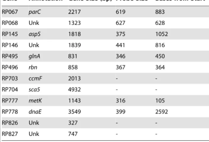
Transcriptional termination and convergent gene pairs To analyze rickettsial transcriptional termination, we focused on the most likely location for termination, the intervening sequences between convergent genes [12]. Of the 104 convergent gene pairs annotated in theR. prowazekii genome [3], we selected 12 genes (Table 1), representing 6 well-separated gene pairs that, with two exceptions, met the following selection criteria. First, each gene is transcribed at detectable levels. This was evaluated using microarray data [4] or by direct measurements using RPA. In addition, in the current study intragenic positive control probes were included to confirm gene transcription (Table 1). Secondly, the gene products were detected by proteomic analysis, with the exception of RP826 and RP777. The latter gene was listed as a pseudogene in Madrid E and therefore not annotated or screened in proteomic analyses [15,16] (unpublished results). Two of these gene pairs (RP703-RP704 and RP826-RP827) were also included due to the prediction by TransTermHP of a strong, bidirectional, intrinsic terminator within the intervening regions [12].
of differing sizes will be visualized. In Table 2, the sizes of the intergenic regions, the probe size, and the amount the probe overlaps the coding regions of the genes are presented for the probes used in this study.
We assayed rickettsial RNA extracted from rickettsiae grown in hen egg yolk sacs and in L929 mouse fibroblast cells. Previous studies had indicated that mRNA assayed at 34uC from rickettsiae grown in L929 cells had a half-life of approximately 15 minutes [6], a property that would preclude the isolation of mRNA from yolk sacs due to the 4–5 hours required to isolate rickettsiae from this source. However, we found that rickettsial mRNA is present in the egg yolk sac rickettsial RNA preparation and can be detected at levels comparable to mRNA isolated from rickettsiae grown in L929 cells. The recovery of mRNA from yolk sac grown rickettsiae is most likely due to performing all manipulations at 4uC during rickettsial purification. This permitted us to assay rickettsial RNA from different rickettsial host backgrounds.
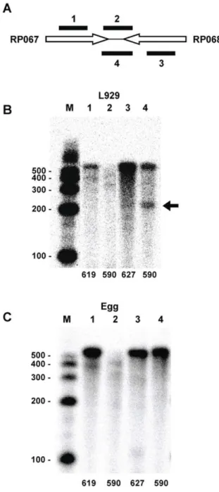
RPA analysis of transcripts specific for three of the gene pairs is presented in Figure 1. The results reveal that the lack of specific termination sites is not an uncommon occurrence inR. prowazekii.
For example, the RP145-RP146 convergent gene pair is transcribed as evidenced by the fully protected intragenic probes that are targeted to sites within the two genes (Fig. 1B, Lanes 1 and 3). However, RP145 transcripts do not exhibit a specific termination site within the intervening region (Fig. 1B, Lane 2). Since there is a 106 base pair overlap of the probe with RP145 (Table 2), this indicates the absence of a transcriptional stop immediately following the RP145 stop codon (a 106 base protected fragment would have been visible by gel electrophoresis). Rather, the presence of a diffuse banding pattern suggests non-site-specific termination throughout the intervening region. In contrast to the RP145 transcript, the probe targeted to the intervening region downstream of RP146 was fully protected (Fig. 1B, Lane 4) demonstrating that the RP146 transcript extends into the RP145 coding region generating antisense RNA to RP145 transcripts (the probe extends 116 bp into the coding region). The RP495-RP496 gene pair exhibited a similar termination profile: RP495 transcripts exhibit no defined termination site (Fig. 1C, Lane 2) and the probe specific for RP496 was fully protected (Fig. 1C, Lane 4). Once again this demonstrates that RP496 transcripts are extending into the RP495 coding region generating antisense RNA (the probe extends 281 bp into the coding region). The protection profile observed with the RP777-RP778 gene pair demonstrated that both gene transcripts exhibited a diffuse pattern indicative of non-site-specific termination throughout the inter-vening region (Fig. 1D, Lanes 2 and 4). Thus, these three convergent gene pairs ofR. prowazekiido not terminate transcripts at specific sites within the intervening region, as might be expected based on gene orientation. In fact, read-through into the opposing gene, generating antisense RNA, is observed for two gene transcripts (Fig. 1, B, Lane 4 and C, Lane 4). These results were obtained whether the RNA was extracted from rickettsiae propagated in hen egg yolk sacs or in L929 tissue culture cells.
Transcriptional termination at predicted terminators To further elucidate rickettsial transcription termination, we selected two rickettsial gene pairs (RP703-RP704 and RP826-RP827) predicted by bioinformatic analysis to exhibit strong intrinsic terminators. Probes were designed to span the predicted intrinsic terminator sites of these gene pairs (Table 1). Analysis of these intrinsic terminator regions revealed a variable pattern of termination based on the source of the RNA.
The RP703-RP704 gene pair exhibits a long intervening region of 1910 bp and a predicted strong, bi-directional terminator Table 1.Targeted genes and intragenic probes.
Gene AnnotationaGene Size (bp) Probe Sizeb Bases from Start
RP067 parC 2217 619 883
RP068 Unk 1323 627 628
RP145 aspS 1818 375 1052
RP146 Unk 1839 441 816
RP495 glnA 831 346 450
RP496 rbn 858 367 364
RP703 ccmF 2013 -
-RP704 sca5 4932 -
-RP777 metK 1143 316 105
RP778 dnaE 3549 399 2592
RP826 Unk 327 -
-RP827 Unk 747 -
-aAnnotations from [3] and ERGOTM(Integrated Genomics, Chicago, IL.). bSize in bases from NCBI.
Unk = Unknown.
doi:10.1371/journal.pone.0016537.t001
Table 2.Gene pairs and intergenic probes.
Gene Pair Annotationa Intergenic Region (bp) Probe Size Overlap Gene A Overlap Gene B
RP067-RP068 parC– Unk 341 593 122 130
RP145-RP146 aspS– Unk 569 791 106 116
RP495-RP496 glnA–rbn 137 512 94 281
RP703-RP704 (1) ccmF–sca5 1910 577 370
-RP703-RP704 (2) ccmF–sca5 1910 833 -
-RP703-RP704 (3) ccmF–sca5 1910 563 -
-RP703-RP704 (4) ccmF–sca5 1910 665 - 293
RP777-RP778 metK–dnaE 366 682 160 156
RP826-RP827 Unk – Unk 253 466 127 86
aAnnotations from [3] and ERGOTM(Integrated genomics, Chicago, IL.). Unk = Unknown.
(59-AAAAAAA GCCCATTTT TTC AAAGTGGGC TTTTTTT-39) located 32 bases from the annotated end of RP704. Interestingly, the RP703 transcripts did not reach this termination site but exhibited a diffuse pattern of termination reminiscent of the transcripts presented in Figure 1 (data not shown). Thus, we were unable to evaluate the efficiency of RP703 transcript termination at the predicted site. However, functional evaluation of this site was possible for the RP704 transcript. The probe for this region was designed to extend 293 bases into RP704 and yield a protected product of 329 bases if the transcript terminated at the predicted site. A representative RPA analysis is presented in Figure 2B. When using RNA isolated from rickettsiae propagated in hen egg yolk sacs, a probe spanning the predicted intrinsic terminator was fully protected with little evidence of a stop at the intrinsic terminator (Fig. 2B, Lane 1). In contrast, the majority of the transcripts terminated at the predicted site when RNA isolated from rickettsiae grown in L929 cells was analyzed (Fig. 2B, Lane 2).
The large intervening region between RP703 and RP704 provided an excellent opportunity to examine the progression of transcription termination of transcripts that read through the predicted terminator as in rickettsiae harvested from hen egg yolk sacs. We used overlapping probes (minimum 42 base overlap) spanning the entire intervening region of 1910 bp and extending 370 bases into the RP703 gene. Probes 1 and 2 were essentially fully protected (Fig. 2D, Lanes 1 and 2) indicating complete
read-through. Less full-length probe was detected for probe 3 (Fig. 2D, Lane 3), and only a negligible amount of full-length probe 4 (Fig. 2D, Lane 4) was detected. This is another example of the absence of a distinct termination site and an incremental, non-site-specific termination.
Differential termination at an intrinsic terminator, based on RNA source, was not unique to the RP703-704 gene pair. RP826-RP827 was an additional convergent gene pair with a predicted strong, bidirectional, intrinsic terminator (59-AAAAA GGGTCTTTA TTAA TAAAGACCC TTTTT) that exhibited a similar termination profile (Fig. 3). Interestingly RPA analysis of RP826 transcripts revealed no difference between RNA obtained from hen egg yolk sac grown rickettsiae and L929 cell grown rickettsiae (Fig. 3B, Lanes 1). The majority of the transcripts correspond to the predicted size for termination at the intrinsic terminator (347 bases). In contrast, there was a dramatic difference in the ratio of terminated versus read-through transcripts for RP827 depending on the rickettsial host (Fig. 3B, Lanes 2). Once again the majority of transcripts detected in RNA from hen egg yolk sac grown rickettsiae completely protected the full-length probe indicating transcription through the termination site and extension of the RP827 transcript into the coding sequence of RP826. The opposite result was observed for RNA isolated from rickettsiae grown in L929 cells where the majority of the RP827 transcripts protected a 187 base portion of the probe indicating termination at the predicted terminator site. Thus,R. prowazekiiexhibits intrinsic
Figure 1. RPA analysis identifying transcripts of three convergent gene pairs that exhibit non-site-specific termination.(A) Schematic indicating the relative positions of the RPA probes for each convergent gene pair (Tables 1 and 2). The probes include two internal probes to confirm gene transcription and two intervening probes of opposite orientation to detect intergenic mRNA. Probes 1 and 2 hybridize to Gene A (left) transcripts while probes 3 and 4 hybridize to Gene B (right) transcripts. (B) RP145-RP146. (C) RP495-RP496. (D) RP777-RP778. Lane numbers correspond to the probe numbers in the schematic. Numbers at the bottom of each lane correspond to the size of the probe in bases. M, sized markers with sizes (in bases) indicated. Lane B3 (underlined) was digitally moved horizontally using Microsoft Office PowerPoint to maintain consistent lane organization.
terminators that appear to function efficiently in rickettsiae grown in L929 tissue culture cells but are predominantly bypassed in rickettsiae growing in hen egg yolk sacs.
Identification of a putative Rho-dependent termination site
The intervening region between RP067 and RP068 does not contain a predicted intrinsic terminator. However, we detected a specific termination site by RPA for the RP068 message when using RNA isolated from rickettsiae grown in L929 cells (Fig. 4B, Lane 4). Similar to the results of gene pairs described in Figure 3 above, RP067 transcripts display a banding pattern indicating non-site-specific termination extending through the intervening region (Fig. 4B, Lane 2). Once again, when we analyzed transcripts from these genes using RNA isolated from rickettsiae grown in hen egg yolk sacs, a very different pattern was observed for the RP068 transcripts (Fig. 4C, Lane 4). No evidence of termination was observed. The entire probe was protected indicating read-through of the transcripts into the RP067 coding region. The lack of a predicted intrinsic terminator and the differentially regulated partial termination at this site suggests that the detected terminator is a Rho-dependent terminator. While we suspect that this is a Rho-dependent terminator, sequences
associated with such terminators are not as easily identifiable as those of intrinsic terminators [17], and we were unable to confirm this identification.
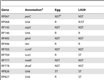
A summary of the transcriptional results for all the genes examined in this study is presented in Table 3.
Discussion
Until now termination of transcription in R. prowazekii has received little attention. Analysis of the R. prowazekii genome reveals a highly reductive genome containing many pseudogenes and a high proportion of non-coding regions. However, the retention of genes involved in the termination process (nusA,nusB,
nusG, mfd, and rho) suggests that transcription termination has a role in rickettsial gene regulation. By targeting multiple areas hypothesized to be regions of termination, this study provides the first comprehensive analysis of transcription termination in this pathogen.
Examination of theR. prowazekiigenome identified the presence of stem loop structures preceding oligo (T) sequences typical of bacterial termination sites. As demonstrated by our results with two predicted terminators found in the intervening regions of the RP703-RP704 and RP826-RP827 gene pairs, such classical terminators do function inR. prowazekii. However, one
istic of rickettsial transcriptional termination evident from these studies is the absence of site-specific termination within many intervening regions. Of the 12 gene transcripts analyzed, all but one (RP826) exhibited either a putative non-site-specific termina-tion or complete read-through of the intervening region that extended into the opposing gene coding sequence. This suggests that specific termination may not be prevalent in this relatively slow-growing, obligate. intracellular bacterium. The presence of protein products from the converging genes demonstrates that the absence of specific termination and the subsequent extension of transcript into the convergent gene do not impede nascent mRNA synthesis to a point affecting the survival of the rickettsiae. The exceptions may be those intrinsic terminators whose efficiencies appear to be regulated.
The efficiency of an intrinsic terminator in E. coli can be modulated as various factors (e.g. NusA) interact and influence the transcription complex [17]. We have identified similar alterations of terminator efficiency in R. prowazekii. RNAs isolated from rickettsiae grown in different host cell environments exhibited regulated transcription termination. Interestingly, rickettsial growth in the hen egg yolk sac generated RNAs that, in all but one case (RP826), failed to terminate at an intrinsic termination site, possibly reflecting a more suitable rickettsial growth
environment. Although increased transcription might be assumed to require more specific termination to prevent a negative impact on convergent genes, the need to prevent wasteful transcription may assume priority if rickettsiae are not in an optimum
Figure 3. RPA analysis identifying transcripts of the RP826-RP827 intervening region. (A) Schematic indicating the relative position of the RPA probes that span the intergenic region. Probe 1 is designed to hybridize to RP826 transcripts while probe 2 hybridizes to RP827 transcripts. (B) RPA analysis of rickettsial RNA isolated from rickettsiae grown in egg yolk sacs or in L929 cells. Lane numbers correspond to probe numbers in the schematic. M, sized markers with sizes (in bases) indicated. The marker lane on the right was digitally moved horizontally using Microsoft Office PowerPoint to maintain consistent lane organization. Arrows identify the band resulting from termination of RP837 transcripts at the predicted terminator. doi:10.1371/journal.pone.0016537.g003
Figure 4. RPA analysis characterizing transcripts of the RP067-RP068 gene pair region. (A) Schematic indicating the relative positions of the RPA probes targeted to the convergent gene pair. The probes include two internal probes to confirm gene transcription and two intervening probes of opposite orientation to detect intergenic mRNA. Probes 1 and 2 hybridize to RP067 transcripts while probes 3 and 4 hybridize to RP068 transcripts. (B) RPA analysis of RNA isolated from rickettsiae harvested from L929 cells. Lanes were digitally moved horizontally using Microsoft Office PowerPoint to maintain consistent lane organization. (C) RPA analysis of RNA isolated from rickettsiae harvested from egg yolk sacs. Lane numbers correspond to probe numbers in the schematic. Numbers at the bottom of each lane in B and C correspond to the size of the probe in bases. M, sized markers with sizes (in bases) indicated. The arrow indicates a band resulting from specific termination of RP068 transcripts.
environment. Alternatively, the need to initiate termination for appropriate gene expression during stress has been demonstrated during oxidative stress in Caulobacter crescentus [18]. Proteomic studies in our laboratory detect more stress-associated proteins in rickettsiae propagated in tissue culture cells than in rickettsiae grown in hen egg yolk sacs (unpublished data).
An alternative explanation for the non-specific termination of transcripts detected between some of the convergent gene pairs is the presence of RNase activity that could fragment a single transcript into many smaller fragments. Our RPA analyses cannot distinguish between transcripts generated by non-specific termi-nation or by RNase digestion. However the presence of specific transcripts generated at predicted termination sites, suggests that RNAse activity on these transcripts is minimal. This coupled with the presence of long transcripts (e.g. RP704) extending far into the intervening region, once again suggests that RNase digestion is not the sole explanation for the observed transcripts.
The presence of rickettsial antisense RNAs generated by the lack of termination between convergent genes was an intriguing finding. The existence of antisense RNA was evident from the complete protection of probes that extended, in some cases, hundreds of bases into convergent genes. While the presence of antisense RNA in bacteria is not uncommon, most such RNAs are associated with specific small regulatory RNAs or with intergenic promotion rather than termination [19,20]. Recently, examination of the Helicobacter pylori transcriptome revealed the widespread occurrence of antisense transcripts leading to the speculation that some of these may be due to imperfect termination [21]. These results are similar to an earlier study in E. coli that detected extensive antisense transcription throughout the genome [22]. Using a whole genome tiling microarray analysis, antisense transcripts have also been identified in the obligate intracellular bacterium,Anaplasma phagocytophilium[23]. Interestingly, in contrast to theE. coliandH. pyloriresults and our identification of several examples of antisense transcripts generated by read-through into convergent genes, the A. phagocytophilum whole genome study identified only one gene, p44, associated with this phenomenon [23]. Bacterial antisense RNAs have been shown to regulate gene expression [20]. The common occurrence of transcriptional read-through into convergent genes and the regulation of terminator function suggests a role in rickettsial intracellular survival and growth.
Supporting Information
Table S1 Primer sequences used to generate RPA probes. (DOC)
Acknowledgments
We thank Lonnie Driskell and Andria Hines for excellent technical assistance and manuscript review. We also thank Amanda Lackey and Aimee Tucker for manuscript review and helpful discussions and Jon Audia and Herbert Winkler for providing a portion of the hen egg yolk sac rickettsial preparations used in this study.
Author Contributions
Conceived and designed the experiments: DOW. Performed the experiments: AW. Analyzed the data: DOW AW. Contributed reagents/ materials/analysis tools: DOW. Wrote the paper: DOW AW.
References
1. Bozeman FM, Masiello SA, Williams MS, Elisberg BL (1975) Epidemic typhus rickettsiae isolated from flying squirrels. Nature 255: 545–547.
2. Reynolds MG, Krebs JW, Comer JA, Sumner JW, Rushton TC, et al. (2003) Flying squirrel-associated typhus, United States. Emerg Infect Dis 9: 1341– 1343.
3. Andersson SGE, Zomorodipour A, Andersson JO, Sicheritz-Ponte´n T, Alsmark UCM, et al. (1998) The genome sequence ofRickettsia prowazekiiand the origin of mitochondria. Nature 396: 133–143.
4. Audia JP, Patton MC, Winkler HH (2008) DNA microarray analysis of the heat shock transcriptome of the obligate intracytoplasmic pathogen Rickettsia prowazekii. Appl Environ Microbiol 74: 7809–7812.
5. Cai J, Pang H, Wood DO, Winkler HH (1995) The citrate synthase-encoding gene ofRickettsia prowazekiiis controlled by two promoters. Gene 163: 115– 119.
6. Cai J, Winkler HH (1993) Identification of tlc and gltA mRNAs and determination of in situ RNA half-life inRickettsia prowazekii. J Bacteriol 175: 5725–5727.
7. Cai J, Winkler HH (1996) Transcriptional regulation in the obligate intracytoplasmic bacteriumRickettsia prowazekii. J Bacteriol 178: 5543–5545. 8. Cai J, Winkler HH (1997) Transcriptional regulation of thegltAand TLC genes
inRickettsia prowazekiigrowing in a respiration-deficient host cell. Acta Virol 41: 285–288.
9. Shaw EI, Marks GL, Winkler HH, Wood DO (1997) Transcriptional characterization of the Rickettsia prowazekii major macromolecular synthesis operon. J Bacteriol 179: 6448–6452.
10. Ding H-F, Winkler HH (1990) Purification and partial characterization of the DNA-dependent RNA polymerase from Rickettsia prowazekii. J Bacteriol 172: 5624–5630.
11. Ding H-F, Winkler HH (1993) Characterization of the DNA-melting function of theRickettsia prowazekiiRNA polymerase. J Biol Chem 268: 3897–3902. 12. Kingsford CL, Ayanbule K, Salzberg SL (2007) Rapid, accurate, computational
discovery of Rho-independant transcription terminators illuminates their relationship to DNA uptake. Genome Biol 8: R22.
13. Shaw EI (1997) Transcriptional Characterization of theRickettsia prowazekii rpoD
Gene Linkage Group. PhD Dissertation University of South Alabama, Mobile, AL.
14. Winkler HH (1976) Rickettsial permeability: an ADP-ATP transport system. J Biol Chem 251: 389–396.
15. Chao CC, Chelius D, Zhang T, Daggle L, Ching WM (2004) Proteome analysis of Madrid E strain ofRickettsia prowazekii. Proteomics 4: 1280–1292. 16. Tucker AM, Pannell LK, Wood DO (2005) Dissecting theRickettsia prowazekii
genome: genetic and proteomic approaches. Ann N Y Acad Sci 1063: 35–46. 17. Nudler E, Gottesman ME (2002) Transcription termination and
anti-termination inE. coli. Genes to Cells 7: 755–768.
18. Italiani VCS, Zuleta LFG, Marques MV (2002) The transcription termination factor Rho is required for oxidative stress survival inCaulobacter crescentus. Molec Microbiol 44: 181–194.
19. Dornenburg JE, DeVita AM, Palumbo MJ, Wade JT (2010) Widespread antisense transcription inExcherichia coli. mBio 1: 1–4.
20. Waters LS, Storz G (2009) Regulatory RNAs in bacteria. Cell 136: 615–628. Table 3.Summary of Gene Termination Events.
Gene Annotationa Egg L929
RP067 parC NSTb NST
RP068 Unk R R/ST
RP145 aspS NST NST
RP146 Unk R R
RP495 glnA NST NST
RP496 rbn R R
RP703 ccmF NST NST
RP704 sca5 R ST
RP777 metK NST NST
RP778 dnaE NST NST
RP826 Unk ST ST
RP827 Unk R ST
aAnnotations from (1) and ERGOTM(Integrated Genomics, Chicago, IL.). bR = Read through into opposing gene, ST = Specific Termination, NST = Non-Specific Termination.
Unk = Unknown.
21. Sharma CM, Hoffmann S, Darfeuille F, Reignier J, Findeib S, et al. (2010) The primary transcriptome of the major human pathogenHelicobacter pylori. Nature 464: 250–255.
22. Selinger DW, Cheung KJ, Mei R, Johansson EM, Richmond CS, et al. (2000) RNA expression analysis using a 30 base pair resolutionEscherichia coligenome arrray. Nature Biotechnol 18: 1262–1268.