i CLAUDINEIA LIZIERI DOS SANTOS
ENVIRONMENTAL ATTRIBUTES IN ASSEMBLING CYANOBACTERIAL COMMUNITIES FROM THE McMURDO SOUND REGION, ANTARCTICA
VIÇOSA
MINAS GERAIS – BRAZIL 2014
Ficha catalográfica preparada pela Biblioteca Central da Universidade Federal de Viçosa - Câmpus Viçosa
T
Santos, Claudineia Lizieri dos, 1982-S237e
2014
Environmental attributes in assembling cyanobacterial communities from the Mcmurdo Sound region, Antarctica : environmental control of cyanobacterial community assembly / Claudineia Lizieri dos Santos. – Viçosa, MG, 2014.
x, 94f. : il. (algumas color.) ; 29 cm.
Orientador: Carlos Ernesto Gonçalves R. Schaefer. Tese (doutorado) - Universidade Federal de Viçosa. Inclui bibliografia.
1. Cianobactéria. 2. Antártica. 3. Lagos. 4. .
I. Universidade Federal de Viçosa. Departamento de Biologia Vegetal. Programa de Pós-graduação em Botânica. II. Título.
ii CLAUDINEIA LIZIERI DOS SANTOS
ENVIRONMENTAL ATTRIBUTES IN ASSEMBLING CYANOBACTERIAL COMMUNITIES FROM THE McMURDO SOUND REGION, ANTARCTICA
APPROVED: March 18, 2014.
___________________________________________ Carlos Ernesto Gonçalves Reynaud Schaefer
(Adviser)
Thesis submitted to Botany Graduate Program of the Universidade Federal de Viçosa, in partial fulfillment of the requirements for the degree of Doctor Scientiae
_________________________ Adriano Nunes Nesi
(Co-adviser) __________________________
Antônio Galvão do Nascimento (Co- supervisor)
__________________________________ Débora Machado Corrêa
ii I dedicate this thesis...
…to the living memories of Rosane Euclydes Aguiar from whom this challenging and beautiful journey started…
AND
iii “In the long history of humankind (and animal kind, too) those who learned to collaborate
iv ACKNOWLEDGMENTS
I want to thank to CAPES, PETROBRÁS, Universidade Federal de Viçosa, Departamento de
Biologia Vegetal, Programa de Pós-graduação in Botânica, PROANTAR-Programa Antártico
Brasileiro, INCT-Criosfera and TERRANTAR for the support given during my PhD.
I am very grateful to the centre of Antarctica studies Gateway Antarctica (“GA”), Canterbury University, New Zealand and especially for Dr. Bryan Storey, who accepted me as a member of the
GA team. Also, I would like to thank New Zealand Antarctic Programme (“AntNZ”) for supporting my Antarctic expedition.
I am very thankful to my supervisor Carlos Ernesto Schaefer for adopting me as member of
his Antarctic team and allowing me to work on Antarctic research. I especially thank you for the
support during difficult times while working on my PhD and for being so friendly to me during these
years.
I would like to express the deepest appreciation to Professor Ian Hawes from whom I have
learned so much about this wonderful continent, ANTARCTICA and its GORGEOUS
CYANOBACTERIA! Also, I want to thank Dr. Hawes for has provided me with opportunities to
travel to the South Pole and his guidance during this research development. Many thanks, Ian.
I am also very grateful to Dr. Paul Broady for have received me and heard all of my doubts so
kindly, even with my horrible English at the beginning and for the valuable suggestions for this work.
I'm so thankful for having had this adorable opportunity to meet such a nice person as Paul Broady!
I want to give a special mention for Professor Antônio Galvão do Nascimento and Professor
Adriano Nunes Nesi, my co-supervisors. Furthermore, the Professor Francisco Barbosa from
Universidade Federal de Minas Gerais and Professor Débora Corrêa from Universidade Federal de
Goiás for their considerations on this work.
I thank in loving memories my past supervisor, Rosane Aguiar with whom I had the immense
pleasure to meet and to share a few years of my life; thanks for teaching me so many things about
science and life.
I also would like to thank the lab members of NIWA (especially Dr. Cathy Kilroy) and
Biology lab of Canterbury University (Reijel and Gregy) for also being friendly and helping whenever
I needed it.
I am very grateful to Susanna Wood from Cawthron Institute, Nelson, New Zealand for
providing Antarctic cyanobacterial strains to carry on a part of this research.
Thanks to Claudia Maria Gonçalves, Ângelo Valentim Lopes and Rogério Gomide
(secretaries and technicians) for all their help during my PhD journey.
Thanks to Daniel Arruda, Carlos Nick, and Helton Silva for the help and suggestions on the
statistical analysis.
v me so kindly and for adopting me as a member of the team, besides providing such lovely company
during my time in NZ.
I would like to express my special thanks for two wonderful girls: Gabriela Carey and Hana
Christenson for being so close to me and friendly while carrying out this research. Words cannot
express my gratitude for you Gabriela Carey. I thank you for each jacket that you brought to me when
the day got colder, each piece of advice, walking on the most wonderful place that I have been so far,
and every little thing that you did for me. Many thanks Gaby; I will not forget you and Peter Carey,
ever. And special thanks to such a kind person Hana Christenson, who received me at your home and gave me the first directions in NZ when I still couldn’t understand even one kiwi English word and thanks for the lovely Antarctic companionship.
I do not want to miss this opportunity to thank people who made my stay in New Zealand one
of the most wonderful experiences of my life, without which I would not have been able to
concentrate on my research. I would like to thank my friend Deepak Sharma, who heard me
complaining so many times about my tiredness and missing home. Thank you for your company and
for having given me so many reasons to smile. Also, I would like to thank Elisabeth Gingrich for all
my English classes and for being so kind to me.
I am also grateful to all of my friends back in Brazil for their words of encouragement and for
directly or indirectly helping me to finish this work - Alberto Abrantes, Kacilda Naomi, Mychelle
Carvalho, Adriana Magalhães, Victor Coelho, Maione Franco, and Gracielly Alcantara. In addition, I
want to thank my flatmates, Cida, Isabela and Laura for being enjoyable with me even on my most
stressful days.
I would like to thank JJ Lelis and Douglas Lindemann for the best Antarctic companionship.
Also, I want to thank Rafael Bastos e João A. Vieira for the help in previous works.
My thanks to a special friend, Bob, for keeping me informed about all events in Brazil in his
own special way while I was away from here and for being a part of a beautiful piece of my story.
My special thanks for those who are the most special people in my life, my two sisters,
Andrea and Adriana Lizieri dos Santos, my brother-in-law, Edson Benito, and my sweet niece,
Liandra Lizieri Benito. I offer my many thanks for being patient with me during these long years that I
have been far away from home and especially to my mother, Sirley Lizieri, who I infinitely love.
Thank you for your love and unwavering belief in me. I also want to mention my father, Sebastião
Ribeiro dos Santos, not for directly helping me on this journey, but for giving me the reasons for my
efforts and for making me stronger, all of which pushed me until this point.
Finally, I thank God, whose existence, although incomprehensible, I do not doubt, and his
presence was made real every day of my life giving me many blessings.
To everyone who has directly or indirectly participated in my studies,
vi TABLE OF CONTENTS
RESUMO... vii
ABSTRACT... ix
1. GENERAL INTRODUCTION... 1
1.2. General Description of sample sites……….. 4
2. References………... 8
CHAPTER 1. Descriptions of morphospecies of cyanobacteria from benthic microbial mats of ponds in the McMurdo Sound region
………...
………….... 121. Introduction... 13
2. Materials and Methods... 14
2.1. Sampling sites... 14
2.2 Sample analysis... 14
3. Results and Discussion………..… 16
4. Concluding remarks……….. 32
5. References... 33
CHAPTER 2. The role of physical-chemical properties of Antarctic ponds in determining the composition and distribution of cyanobacterial assemblages in Antarctic systems………... 37
1. Introduction... 38
2. Materials and Methods..……… 41
2.1 Sample sites... 41
2.2. Pond sampling... 41
2.2.1Measurements of temperature, pH, conductivity, DO and depth... 41
2.2.2 Determination of dissolved inorganic carbon... 41
2.2.3Determination of phosphorus and nitrogen content... 41
2.2.4Determination of plankton chlorophyll-a... 42
2.2.5 Determination of benthic pigments, EPS and organic matter….……….. 42
2.2.6 Benthic cyanobacterial mat composition... 43
2.3 Statistical analysis of data... 45
3. Results... 46
4. Discussion... 59
5. Final remarkes and consideration ... 63
6. References... 64
CHAPTER 3. A comparison of the mat-building ability of five strains of Antarctic cyanobacteria………. 71
1. Introduction... 72
2. Materials and Methods..……… 73
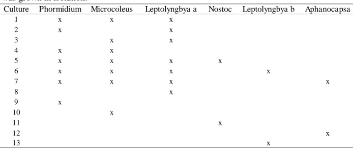
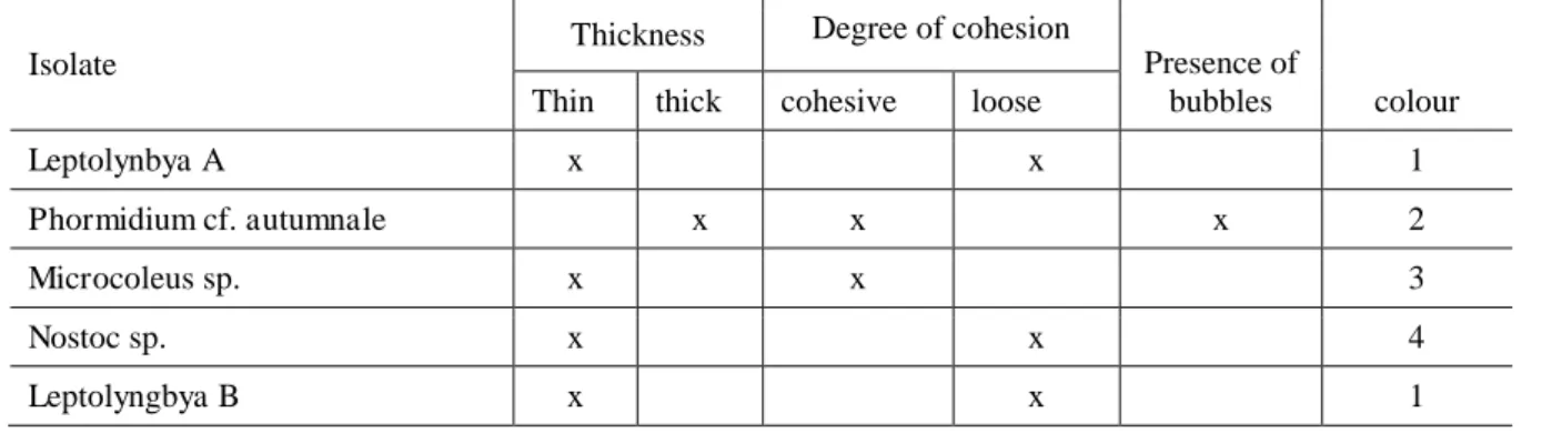
2.1. Acquisition and Acclimation of the cyanobacteria strains... 73
2.2 Experimental Setup... 74
2.3 Measurement of developed mat... 74
2.3.1 Chlorophyll-a pigment... 75
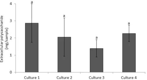
2.3.2 Total EPS... 75
2.3.3 Organic matter... 75
2.4 Statistical Analyses... 75
3. Results... 76
4. Discussion... 85
5. Concluding remarks ... 87
6. References... 88
vii RESUMO
SANTOS, Claudineia Lizieri dos, D.Sc., Universidade Federal de Viçosa, março de 2014. Atributos ambientais na montagem da comunidade de cianobactérias da região do McMurdo Sound, Antártica. Orientador: Carlos Ernesto Gonçalves Reynaud Schaefer. Co-orientadores: Antônio Galvão do Nascimento e Adriano Nunes Nesi.
ix ABSTRACT
SANTOS, Claudineia Lizieri dos, D.Sc., Universidade Federal de Viçosa, March of 2014. Environmental attributes in assembling cyanobacterial communities from the Mcmurdo Sound region, Antarctica. Advisor: Carlos Ernesto Gonçalves Reynaud Schaefer. Co-Advisors: Antônio Galvão do Nascimento and Adriano Nunes Nesi.
1 1. GENERAL INTRODUCTION
Antarctica is a continent locked in ice, with almost 99.7% of current terrain covered by permanent ice and snow (Convey et al., 2008). The continent has been relatively isolated from the rest of the world since its separation from Gondwanaland about 85 Ma years ago, and final separation from South America about 35 Ma years ago (McLoughlin, 2001; Lewis et al., 2008.). As Antarctica became isolated from warmer waters and greenhouse gases declined in the atmosphere, the continent cooled down and glaciers began to form (Tripati et al., 2005; Fernández-Carazo et al., 2012). As climate cooled, the continent lost much of its temperate biota and, for the last 10 million years, has largely been a cold desert (Lewis et al., 2008). Since the ice sheet formation, Antarctica has remained isolated from other continents, but has continued to support a range of habitats, which are home for a variety of species, most of which are microorganisms (Convey et al., 2008; Fernández-Carazo et al., 2012).
Today ice-free areas of Antarctica cover a tiny proportion (0.32%) of the continent (British Antarctic Survey, 2004; Convey et al., 2008). All these habitats, including terrestrial and aquatic, face extended seasonal snow and/or ice cover mainly during winter, which restricts periods of biological activity, but also can protect the biota from extremes of temperature and wind abrasion (Convey et al., 2008).
Antarctic biota encounters a range of environmental stresses which has been described as one of the most remote, harsh and challenging for terrestrial life (Convey, 2006). Besides the extreme temperature and wind abrasion, these include lack of liquid water leading to desiccation, nutrient limitation due to poorly developed soils and catchments, repeated freeze thaw cycles, bright sunlight exposure during summer and prolonged darkness during winter (Moorhead et al., 2005; Mueller, 2005; Vincent, 2007; Convey et al., 2008). Antarctic contains the coldest and driest deserts on earth (for example the McMurdo Dry Valleys) (Vincent, 2004).
2 Although harsh climate conditions predominate on the Antarctic continent, there is still a great diversity of aquatic habitats in Antarctica, ranging from the benthos, water-column and fluctuating sea-ice cover of the circumpolar Southern Ocean to diverse lakes and ponds, and intermittently wet soils which sustain Antarctic biodiversity (Wynn-Williams, 1996). Even the most arid regions, such as the McMrudo Dry Valleys, are home to perennially ice-covered, endorheic, meromictic lakes and ponds, which are home to microbially-dominated communities (Taton et al., 2003; Sutherland & Hawes, 2008). Antarctica is the only continent that is dominated by microbial communities, mostly based on cyanobacteria, algae, mosses and lichens). Only two vascular plants are known from the Antarctic, both restricted to coastal regions of the Antarctic Peninsula (Convey, 2006). Due cyanobacteria being highly tolerant of extreme conditions and quickly responsive to sudden moisture availability after desiccation and/ or freezing in cold deserts, they often dominate these Antarctic habitats (Hawes et al., 1992).
Cyanobacteria are photosynthetic bacteria that require light, liquid water, air and some mineral nutrients for growth. They serve as primary colonizers of soils newly exposed by glacial retreat. Some taxa (e.g. Nostoc sp.) can fix atmospheric nitrogen that can locally enrich the predominantly oligotrophic biotopes (Vincent, 2000). The occurrence of cyanobacteria in Antarctica has been recorded since one of the first research expeditions conducted on the continent in the early twentieth century (as reported in Broady & Kibblewhite, 1991). Cyanobacteria are uniquely acclimated to the extreme conditions that Antarctic places offer (Taton et al., 2003; Sutherland & Hawes, 2008) with many ecophysiological and strategic adaptations that enable them to survive, colonize and even flourish in what is often considered to be a biologically hostile environment (Wynn-Williams, 1996). The abundance of cyanobacteria in Antarctic ecosystems has been related to both adaptive capacity, with high tolerance to extreme environment, as well as, the lack of predators and competing species which are eliminated by the effect of low temperature and freezing environment (Nadeau & Castenholz, 2000).
3 capable of synthesizing exocellular polysaccharides as an additional structure of surface that differs in thickness, consistency and appearance (Phillipis & Vincenzini, 1998).
The production of EPS is generally considered to be directly related to environmental restraints on the microorganism and it is involved in the protection of a number of factors such as desiccation, freezing and ultraviolet irradiation (Phillipis & Vincenzini, 1998; Tamaru et al., 2005). The main function assigned to capsules or other investments of polysaccharide origin is to serve as a barrier between the organism and its immediate contact with the environment (Phillipis & Vincenzini, 1998). EPS production is also involved in the organization of cyanobacteria into mats (also termed biofilm), which might also provide a strategy that favors survival under adverse conditions (Rickard et al., 2003; Garcia-Meza et al., 2005). Mats are typically found attached to a solid substratum in a moist or liquid environment from which those nutrients are obtained (Rickard et al., 2003).Mats are functional consortia between cells (aggregate) of different microbial species, which present greater metabolic activity than the isolated species, and togerthe modify the light, nutrient and moisture regime within the mat in ways that can benefit individual growth and survival. The coaggregation of species and the specific interactions in the biofilm environment are an important mechanism to the formation of communities that optimizes cellular interactions, colonization of organisms and allows the set of strains to survive and proliferate under conditions where cells (species) isolated would show reduced growth (Rickard et al., 2003).
Several studies have recorded that the maritime and coastal microbiota of Antarctica has relatively high species diversity, but this becomes depauperate further inland and south where life exists near its limits (Wynn-Williams, 1996). The extreme stresses imposed by winter freezing on further inland must play a role in structuring biological communities (Hawes et al., 1999).
4 made. We also investigated the process (e.g. random e/or deterministic) that can be involved in determining the presence and/or absence of cyanobacteria communities at any one site. A quick method of assessing mat formation to demonstrate the effect of certain cyanobacterial strains on the mat-building also was developed through this research.
1.2. General description of sampling sites
The sampled ponds for this work are spread in the McMurdo Sound region of the Antarctic continent, including ponds on Ross Island, the McMurdo Ice Shelf and McMurdo Dry Valleys (Lower and Upper Wright Valley) (Figure 1 and 2).
6 McMurdo Dry Valleys
McMurdo Dry Valleys which experiences some of the most adverse environmental conditions in Antarctica is located in Southern Victoria Land, on the Wesern coast of the Ross Sea. The McMurdo Dry Valleys became a hyper-arid desert about 14 million years ago, and has remained dry ever since, despite the coming and going of the Ross Ice Shelf (Lewis et al., 2008). This makes the McMurdo Dry Valley region a very old habitat, with plenty of time to accumulate and adapt species.
Wright Valley
The Wright Valley is one of the conspicuous ice-free areas and it is located in the McMurdo Dry Valleys. The Wright Valley is 45 km long and 7 km wide and is an enclosed basin. The Olympus and Asgard mountain ranges lie to the north and south respectively, and stand up to 2000 m above the valley floor. The Upper Wright and Lower Wright glaciers terminate at either end of the valley. The valley lies west to east, and perpendicular to the sea. Much of the valley floor is covered in glacial moraine and till, and soils are derived from this and bedrock. Major materials in these are sandstone, granite, diorite, dolerite and basalt. The dry valleys are too far inland to see marine animals; however mummified seals do occur (Campbell & Claridge, 1987).
Upper Wright Valley
Also known as The Labyrinth is an area at the west end of Wright Valley, furthest from the sea, and is so named because large troughs have been carved through the valley by sub-glacial water flow (Healy 2005). The bed rock is mostly composed of dolerite, and little detritus or sediment is present (Torii et al., 1989). The area is at 800 - 1000 m elevation, and lies at the base of the Upper Wright Glacier (Healy, 2005). More than 60 ponds exist in depressions of the troughs, and these are fed by melt from snow and the glacier. The geochemistry of the ponds has been documented, and ponds vary from fresh to saline, with salts accumulated by atmospheric deposition, and concentrated by evaporation.
Lower Wright Valley
7 by glacial melt and the limited precipitation that reaches the valley, including those around Bull Pass (Webster et al., 1994).
Ross Island (Hut Point),
Ross Island is an island formed by four volcanoes in the Ross Sea near the continent of Antarctica, off the coast of Victoria Land in McMurdo Sound. Hut Point Peninsula is about 20 kilometers long and 2 to 4 kilometres wide. It consists of a series of en echelon lines of volcanic cones that extend in a south-southwest direction from Mount Erebus, Ross Island. The cones are composed of basanite and basanitoid lavas with lesser amounts of hawaiite and phonolite (Kyle & Treves, 1974). The Hut Point site is one of the principal sites of early human activity in Antarctica. It is an important symbol of the Heroic Age of Antarctic exploration and, as such, has considerable historical significance. Some of the earliest advances in the study of earth sciences, meteorology, flora and fauna in Antarctica are associated with the Discovery Expedition based at this site (Management Plan for Antarctic Specially Protected Area 2010).
McMurdo Ice Shelf, Bratina Island
8 2. References
British Antarctic Survey. 2004. Antarctica, 1:10 000 000 scale map. BAS (Misc) 11. (http://www.antarctica.ac.uk/Resources/schoolzone/resources/Factsheets/index.html). British Antarctic Survey, Cambridge, UK.
Broady, P. A. & Kibblewhite, A. L. 1991. Morphological characterization of Oscillatoriales (Cyanobacteria) from Ross Island and southern Victoria Land, Antarctica. Antarctic Science 3: 35-45.
Campbell, I. B.; Claridge, G. G. C. 1987. Antarctica: Soils, Weathering Processes and Environment. Elsevier: Amsterdam, 368p.
Convey, P. 2006. Antarctic terrestrial ecosystems: responses to environmental change. Polarforschung 75: 101-111.
Convey, P.; Gibson, J. A. E.; Hillenbrand, Claus-Dieter; Hodgson, D. A.; Pugh, P. J. A.; Smellie, J. L. & Stevens, M. I. 2008. Antarctic terrestrial life - challenging the history of the frozen continent? Biology Reviews 83:103-117.
Debenham, F. 1920. A new mode of transportation by ice: The raised marine muds of South Victoria Land (Antarctica). Quarterly Journal of the Geological Society of London 75: 51-76.
Fernández-Carazo, R.; Namsaraev, Z.; Mano, Marie-Jose; Ertz, D. & Wilmotte, A. 2012. Cyanobacterial diversity for an anthropogenic impact assessment in the Sør Rondane Mountains area, Antarctic. Antarctic Science 24: 229-242.
Friedmann, E.I. 1982. Endolithic microorganisms in the Antarctic cold desert. Science 215: 1045-1053.
9 Hawes, I.; Howard-Williams, C. & Vincent, W.F. 1992. Desiccation and recovery of
cyanobacterial mats. Polar Biology 12: 587-94.
Hawes I. & Schwarz A.-M. 1999. Photosynthesis in an extreme shade environment: Benthic microbial mats from Lake Hoare, a permanently ice-covered Antarctic lake. Journal of Phycology 35: 448-459.
Hawes, I. & Howard-Williams, C. 2003. Pond life on the McMurdo Ice Shelf, one of the world’s strangest ecosystems. Water & Atmosphere 11: 18-19.
Healy, M. 2005. The Physical and Geochemical Properties of Glacial Meltwater Ponds in the Dry Valleys, Antarctica. University of Auckland, Auckland.
Kyle, P. R. and Treves, S.B. 1974. Geology of Hut Point Penisula, Ross Island. Antarctica Jornal 9: 232-234.
Lewis, A.R.; Marchant, D.R.; Ashworth, A.C.; Hedenäs, L.; Hemming, S.R.; Johnson, J.V.; Leng, M. J.; Machlus, M. L.; Newton, A. E.; Raine, J. I.; Willenbring, L. K.; Williams, M. & Wolfe, A. P. 2008. Mid-Miocene cooling and the extinction of tundra in continental Antarctica. PNAS 105: 10676 - 10680.
Management Plan for Antarctic Specially Protected Area. 2010. Hut Point, Ross Island, Measure 10, Annex. No. 158.
McLoughlin, S. 2001. The breakup history of Gondwana and its impact on pre-Cenozoic floristic provincialism. Australian Journal of Botany 49:271-300.
Moorhead, D.; Schmeling, J. & Hawes, I. 2005. Modelling the contribution of benthic microbial mats to net primary production in Lake Hoare McMurdo Dry Valleys. Antartic Science 17: 33-45.
10 Nadeau, T. L. & Castenholz, W. F. 2000. Characterization of psychrophillic oscillatorians
(Cyanobacteria) from antarctic meltwater ponds. Journal of Phycology 36: 914-923.
Phillipis, R. & Vincenzini, M. 1998. Exocellular polysaccharides from cyanobacteria and their possible applications. FEMS Microbiology Reviews 22: 151-175.
Rickard, A. H.; Gilbert, P.; High, N. J.; Kolenbrander, P. E. & Handley, P. S. 2003. Bacterial coaggregation: an integral process in the development of multi-species biofilms. TRENDS in Microbiology 11: 94-100.
Rodrigues, D. F. & Tiedje, J. M. 2008. Coping with our cold planet. Applied and Environmental Microbiology 74: 1677-1686.
Simões, J. C.; Arigony-Neto, J. & Bremer, U. F. 2004. O uso de mapas antárticos em publicações. Pesquisa Antártica Brasileira 4: 191-197.
Sutherland, D. L. & Hawes, I. 2008. Annual growth layers as proxies of past growth conditions for benthic microbial mats in a perennially ice-covered Antarctic lake. FEMS Microbiology, Ecology 67: 279-292.
Swithinbank, C. W. M. 1970. Ice movement in the McMurdo Sound area of Antarctica. IAHS SCAR Publication 86: 472-487.
Tamaru, Y.; Takani, Y.; Yoshida, T. & Sakamoto, T. 2005. Crucial role of extracellular polysaccharides in desiccation and freezing tolerance in the terrestrial cyanobacterium Nostoc commune. Applied and Environmental Microbiology 71: 7327-7333.
Taton, A.; Grubisic, S.; Brambilla, E.; De Wit, R. & Wilmotte, A. 2003. Cyanobacterial diversity in natural and artificial microbial mats of Lake Fryxell (McMurdo Dry Valleys, Antarctica): a morphological and molecular approach. Applied and Environmental Microbiology 69: 5157-5169.
11 Tripati, A.; Backman, J.; Elderfield, H. & Ferretii, P. 2005. Eocene bipolar glaciation
associated with global carbon cycle changes. Nature 436: 341-346.
Varin, T.; Lovejoy, C.; Jungblut, A. D.; Vincent, W. F. & Corbeil, J. 2012. Metagenomic Analysis of Stress Genes in Microbial Mat Communities from Antarctica and the High Arctic. Applied and Environmental Microbiology 78: 549-559.
Vincent, W. F. 2000. Cyanobacterial dominance in the polar regions, p. 321-340. In: Whitton, B. & Potts, M. (eds.). Ecology of the Cyanobacteria: their diversity in space and time. Kluwer Academic Press, Dordrecht, The Netherlands.
Vincent, W. F.; Mueller, D. R. & Bonilla, S. 2004. Ecosystems on ice: the microbial ecology of Markham Ice Shelf in the high Artic. Crybiology 48: 103-112.
Vincent W.F. 2007. Cold tolerance in cyanobacteria and life in the cryosphere, p 287-301. In: Seckbach J (ed), Algae and cyanobacteria in extreme environments. Springer, Heidelberg, Germany.
Zhang, T. & Fang, H. H. P. 2001. Quantification of extracellular polymeric substances in biofilms by confocal laser scanning microscopy. Biotechnology Letters 23: 405-409.
Zhao, D.; Chen, X.; He, H.; Shi, M. & Zhang, Y. 2007. Gene cloning and sequence analysis of the cold-adapted chaperones DnaK and DnaJ from deep-sea psychrotrophic bacterium Pseudoalteromonas sp. SM9913. Acta Oceanologica Sinica 26:91-100.
Webster, J. G.; Brown, K. L. & Vincent, W. F. 1994. Geochemical processes affecting meltwater chemistry and the formation of saline ponds in the Victoria Valley and Bull Pass region, Antarctica. Hydrobiologia 281: 171-186.
12
CHAPTER 1
Descriptions of morphospecies of cyanobacteria from benthic microbial
13 1. Introduction
The Cyanobacteria (Cyanophyceae, blue-green algae) are an ancient group of micro-organisms that comprises unicellular to multicellular species which possess chlorophyll a and perform oxygenic photosynthesis (Schopf, 2000; Frey, 2012). They are found in almost all terrestrial and aquatic biotopes (Whitton & Potts, 2000) and they are well known for their ability to dominate habitats in temperate and extreme environments (Pearl & Huisman, 2008; Wood et al., 2010).
In Antarctica, cyanobacteria are widespread in lakes, ponds, streams, soils and on the surfaces and within rocks, where they can form macroscopically visible crusts or thin biofilms (Friedmann, 1982; Vincent, 2000; Andersen et al., 2011; Hawes et al., 2011). They often dominate the biomass and biological productivity of Antarctic regions due to their adaptation to the polar environment (Vincent, 2000; Jungblut et al., 2009).
Antarctic cyanobacteria tolerate a wide range of climatic conditions and harsh physicochemical parameters (e.g. high salinities, UV radiation, extended periods of darkness, freezing and desiccation) (Zakhia et al., 2009). They have developed several stress response mechanisms and they are highly responsive to sudden moisture availability after desiccation and freezing (Hawes et al., 1992; Vincent, 2007) which gives them the impressive ability to colonise and dominate these regions.
14 The present research presents descriptions of the morphology of cyanobacteria from pond mat communities sampled from ponds along inland to coastal gradients in the McMurdo Sound region. Twenty five ponds were sampled, including many never previously sampled.
This work will help extend knowledge of the distribution of Antarctic cyanobacteria morphospecies and these data can be compared with future molecular analyses to increase the understanding of cyanobacteria identities.
2. Materials and Methods
Sampling sites
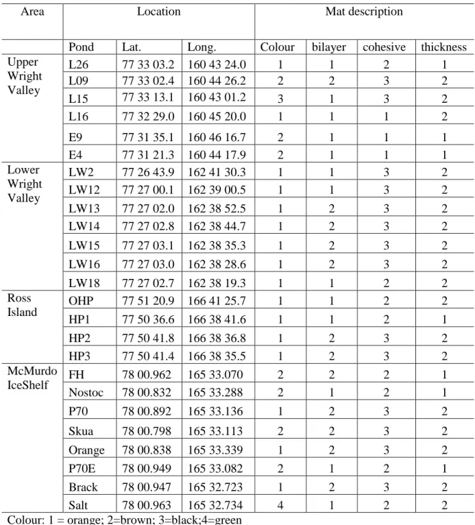
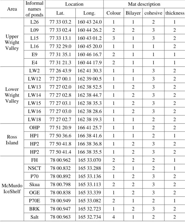
Samples of benthic microbial mats from 25 Antarctic ponds distributed along inland-coastal gradients in the McMurdo Sound region (Ross Island, McMurdo Ice Shelf, Lower Wright Valley and Upper Wright Valley) were collected, in summer season 2012. The area location, nominal longitudes and sampling description for each pond were described in the Table 1. Samples were collected from the pond margins using a cut-off 20 mL syringe. Cores were taken with care to collect the mat only and no underlying sediment. The resulting mat core was carefully lifted from the sediment and transferred to a plastic container and kept chilled while in the field and subsequently frozen (-20°C) for return to New Zealand.
Sample analysis
For morphological identification, benthic mat samples were thawed and a subsample was directly observed by light microscopy. Remaining sample material was placed in 50 ml sterile polycarbonate bottles (Biolab, NIWA) containing the mineral nutrient medium MLA (Bolch & Blackburn, 1996) for future observation and maintained at laboratory temperatures of about 21°C. No attempt was made to isolate cyanobacterial morphospecies into unialgal cultures.
15 The percentage of frequency of occurrence of the morphospecies for each studied area was calculated by: FO = (Npi x 100)/Ntp; where FO= Frequency of occurrence, Npi= number of pond that the morphospecie was found and, Ntp= total number of studied pond.
Table 1. Locations of study sites and mat description
Area Location Mat description
Pond Lat. Long. Colour bilayer cohesive thickness Upper
Wright Valley
L26 77 33 03.2 160 43 24.0 1 1 2 1
L09 77 33 02.4 160 44 26.2 2 2 3 2
L15 77 33 13.1 160 43 01.2 3 1 3 2
L16 77 32 29.0 160 45 20.0 1 1 1 2
E9 77 31 35.1 160 46 16.7 2 1 1 1
E4 77 31 21.3 160 44 17.9 2 1 1 1
Lower Wright Valley
LW2 77 26 43.9 162 41 30.3 1 1 3 2
LW12 77 27 00.1 162 39 00.5 1 1 3 2
LW13 77 27 02.0 162 38 52.5 1 2 3 2
LW14 77 27 02.8 162 38 44.7 1 2 3 2
LW15 77 27 03.1 162 38 35.3 1 2 3 2
LW16 77 27 03.0 162 38 28.6 1 2 3 2
LW18 77 27 02.7 162 38 19.3 1 1 2 2
Ross
Island OHP 77 51 20.9 166 41 25.7 1 1 2 2
HP1 77 50 36.6 166 38 41.6 1 1 2 1
HP2 77 50 41.8 166 38 36.8 1 2 3 2
HP3 77 50 41.4 166 38 35.5 1 2 3 2
McMurdo
IceShelf FH 78 00.962 165 33.070 2 2 2 1
Nostoc 78 00.832 165 33.288 2 1 2 1
P70 78 00.892 165 33.136 1 2 3 2
Skua 78 00.798 165 33.113 2 2 3 2
Orange 78 00.838 165 33.339 1 2 3 2
P70E 78 00.949 165 33.082 2 1 2 1
Brack 78 00.947 165 32.723 1 2 3 2
Salt 78 00.963 165 32.734 4 1 2 2
Colour: 1 = orange; 2=brown; 3=black;4=green Bilayer: 1= no; 2= yes
16 3. Results and Discussion
On the basis of microscope observations, twenty nine cyanobacterial morphospecies were distinguished in the samples. Four were assigned to the order Chroococcales, 22 to Oscillatoriales and three to Nostocales. No members of the Stigonematales were encountered. The morphotypes are designated 1-29 and a summarized morphological and morphometric description with data on site of occurrence of each morphotype are shown in Table 2. Photomicrographs are shown in Figures 1, 2 and 3.
OSCILLATORIALES
Morphotype 1 (Fig.1 A1-A2)
Description: Trichome which can form short filament (4 to 16 celled) or rarely
trichome solitary. Cells are cylindrical or up to barrel-shaped, ± isodiametric by fragmentation of trichome, sheaths lacking, immotile, and constricted at the cross-walls.
Remarks: This morphotype was assigned to genus Borzia and appeared in 11 of the
studied samples. Borzia sp. was previous documented in continental Antarctic systems by Hodgson et al. (2001) and Broady (2005). Despite of the increasing availability of information about Antarctic cyanobacteria diversity, it was not found literature that has reported this morphospecie on the Antarctica Peninsula habitats, suggesting that this morphotype has restricted distribution.
Morphotye 2 (Fig.1 B)
Description: Trichome forming fine filament, a little flexuous, elongated, broadly
constricted at the cross-walls, immotile, without firm sheaths, simple. Necridic cells, akinetes, aerotopes and heterocytes were not observed. Cells barred-shaped or sometimes almost isodiametric.
Remarks: The morphotype 2 was found at only one sample and based on the classical
literature we suggest that this strain contains resemblance with genus Komphovorum. Although the width description to this genus made by Komárek & Anagnostidis (2005) is a little wider than the width observed to the morphotype recorded in the present work.
Morphotype 3 (Fig.1C)
Description: Filamentous trichome, thin, cylindrical, straight or slightly flexuous,
17 the ends. Trichome deeply motile, with intense gliding in the direction of the longitudinal axis forwards and backwards or waving (oscillation). Cells longer than wide, sometimes with large prominent granules. Apical cells usually conical, hooked or bent.
Remarks: The possession of consistent characters observed in this strain allowed the
assignment to Geitlerinema cf. ionicum and the morphotype was observed at nine samples. Studies from Antarctic Peninsula (Komárk, 1999) and Antarctic continental (Taton et al., 2006a) have documented the presence of Geitlerinema sp., which show wide distribution of this morphospecie across Antarctica.
Morphotype 4 (Fig.1 D1-D2)
Description: Trichomes usually straight or a little waved, consisting of few to several
cells, with broadly constrictions at cross-walls. Trichomes without sheaths. Motility lacking. Cells usually cylindrical with rounded ends, sometimes almost barred-shaped, apical cells not differentiated.
Remarks: This morphotype would separate in two morphospecies by using the length
of the trichome, which sometimes possessed long filament (Fig.1 D1) and occasionally short trichome (Fig.1 D2). However, we have not found consistent character to separate these morphospecies. Morphotype 4 was recorded at 14 samples and it was assigned to genus Pseudanabaena sp.
Morphotype 5 (Fig.1 E1-E2)
Description: Filament very short, few-celled (3 – 6 celled), thin, straight or slightly curved. Cell cylindrical, constantly longer than wide, sometimes granules at the ends were present. Trichome constricted, not attenuated, motility, trembling-like.
Remarks: This strain also contains similarity to genus Pseudanabaena and appeared
at eight of the studied samples.
Morphotype 6 (Fig.1 F1-F3)
Description: Trichome very thin, not attenuated at the ends, with firm, thin sheaths
18 Remarks: Filaments allocated to morphotype 6 were thinner than morphotype 4 but
similar to width of filaments belonging to morphotype 5. However, morphotype 6 forms long filament while the possession of morphotype 5 is characterized by constantly short filament. The features found in morphotype 6 are also acceptable to genus Pseudanabaena and was recorded at 14 samples.
19 Morphotype 7 (Fig.1 G1-G4)
Description: Filamentous trichome, straight or flexuous, finely waved, long, with
sheath (Fig.1 G3) or not (Fig.1 G2), the most not attenuated at the ends but attenuation present in some filaments (Fig.1 G1). Trichomes immotile or with indistinct trembling. Cells the most cylindrical, however, also isodiametric or a little longer than wide.
Remarks: Morphotype 7 was assigned to genus Leptolyngbya sp. and this
morphotype also could include more than one morphospecies by the possession of trichomes very densely spirally coiled, which was occasionally observed in this work (Fig. G4). Komárek (2007) has separated and classified the spirally coiled trichome belonging to genus Leptolynbya as Leptolyngbya borchgrevinkii. Morphotypes 7 were found at all samples studied.
Morphotype 8 (Fig. H1-H2)
Description: Filaments nearly straight, sometimes slightly curved, pale blue-green,
not constricted at cross-walls. Cells of various lengths, sometimes longer than wide but also wider than long. Apical cell somewhat elongated, sometimes rounded.
Remarks: This strain was assigned to genus Leptolynbya sp. as well. The major
characteristic separating the morphotype 7 from 8 was the cells shape, which is nearly square at the morphotype 8 and slightly cylindrical or isodiametric at the morphotype 7. Morphotype 8 was observed in 22 samples.
Morphotype 9 (Fig.2 A)
Description: Trichome long, curved, with sheath, thin, straight at the ends, not
attenuated, and not constricted.
Remarks: This morphotype was distinguished from 6, 7 and 8 morphotypes by cell
length. Morphotype 9 contains cells constantly longer than wide (approximately 5x longer than wide). This strain might belong to genus Leptolynbya and we recorded this morphotype at only one of the studied samples.
Morphotype 10 (Fig.2 B1-B3)
Description: comprised filaments solitary or in small free clusters, thin, slightly
20 constricted at cross-walls, not attenuated towards the ends. Cells cylindrical, ± isodiametric or slightly longer or shorter than wide, end cells rounded.
Remarks: This morphotype was characterized by the presence of false branching and
the characters observed at this strain allowed to assignment to Plectolyngbya cf. hodgsonii, which has been recorded by Taton et al. (2011) in Antarctic ecosystems as well. We have recorded morphotype 10 at five samples.
Morphotype 11 (Fig.2 C1-C4)
Description: Filaments long, flexuous or slightly curved, sometimes irregularly
coiled, richly and repeatedly branched, somewhat constricted at the cross walls. Sheath thick, slightly widened from trichomes. Cells always shorter than wide.
Remarks: This strain also could belong to genus Plectolyngbya and the major
characteristic to distinguish morphotype 11 from morphotype 10 was the cell width, which is wider at morphotype 11. We recorded this strain at only two samples.
Morphotype 12 (Fig.2 D)
Description: Trichome straight, distinctly constricted at the cross wall, cell rounded
up to barrel-shaped, shorter than wide to isodiametric, with prominent granules, apical cells widely rounded.
Remarks: This morphospecie might be assigned to genus Leptolyngbya with
resemblance to Phormidum priestley as described by Komárek & Anagnostidis (2005) and it was observed at 17 of the studied samples.
Morphotype 13 (Fig.2 E)
Description: Filaments densely joined and entangled with one another with irregularly
wavy forming tallus. Trichome not attenuated to the ends, slightly constricted at the cross-walls. Cells cylindrical, mostly longer than wide, apical cells mainly conical.
Remarks: We have suggested that this strain contains characteristic which could be
assigned to genus Schizothrix sp. and it was found at three samples.
Morphotype 14 (Fig.2 F)
Description: Trichome cylindrical, straight or slightly waved, sometimes a little
21 Remarks: The observed characteristics allowed the assignment of this morphotype to
genus Ocillatoria sp. and appeared at three of the studied samples.
Morphotype 15 (Fig.2 G)
Description: Trichome straight, cylindrical, not constricted, slightly attenuated at the
end (outer cell). Filaments cells were always shorter than wide, somewhat thickened outer cell wall.
Remarks: The possession of consistent characters in this morphotype also allowed
the assignment to genus Ocillatoria. Although the width of morphotype 15 being only a little broader than morphotype 14, the cell length of morphotype 15 is constantly longer than morphotype 14. Moreover, the shape of apical cell also differs comparing the two morphotypes. Apical cell of morphotype 15, range to round to slightly elongated while in morphotype 14 is broadly rounded. We have recorded morphotype 15 at only one sample.
Morphotype 16 (Fig.2 H)
Description: Tallus olive green, thick, weakly curved, not constricted at the
cross-walls, not attenuated at the ends. Filament cell always wider than long. Apical cell rounded with slightly thickened outer cell wall.
Remarks: This morphotype contains characteristics which could be assigned to genus
Oscillatoria as well. The main feature that separate morphotype 16 from the other morphotypes designated to this genus in this study was the cell width. Morphotype 16 possessed cells wider than morphotypes 14, 15 and 17. This morphospecie was recorded at only two samples.
Morphotype 17 (Fig.2 I)
Description: Filament straight, not constricted, slightly attenuated and somewhat
hooked at the ends. Cell were always shorter than wide.
Remarks: This morphotype also was assigned to Oscillatoria genus and was
23 Morphotype 18 (Fig.2 J)
Description: Filament single, trichome straight or slightly wavy, cylindrical, not
attenuated towards ends, slightly constricted at cross-walls, cells very shorter, always shorter than wide, terminal cells widely-rounded.
Remarks: The strain was assigned to Crinalium cf. glaciale and we have observed
morphospecie 18 at only one sample. Crinalium sp. was earlier described by Broady & Kibblewhite (1991) dwelling Antarctica glacier.
Morphotype 19 (Fig.3 A)
Description: Filament long, variously curved but sometimes straight, not constricted,
sheath present. Cell of filament were short, constantly wider than long with rounded apical cell.
Remarks: The presence of hyaline and thick sheath at the morphotype 19 suggests
that this strain could belong to Lyngbya genus. The strain was recorded at 3 samples.
Morphotype 20 (Fig.3 B1-B3)
Description: Filamentous trichomes, straight or slightly curved, not or only slightly
constricted at cross walls, presence of sheath (Fig.3 B3) or absence (Fig.3 B1-B2). Cells quadratic or longer than wide, occasionally with large granules. Terminal cells rounded at the ends (Fig.3 B2) or conical-shaped (Fig.3 B1).
Remarks: The regular occurrence of characters observed in this morphotype allowed
the assignment to Phormidium cf. murrayi. This morphospecie was previous reported by Mataloni et al. (2005) and Komárek & Elster (2008). However, recently research has separated Antarctic strains which have been previously identified as Phormidium cf. murrayi to a new genus and classified as Wilmottia cf. murrayi (Strunecký et al., 2011). Additionally, by the position in phylogenetic trees the new genus is separated genetically from all related oscillatorialean genera and comprising only one species up to now (Strunecký et al., 2011). The presence of the morphotype 20 was detected at 14 samples.
Morphotype 21 (Fig.3 C)
Description: Thick filament, mostly straight but sometimes slightly curved (especially
24 Remarks: This strain was assigned to Phormidium cf. autumnale and it is very often
documented from many Antarctic systems (Broady & Kibblewhite, 1991; Mataloni et al., 2005; Komárek & Elster, 2008; Corrêa, 2012). In this study we have identified Phormidium cf autmnale at 17 of studied sites.
Morphotype 22 (Fig.3 D)
Description: Filament straight or slightly curved, not constricted, slightly attenuated
at the ends and also was assigned to genus Phormidium.
Remarks: The distinction of the morphotypes 21 and 22 were made by using cells
shape and size. Filaments designated to morphotype 22 were mostly thinner than filaments assigned to morphotype 21. Additionally, morphotype 22 contains cells nearly quadratic or sometimes longer than wide while morphotype 21 possessed cells mostly wider than long. The characters observed in the morphotype 22 suggest the assignment of this strain to Phormidium cf. setchellianaum. The presence of the morphospecie was detected at six samples.
NOSTOCALES
Morphotype 23 (Fig.3 E)
Description: Filamentous, isopolar, more or less straight or curved. Trichomes
uniserial, cylindrical, constricted at cross walls, presence of heterocytes in more or less regular distances from one another. Cells slightly barrel-shaped which the length never exceeds the width. Heterocytes somewhat differ in their size from vegetative cells (slightly larger than vegetative cells).
Remarks: This strain was assigned to genus Nodularia sp. Different morphospecies
belonging to Nodularia have been describe from different regions of the Antarctica by Broad (2005); Taton et al. (2006a), Komárek & Elster (2008). In the present work, morphotypes allocated to genus Nodularia were recorded at six of the studied samples.
Morphotype 24 (Fig.3 F1-F4)
Description: Filamentous tallus, widely constricted with intercalary heterocytes,
25 during development. The mucilage of the colony is firm, wide and sometimes yellowish green to brownish. Cells are barrel shaped with a uniform shape and size along trichome bright blue-green (after growing in culture medium, Fig. 3 F1- F4) or olive green (field sample Fig.3 F2-F3).
Remarks: By the trichome characterization the morphotype was assigned to genus
Nostoc and appeared at nine sites. Despite we have identified morphotypes belonging to genus Nostoc at only nine of the 25 studied ponds, this morphospecie has been documented in a wide range of Antarctic habitats (Broad, 2005; Taton et al., 2006a, Komárek & Elster, 2008, Fernández-Carazo et al., 2012; Corrêa, 2012).
Morphotype 25 (Fig.3 G1-G2)
Description: Filaments heteropolar, differentiated into basal and apical parts, simple,
solitary or in small groups but not in common mucilage. Trichome unbranched, constricted at the cross wall with terminal heterocytes and widened basal part. Apical part composed from narrow, long, hyaline cells. Hair formation at the apical ends of filaments was observed. Sheaths always present. Cells slightly barrel-shaped.
Remarks: The characters observed in this strain suggest that the morphotype belong
to genus Calothrix and we recorded this morphospecie at only one site. This morphospecie also has been previous identified from different Antarctic regions (Broad, 2005; Komárek & Elster, 2008; Martineau et al., 2013), however, with distinct designation of morphospecies.
CHROOCOCCALES
Morphotype 26 (Fig. 3 H)
Description: Groups of cells (only few-celled) surrounded by mucilagionous
envelopes; colonial slime fine, diffluent, homogeneous and colourless. Cell widely oval, bright blue-green with homogeneous or granular content.
Remarks: The persistent characteristics observed in the strain have led us to assign
26 Morphotype 27 (Fig. 3 I)
Description: Group of cells forming colonies with numerous, sparsely arranged cells;
colonial mucilage colourless, indistinct margin, formless, cells with individual gelatinous envelopes. Cells widely rounded.
Remarks: By the features observed, this morphotype could be assigned to genus
Aphanocapsa and was identified at seven samples.
Morphotype 28 (Fig. 3 J)
Description: Group of cell forming colonies, mucilage colourless, indistinct margin,
formless, cells with individual gelatinous envelopes. Cells rounded.
Remarks: The morphotype 28 was distinguished from morphotype 27 mainly by
colony shape. Morphotype 28 possessed colony with densely arranged cells while morphotype 27 comprising sparse colony. We have suggested that this strain also belongs to genus Aphanocapsa and was recorded at five of the studied samples.
Morphotype 29 (Fig. 3 L)
Description: Group of agglomerated cells forming colonies, colonial mucilage
colourless, indistinct margin, formless. Cells range to round to slightly elongated.
Remarks: The morphotype 29 also might belong to genus Aphanocapsa, and the
29 Table 2. Characteristic of the morphotypes from benthic microbial, along inland-coastal gradients in the McMurdo Sound region
Mph. Trichome shape Number of trichomes in sheath
Terminal attenuation of trichome
* Presence of calyptra Shape of apical cell Constrictions at transverse walls
Granules Branching Range in width of trichomes ( µm)
Range of cell length ( µm)
Sites of occurrence Designation Figure
1 Straight or slightly curved
Sheathless
- - Rounded up to barrel-shaped
+ + - 2.23 – 3.33 2.33 – 4.0 L9; L15; L16; L26; LW2; LW12; LW13; LW14; LW15;LW16; LW18
Borzia sp. 1 A1-2
2 Slightly waved - - Broadly
rounded
+ + - 1.50-1.85 1.13-1.85 OGE cf. Konvophorum 1 B
3 Straight or slightly screw-like coiled
Slightly attenuated or bent
- Conical up to hooked
- + - 1.04-1.53 3.47-5.23 HP3;BRK;NSTC; OGE; FH; LW12; LW14;LW16;LW18 Geitlerinema cf. ionicum 1 C 4 Straight or slightly curved
- - Slightly barrel-shaped
+ + - 1.08-1.61 1.50-4.72 HP2;HP3;OGE;NSTC; FH; E4; E9; LW2;
LW12;LW13;LW14; LW15;LW16;LW18
Pseudanabaena 1 D1-2
5 - - Cylindrical
Slightly constricted
+ (cell ends)
- 0.81-1.08 4.08-5.94 OHP; SALT; P70-E; L9;L26;LW2; LW13;LW16
cf.Pseudanabaena 1 E1-2 6
1
- - Cylindrical +
(cell ends)
- 0.93-1.07 2.14-5.18 OHP;OGE;P70-E;E4; L9; L15; L16; L26; LW12; LW13; LW14; LW15; LW16; LW18
cf. Pseudanabaena 1 F1-3
7
Facultative
- Cylindrical or conical-rounded
+ - 1.19-1.89 (0.76 apical cell)
1.42-5.04 HP1;HP2;HP3; OHP; BRK; NSTC; OGE; SALT; P70; P70-E; FH; SKUA; E4;E9;L9;
L15;L16;L26;LW2;LW12;L W13;LW14; LW15; LW16; LW18
Leptolyngbya sp.
1 G1-4
8 - - Rounded
or Hemispheric al
- - - 1.22-1.53 0.76-2.40 HP1;HP2;HP3; OHP; BRK; NSTC; OGE; P70; P70-E; FH;
SKUA;E4;E9;L9;L15;L16;L 26;LW12;LW13;LW14; LW15; LW16
Leptolyngbya sp. 1 H1-2
9 Curved - - Cylindrical - - - 2.5 11 – 12.8 LW16 cf. Leptolgynbya 2 A
10 Straight or
slightly curved
- -
Rounded Slightly constricted
- False branching
1.34-.1.62 1.15-1.89 BRK; SALT; LW13; LW14; LW18
Plectolyngbya cf. hodgsonii
2 B1-3
11 Slightly curved Not or slightly attenuation
- - False
branching
1.89-2.66 1.11-1.70 L15, L16, cf. Plectolyngbya 2 C1-4
12 Straight - - + - 1.50-1.85 1.50-2.0
HP2;HP3; BRK; NSTC; OGE; SALT; P70;P70-E; FH; E9;L15;LW2;LW12; LW14; LW15; LW16; LW18
cf. Leptolyngbya with resemblance to Phormidum prestleyei
30 *Presence of calyptra = thickened membrane on mature apical cell
13 Slightly irregularly wavy
More than one
- - Cylindrical or conical-rounded
Slightly constricted
+ - 1.51-1.60 4-5.3 E4; E9; LW12 cf. Schizothrix 2 E
14 Straight
Sheathless
Slightly attenuation slightly
+ Broadly rounded
- Finely granulated
- 8.23-10.5 1.76-3.52 NSTC;FH;OGE Oscillatoria sp. 2 F
15 Straight + rounded to
slightly elongated
- - - 9.41 3.52-4.70 HP3 Oscillatoria sp. 2 G
16 Slightly curved +
rounded
-
Finely granulated
- 10.6-12.0 3.0 -4.23 HP2;HP3 cf Oscillatoria 2 H
17 Straight + - - 5.5-6.42 1.42-2.2 OGE;FH Oscillatoria sp. 2 I
18 Slightly curved - - Widely
rounded
Slightly constricted
- - 11-15 1.87-2.85 E9 Crinalium sp. 2 J
19
Straight or slightly curved
1 - - Rounded - + - 7.8 3.2 HP2; P70; NSTC cf. Lyngbya 3 A
20 1 (facultative sheath) + (slightly attenuation at the apical cell)
- Cylindrical up to tapering
Slightly constricted
+ - 2.22-3.33 4.81-7.40 HP2; OHP;BRK; NSTC; OGE; FH; SKUA;P70-E; P70; E4; LW12; LW14; LW15; LW16
Phormidum murrayi also termed as Wilmottia murrayi) 3 B1-3 21 Straight + (Thickened outer cell wall)
Elongated, rounded or hooked
- + - 5.0-7.0 2.5-4.73 HP1;HP2; HP3; OHP;BRK; NSTC; OGE; FH; SKUA; SALT;P70-E; P70; L16; LW12;LW13; LW14; LW16
Phormidum cf. autumanale
3 C
22 +
(thickened outer cell wall)
Elongated to conical-rounded
- + - 4.54-5.0 3.8-6.66 HP1; OHP;FH;SKUA; NSTC;P70;
Phormidium cf. setchellianaum
3 D
23 Straight or slightly curved
1 - - Barrel
(shorter than wide)
+ Finely granulated
- 4.94-5.88 (7.0 heterocysts)
2.7 HP2;FH;OGE;BRK; NSTC;SKUA
Nodualaria sp. 3 E
24 Irregularly coiled Densely agglomerate forming colony
- - Broadly
rounded
+ - - 4.34 3.26-4.3 HP2;HP3; OHP;FH; OGE;NSTC; P70-E;P70;L15
Nostoc sp. 3 F1-4
25 Straight or slightly curved
1 + - Narrow and
elongated
+ - - 4.70 -5.88 4.11 L15 Calotrix sp. 3 G1-2
26
-
Colony (few cells)
- -
all cells are broadly rounded
- - - 2.42-2.85
(diameter)
L15; LW16 Chroococcus sp. 3 H 27
Colony (densely packed cell)
- - - 2.0 – 2.5
(diameter)
L9;L26;LW12; LW13;LW14;LW15; LW16
cf. Aphanocapsa 3 I
28 - - - 2.4 -3.8
(diameter)
HP2; SALT; L9;L15;LW18 cf. Aphanocapsa 3 J
29 - - Elongated - - - 1.6-1.8
(diameter)
31 Distribution of morphospecies
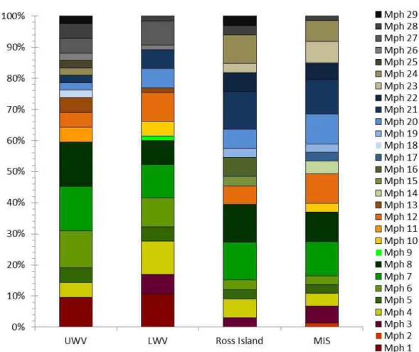
In general, Oscillatoriales cyanobacteria, mainly morphotypes assigned to genus Leptolynbya (morphotypes 7 and 8) were widely spread in Upper and Lower Wright Valleys, Ross Island and MIS and showed the highest frequency of occurrence in the four studied areas (Fig. 4). Conversely, morphospecies assigned to order Nostocales were mostly recorded on Ross Island and MIS while only one of the studied sites from Upper Wright Valley and none from the Lower Wright Valley contained a strain belonging to this order (Fig. 4). Conversely, representatives of the order Chroococcales were most frequent in Upper and Lower Wright Valleys and were recorded at only two ponds from Ross Island and one site of the MIS area (Fig. 4).
32 Molecular researches have recognised that the genotype analyses from different biotopes yield wider diversity than is recognisable from phenotype identification (Taton et al., 2003; 2006a). However, according to Komárek (2007) the traditional definition and naming of species in agreement to the botanical nomenclatoric rules is still necessary and the only acceptable method for characterisation of cyanobacterial taxonomic units (generic and subgeneric; ecologically as well as morphologically).
We considerate that for the future work a complementary molecular research on these studied areas is important for connect the genotype with phenotypes characters and give a reliable designation of names (genus and species) for the morphotypes recorded through this work. Moreover, we do not have isolated the strains and all the observation and description were made from the field samples which sometimes contained numerous sediments and strong aggregation of cyanobacteria filament. These facts may have contributed to an overlook of the sample omitting the presence of the others morphospecies.
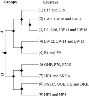
On the basis of the ease with which cyanobacteria tolerate desiccation, and can be expected to be well dispersed within the area by strong winds, there is an expectation that there is an equal opportunity for all taxa to colonize all water bodies and that the sites can be considered to form a metacommunity (Wilson 1992). This relates to the “everything is everywhere” hypothesis, which is challenged when connectivity between habitats within a metacommunity is low (Verleyen et al., 2009). The finding that the four sites contain different taxa rasies a number of questions. The strong similarities found in the cyanobacteria distribution amongst the four studied areas suggest that they are part of a metacommunity, but are the differences due to species sorting along the variations of the environmental, physical-chemical properties of the water body, of the climatic characteristics of each site, or is the dispersal of propagules less effective within the McMurdo Dry Valleys than expected? Is everything equally everywhere? It has been argued that the forces that define the cyanobacterial communites include both deterministic and stochastic processes (Sloan et al., 2006). The factors that are involved in determining the cyanobacterial community has been a concern for microbial ecology and it is a challenge for future work. The next chapter of this thesis will explore this question.
4. Concluding remarks
33 that each site contains distinct floristic elements that are rare or absent from others. The expected high degree of airborne connectivity between the sites, the tolerance of cyanobacteria to desiccation, and the similarities of the community suggests that the four sites may approach a metacommunity. Distinct site-specific differences between sites, which are consistent across ponds within each site, suggest that either growth conditions within sites are selecting different taxa or dispersal of some taxa is constrained by dispersal.
5. References
Andersen, D. T.; Sumner, D. Y., Hawes, I.; Webster-Brown, J. & Mckay, C. P. 2011. Discovery of large conical stromatolites in Lake Untersee, Antarctica. Geobiology 9: 280-293.
Bolch, C. J. S. & Blackburn, S. I. 1996. Isolation and purification of Australian isolates of the toxic cyanobacterium Microcystis aeruginosa. Journal of Applied Phycology 8: 5-13.
Broady, P. A. & Kibblewhite, A. L. 1991. Morphological characterization of Oscillatoriales (Cyanobacteria) from Ross Island and southern Victoria Land, Antarctica. Antarctic Science 3: 35-45.
Broady, P. A. 2005. The distribution of terrestrial and hydro-terrestrial algal associations at three contrasting locations in southern Victoria Land, Antarctica. Algological Studies 11: 95-112.
Comte. K., Sabacka, M., Carré-Mlouka A, Elster J, Komárek J. 2007. Relationships between the Arctic and the Antarctic cyanobacteria; three Phormidium-like strains evaluated by a polyphasic approach. FEMS Microbiology, Ecology 59: 366-376.
Fernández-Carazo, R.; Namsaraev, Z.; Mano, Marie-Jose; Ertz, D. & Wilmotte, A. 2012. Cyanobacterial diversity for an anthropogenic impact assessment in the Sør Rondane Mountains area, Antarctic. Antarctic Science 24: 229-242.
34 Friedmann, E. 1. 1982. Endolithic microorganisms in the Antarctic cold desert. Science 215:
1045-1053.
Garcia-Pichel, F. 2009. Cyanobacteria. – In: Schaechter, M. (ed.): Encyclopedia of Microbiology. Pp. 107-124. – Academic Press, Oxford.
Hawes, I.; Howard-Williams, C. & Vincent, W.F. 1992. Desiccation and recovery of cyanobacterial mats. Polar Biology 12: 587-94.
Hawes, I.; Sumner, D. Y.; Andersen, D. T. & Mackey, T. J. 2011. Legacies of recent environmental change in the benthic communities of Lake Joyce, a perennially ice-covered Antarctic lake. Geobiology 9: 394-410.
Hodgson D.A.; Vyverman W. & Sabbe K. 2001. Limnology and biology of saline lakes in the Rauer Islands, Eastern Antarctica. Antarctic Science 13: 255-270.
Jungblut A.-D.; Hawes I.; Mountfort, D.; Hitzfeld, B.; Dietrich, D.R.; Burns, B.P. & Neilan, B.A. 2005. Diversity within cyanobacterial mat communities in variable salinity meltwater ponds of McMurdo Ice Shelf, Antarctica. Environmental Microbiology 7: 519-529.
Jungblut, A.-D.; Lovejoy, C. & Vincent, W. F. 2009. Global distribution of cyanobacterial ecotypes in the cold biosphere. The International Society for Microbial Ecology Journal 4: 191-202. Friedmann, E.I. 1982. Endolithic microorganisms in the Antarctic cold desert. Science 215: 1045-1053.
Komárek, J. & Anagnostidis, K. 1989. Modern approach to the classification system of cyanophytes. 4 - Nostocales. Archiv für Hydrobiologie Supplement 56: 247-345.
Komárek J. 1999. Diversity of cyanoprokaryotes (cyanobacteria) of King George Island, maritime Antarctica-a survey. Archiv fuer Hydrobiologie 94: 181-193.
35 Komárek, J. & Anagnostidis, K. 2005. Cyanoprokaryota. 2. Teil, Oscillatoriales. In Budel, B., Krienitz, L., Gartner, G. & Schagerl, M. (eds), Süßwasserflora von mitteleuropa, Band 19/2 Spektrum Akademischer Verlag, Elsevier GmbH. München, German.
Komárek, J. 2007. Phenotype diversity of the cyanobacterial genus Leptolyngbya in the maritime Antarctic. Polish Polar Research 28: 211-231.
Komárek, J. & Elster, J. 2008. Ecological background of cyanobacterial assemblages of the northern part of James Ross Island, Antarctica. Polish Polar Research 29: 17-32.
Martineau, E.; Wood, S.A.; Miller, M.R.; Jungblut, A. D.; Hawes, I.; Webster-Brown, J. & Packer, M.A. 2013. Characterisation of Antartic cyanobacteria and comparison with New Zealand strains. Hydrobiologia 711: 139-154.
Mataloni, G.; Vinocur, A. & Pinto, P. D. T. 2005. Abiotic characterization and epilithic communities of a naturally enriched stream at Cierva Point, Antarctic Peninsula. Antarctic Science 17: 163-170.
Nadeau, T. L., Milbrandt, E. C. & Castenholz, R. W. 2001. Evolutionary relationships of cultivated Antarctic Oscillatoriaceans (cyanobacteria). Journal of Phycology 37:650-654.
Pearl, H. W. & J. Huisma. 2008. Blooms like it hot. Science 320: 57-58.
Schopf, J.W. 2000. The fossil record: tracing the roots of the cyanobacterial lineage. – In: Whitton, B.A. & Potts, M. (eds.): The Ecology of Cyanobacteria. Their Diversity in Time and Space. Pp. 13-35. - Kluwer Acad. Publ., Dordrecht.
Sloan, W. T.; Lun, M.; Wooddcock, S.; Head, I.M.; Nee, S. & Curtis, T. P. 2006. Quantifying the roles of immigration and chance in shaping prokaryote community structure. Environmental Microbiology 8: 732-740.
Strunecký, O.; Elster, J. & Komárek, J. 2011. Taxonomic revision of the freshwater cyanobacterium „Phormidium“ murrayi = Wilmottia murrayi. Fottea 11: 57-71.
36 Antarctica): a morphological and molecular approach. Applied and Environmental Microbiology 69: 5157-5169.
Taton, A.; Grubisic, S.; Balthazart, P.; Hodgson, D.A.; Laybourn- Parry, J. & Wilmotte, A. 2006a. Biogeographical distribution and ecological range of benthic cyanobacteria in East Antarctic lakes. FEMS Microbiology, Ecology 57: 272-289.
Taton, A.; Grubisic, S.; Ertz, D.; Hodgson, D. A.; Piccardi, R.; Biondi, N.; Tredici, M.R.; Mainini, M.; Losi, D.; Marinelli, F. & Wilmotte, A.. 2006b. Polyphasic study of Antarctic cyanobacterial strains. Journal of Phycology 42: 1257-1270.
Taton, A.; Wilmotte, A.; Šmarda, J.; Elster, J.; Komárek, J. 2011. Plectolyngbya hodgsonii: a novel filamentous cyanobacterium from Antarctic lakes. Polar Biology 34: 181-191.
Vincent, W. F. 2000. Cyanobacterial dominance in the polar regions, p. 321-340. In: Whitton, B. & Potts, M. (eds.). Ecology of the Cyanobacteria: their diversity in space and time. Kluwer Academic Press, Dordrecht, The Netherlands.
Vincent W.F. 2007. Cold tolerance in cyanobacteria and life in the cryosphere, p 287-301. In: Seckbach J (ed), Algae and cyanobacteria in extreme environments. Springer, Heidelberg, Germany.
Zakhia, F.; Jungblut, A.-D.; Taton, A.; Vincent, W. F. & Wilmotte, A. 2009. Cyanobacteria in cold environments. In: Margesin, R., F. Schinner, J. C. Marx & C. Gerday (eds), Psychrophiles: from biodiversity to biotechnology. Springer, New York: 121-135.
Whitton, B. A. & Potts, M. (eds). 2000. The ecology of cyanobacteria [electronic resource]: their diversity in time and space. Kluwer Academic, Boston.
Wilson, D. S. 1992. Complex interactions in metacommunities, with implications for biodiversity and higher levels of selection. Ecology 73: 1984-2000.
37