Production and Functional Characterization
of Murine Osteoclasts Differentiated from
ER-Hoxb8-Immortalized Myeloid Progenitor
Cells
Frank Zach, Alexandra Mueller, André Gessner*
Institute of Clinical Microbiology and Hygiene, University Hospital Regensburg, Regensburg, Germany
*andre.gessner@ukr.de
Abstract
In vitrodifferentiation into functional osteoclasts is routinely achieved by incubation of embryonic stem cells, induced pluripotent stem cells, or primary as well as cryopreserved spleen and bone marrow-derived cells with soluble receptor activator of nuclear factor kappa-B ligand and macrophage colony-stimulating factor. Additionally, osteoclasts can be derived from co-cultures with osteoblasts or by direct administration of soluble receptor acti-vator of nuclear factor kappa-B ligand to RAW 264.7 macrophage lineage cells. However, despite their benefits for osteoclast-associated research, these different methods have sev-eral drawbacks with respect to differentiation yields, time and animal consumption, storage life of progenitor cells or the limited potential for genetic manipulation of osteoclast precur-sors. In the present study, we therefore established a novel protocol for the differentiation of osteoclasts from murine ER-Hoxb8-immortalized myeloid stem cells. We isolated and immortalized bone marrow cells from wild type and genetically manipulated mouse lines, optimized protocols for osteoclast differentiation and compared these cells to osteoclasts derived from conventional sources.In vitrogenerated ER-Hoxb8 osteoclasts displayed typi-cal osteoclast characteristics such as multi-nucleation, tartrate-resistant acid phosphatase staining of supernatants and cells, F-actin ring formation and bone resorption activity. Fur-thermore, the osteoclast differentiation time course was traced on a gene expression level. Increased expression of osteoclast-specific genes and decreased expression of stem cell marker genes during differentiation of osteoclasts from ER-Hoxb8-immortalized myeloid progenitor cells were detected by gene array and confirmed by semi-quantitative and quan-titative RT-PCR approaches. In summary, we established a novel method for the quantita-tive production of murine bona fide osteoclasts from ER-Hoxb8 stem cells generated from wild type or genetically manipulated mouse lines. These cells represent a standardized and theoretically unlimited source for osteoclast-associated research projects.
a11111
OPEN ACCESS
Citation:Zach F, Mueller A, Gessner A (2015) Production and Functional Characterization of Murine Osteoclasts Differentiated from ER-Hoxb8-Immortalized Myeloid Progenitor Cells. PLoS ONE 10(11): e0142211. doi:10.1371/journal.pone.0142211
Editor:Dominique Heymann, Faculté de médecine de Nantes, FRANCE
Received:August 17, 2015
Accepted:October 19, 2015
Published:November 3, 2015
Copyright:© 2015 Zach et al. This is an open access article distributed under the terms of the
Creative Commons Attribution License, which permits unrestricted use, distribution, and reproduction in any medium, provided the original author and source are credited.
Data Availability Statement:All relevant data are within the paper and its Supporting Information files.
Funding:This study was supported by grants from the Bayerische Forschungsstiftung (www. forschungsstiftung.de/; AZ 1070-13 to AG) and the Deutsche Forschungsgemeinschaft (www.dfg.de/; 1656, Ref. No. GE671/14-1 to AG). The funders had no role in study design, data collection and analysis, decision to publish, or preparation of the manuscript.
Introduction
Homeostasis and controlled remodeling of bone tissues are maintained by the coupled and bal-anced action of bone resorbing osteoclasts (OCs) and bone forming osteoblasts [1–3]. The dis-ruption of OC differentiation or activity processes has been described as a key feature in the development of pathological bone abnormalities seen in Paget’s disease of bone (PDB) [4], osteoporosis [5], inflammatory arthritis [6], periodontitis [7], or cancer metastasis to bone [8,9]. For example, patients suffering from PDB display a disturbed OC activity, which is believed to be caused by environmental as well as genetic factors [4]. Thus, familial PDB is associated with mutations in the ubiquitin associated (UBA) domain of sequestosome 1 which encodes p62, a scaffold protein known to be involved in cytokine signaling, and that can serve as a cargo adaptor for polyubiquitinated proteins [4].
OCs are highly differentiated and polarized cells originating from the monocyte-macro-phage lineage [10]. Mature OCs can be identified by different biological markers such as tar-trate-resistant acid phosphatase (TRAP) staining, multi-nucleation, F-actin ring formation and their unique bone resorbing capacity [10,11]. Besides their primary function in the regulation of bone resorption, OCs are important orchestrators of several other processes, e.g. the regula-tion of hematopoiesis, bone formaregula-tion and angiogenesis of blood vessels during bone develop-ment [12,13].
In contrast to bone forming osteoblasts, which are mesenchymal-derived cells, OCs are of myeloid origin [13].In vitrodifferentiation into OCs has successfully been achieved by 1) direct supplementation of primary or cryopreserved spleen and bone marrow (BM)-derived OC pre-cursors, embryonic stem cells (ESCs) or induced pluripotent stem cells (iPSCs) with macro-phage colony-stimulating factor (M-CSF) and soluble receptor activator of nuclear factor kappa-B ligand (sRANKL), or 2) by the use of osteoclast-osteoblast co-cultures systems [14– 20]. Nevertheless, it is not feasible to cultivate and expand OCs for longer periods of time. In addition, available cell numbers from single differentiation experiments are limited and experi-mental outcome may be variable. Furthermore, the murine myeloid cell line RAW 264.7, which can also be differentiated into OCs in theoretically unlimited amounts by incubation with sRANKL [21], and which is another standard source of OCs, has to be further manipu-lated, e.g. by siRNA-treatment in order to perform gene knockdown experiments.
Due to their myeloid origin, OCs might potentially be generated by differentiation of immortalized myeloid progenitors ectopically expressing the homeodomain containing tran-scription factorHoxb8. Expression ofHoxb8(formerly namedHox-2.4) has long been known to alter growth, differentiation and survival of myeloid cells [22]. Thus, ER-Hoxb8 cells have been developed and characterized as retroviraly transduced murine bone marrow myeloid (BMM) cells, which can be used as conditionally immortalized monocyte-macrophage progen-itors [23]. Under the control ofβ-estradiol, these cells produce Hoxb8 which subsequently inhibits myeloid differentiation and arrests BMMs in an immortalized and self-renewing stem cell (SC) state [23]. In recent years, ER-Hoxb8 SCs have successfully been differentiated into macrophages, neutrophil granulocytes or dendritic cells upon removal ofβ-estradiol and stem cell factor (SCF) and respective supplementation with M-CSF, granulocyte colony-stimulating factor (G-CSF) or granulocyte macrophage colony-stimulating factor (GM-CSF) [23,24]. Since macrophages and OCs have a common myeloid progenitor and macrophages can been pro-duced in theoretically unlimited amounts using the ER-Hoxb8 SC technique, we intended to investigate the OC differentiation potential of ER-Hoxb8 SCs, which has not been addressed and characterized so far.
and furthermore compares these cells to OCs derived from other conventional sources. Our study describes the differentiation and functional characterization of OCs from immortalized myeloid progenitor cells that were isolated from BM of different wild type (WT) and geneti-cally modified mice. Successfully differentiated OCs show excessive TRAP staining and multi-nucleation as well as functional OC characteristics like F-actin ring formation and resorption activity on dentin discs or a calcium phosphate (CaP) substrate. Thus, immortalized ER-Hoxb8 cells represent a novel and valid source for the potentially unlimited production of functional and biologically activein vitroOCs. These cells are a useful tool for enabling and simplifying basic research on OC-associated diseases, as well as for the development of new drugs to manipulate differentiation from OC precursors or functions of mature OCs derived from WT or genetically modified mice.
Materials and Methods
Reagents and Media
Recombinant murine M-CSF, sRANKL, GM-CSF, TNF-α, IL-3, IL-4 and IL-15 were pur-chased from Peprotech Inc. (Rocky Hill, NJ, USA). Other cytokines were obtained from Immu-noTools (Friesoythe, Germany; IL-6) or R&D Systems (Minneapolis, MN, USA; INF-γ). Unless stated otherwise, reagents were bought from Sigma-Aldrich (St. Louis, MO, USA; CaCl2, DMSO,β-estradiol, FBS, fibronectin, FITC-phallodin,β-mercaptoethanol, PBS,
poly-brene, toluidine blue, Triton™X-100), Merck (Darmstadt, Germany; HCl, MgCl2, NH4Cl) or
Roth (Karlsruhe, Germany; AgNO3, KHCO3, NaCl, Na2EDTA, NaHCO3, Na2HPO4, NaOCl).
Cell culture media, except forα-MEM (Sigma-Aldrich), were purchased from Life Technolo-gies (Carlsbad, CA, USA). Cell culture supplements penicillin/streptomycin and L-glutamine were obtained from Pan-Biotech (Aidenbach, Germany).
Animals
Male C57BL/6, and p62/sequestosome 1-deficient C57BL/6 mice [25], BALB/c, and IL-4 recep-tor- (IL-4R) deficient BALB/c mice [26], and C3H/HeJ mice were kept at the animal facility of the University of Regensburg in a specific pathogen-free environment. Unless stated otherwise, WT mice used for experimental approaches, as well as for propagation of KO mice mentioned above, were obtained from Charles River Laboratories (Wilmington, MA, USA). Mice were sacrificed by CO2asphyxiation at the age of 10–20 wk. Procedures including animals were
per-formed according to the animal guidelines of the animal facility of the University of Regens-burg. All animal experiments were approved by the local veterinary authorities and the ethics committee of the District Government of Upper Palatinate (reference 54–2532.1-38/12).
Isolation of BMCs, generation and cryopreservation of immortalized
ER-Hoxb8 SCs
ER-Hoxb8 SCs were generated as described previously [23]. In brief, femurs and tibiae were cut and cells were flushed into petri dishes with RPMI containing 15% FBS,β -mercaptoetha-nol, penicillin (100 U/ml), streptomycin (100μg/ml) and L-glutamine (2 mM). Filtered cells (sieve: 100μm) were centrifuged after a red blood cell lyses step (150 mM NH4Cl, 0.1 mM
Na2EDTA, 10 mM KHCO3, pH 7.2–7.4), resolved in medium and plated in cell culture dishes.
Transfection of Phoenix™retroviral packing cell line [27] with both ecotropic packaging and 3HA-ERHBH-HoxB8-Neo (“ER-Hoxb8”) plasmids (provided by St. Jude Children’s Research Hospital, Memphis, TN, USA, and obtained from G. Haecker, Freiburg, Germany) and infec-tion of BM cells with viral supernatants was performed as follows: Phoenix™cells were seeded in complete DMEM at a density of 2 x 10⁶cells (4.5 x 10⁴cells per cm2) and grown over night in 100 mm dishes. Medium was replaced 30 min prior to CaCl2transfection. Cells were
trans-fected with 10μg of both plasmids and incubated for 2 d (medium replacement after 16 h). BMMs were then suspended in complete Opti-MEM1containing SCF (1:25) andβ-estradiol (2μM) and seeded at a density of 2 x 10⁵cells per cm2on fibronectin-coated (0.001% in PBS) 6-well plates. Viral infection of BMMs with Phoenix™supernatants (with polybrene [4μg/ml]) was achieved by centrifugation for 3 h (1300 x g) at 32°C. Transduced cells were cultured for 5 d and subsequently split every 2–3 d in densities of 2 x 10⁴cells per cm2into new 6-wells. After three wk of continuous passaging and expansion, cells were harvested and dissolved in culture medium with 20% DMSO for long-term storage in liquid N2(-196°C). Cryopreservation was
performed with the computer-controlled freezing system IceCube 15 M from SY-LAB (Neu-purkersdorf, Austria).
In vitro OC and macrophage differentiation of ER-Hoxb8 progenitor cells
ER-Hoxb8 SCs were harvested after 2–3 d in culture (cell density: ~ 1 x 10⁶cells per 6-well, ~ 1 x 10⁵cells per cm2) by centrifugation (8 min, 300 x g). In order to remove residual SCF andβ -estradiol, cells were washed twice with PBS. Cells were resolved in Opti-MEM1
(orα-MEM)
medium containing either M-CSF (30 ng/ml) alone for macrophage differentiation, or M-CSF (30 ng/ml) together with sRANKL (50 ng/ml) for OC differentiation. For optimal osteoclasto-genesis, cells were seeded in 96- and 24-well plates or 100 mm cell culture dishes at densities between 1.6 and 3.2 x 10⁴cells per cm2and incubated for 5–6 d until the completion of cell fusion and differentiation processes.
In vitro OC differentiation of primary BMMs and RAW 264.7
macrophages
BMCs were isolated by flushing femurs and tibia from BALB/c mice with completeα-MEM. Filtered cells (sieve: 100μm) were washed twice with PBS, centrifuged after a red blood cell lysis step (150 mM NH4Cl, 0.1 mM Na2EDTA, 10 mM KHCO3, pH 7.2–7.4), resolved inα
-MEM with M-CSF (30 ng/ml) and cultured in cell culture dishes for 1d. Non-adherent cells were used as progenitors for osteoclastogenesis. Cells were resuspended inα-MEM, supple-mented with M-CSF (30 ng/ml) plus sRANKL (50 ng/ml) and cultured (1.6 x 10⁴cells per cm2) for 4 d until OC differentiation was completed.
To generate OCs from the macrophage cell line RAW 264.7 (ATCC1TIB-71™), cells were cultured in complete DMEM until confluence, then centrifuged, resuspended in completeα -MEM and plated overnight in respective cell culture plates (5.5 x 10³cells per cm2). On the next day, cells were supplemented with sRANKL (50 ng/ml) and differentiated for 3–4 days. Culture medium was replaced 48 h after the first addition of sRANKL.
OC isolation and transfer
trypsin/EDTA (5–10 min). Trypsin was inactivated by the addition of cell culture medium. Cell suspension was centrifuged at 478 x g for 8 min. Cells were suspended with complete medium (M-CSF [30 ng/ml], sRANKL [50 ng/ml]) and re-plated for different OC assays (uncoated or CaP-coated glass coverslips, CaP-coated 24-well plates, dentin discs placed in 24-well plates). Cells on CaP or dentin discs were incubated for a further 48 h prior to charac-terization or quantification of CaP resorption activity. Isolation of mature ER-Hoxb8-derived macrophages and IL-4-treated OC precursor cells was done in parallel with the isolation of mature OCs as described here.
TRAP supernatant activity assay and TRAP staining of cells
Detection of TRAP in OCs was performed using the TRAP staining kit from B-Bridge Interna-tional (Cupertino, CA, USA) according to the manufacturer’s recommendations. For quantifi-cation of TRAP activity in cell culture medium samples, 30μl of each supernatant were mixed with 60μl of substrate solution and incubated at 37°C. After 3–6 h, optical density was mea-sured at 540 nm with a microplate reader (Bio-Rad Laboratories, Hercules, CA, USA). Absorp-tion values of blank medium samples were subtracted.
Cytospin
1centrifugation and DiffQuik
1staining of mature OCs
To examine morphology and cytoplasmic details of OCs derived from different OC progenitor cell lines, mature OCs were isolated as described above and centrifuged (18 x g, 5 min) onto glass microscope slides via a Cytospin1
centrifuge. Cells were then air dried overnight and sub-sequently stained with the DiffQuik1staining kit (Medion Diagnostics International Inc., Miami, FL, USA) according to the manufacturer’s protocol.
Detection of F-actin ring formation
In order to visualize different podosome and F-actin ring structures in differentiating or mature OCs, untreated or CaP-coated coverslips were used. OCs were washed with PBS and fixed with 4% paraformaldehyde (SERVA, Heidelberg, Germany) for 10 min. Cells were washed with PBS, permeabilized with 0.5% Triton™X-100 for 5 min and washed with PBS. Cells were incubated with FITC-phalloidin (1:1,000) for 30 min. After three washing steps with PBS, coverslips were mounted with SlowFade1Gold Antifade Mountant (Life Technologies). Cells were examined with a BZ-9000 fluorescence microscope (Keyence, Osaka, Japan).
Pit formation assay with dentin discs
Biomimetic CaP coatings and quantification of CaP resorption
CaP coatings were prepared as described previously [28,29]. The detection and quantification of resorption activity was done with a biomimetic CaP assay [28] using either mature OCs or OCs directly differentiated on CaP-coated plates or glass coverslips. Mature OCs were incu-bated at 37°C for 48 h, washed twice with PBS and finally removed from plates with NaCl (1 M, in 0.2% Triton™X-100). Culture plates or coverslips were washed twice with water and stained with 5% aqueous AgNO3solution in combination with UV light treatment for 1 h.
After three washing steps with water, surface areas were analyzed by bright-field microscopy (BZ-9000, Keyence). Resorption areas were quantified in overview images which were gener-ated by merging 49 (7x7) individual microscopic images (original magnification: 20x) taken from a randomly designated initial point. The overview images covered approximately 10 mm2 of the respective 24-well plate. These images were inverted and resulting black dots (threshold size of single area: 100μm2) representing CaP resorption areas were quantified with the BZ-II Analyzer software (Keyence).
Gene expression profiling
RNA isolation (RNeasy1Micro Kit, Qiagen, Hilden, Germany), RNA quality control and gene array experiments with the Mouse Gene 2.0 ST Array (Affymetrix, Santa Clara, CA, USA) were done at the Kompetenzzentrum für Fluoreszente Bioanalytik (KFB; Regensburg, Germany). Gene expression data were generated at the KFB with AffymetrixGeneChip Command Console (AGCC)andExpression Consolesoftware packages.
Extraction of RNA, cDNA synthesis, RT-PCR and qRT-PCR
RNA was extracted from ER-Hoxb8-immortalized SCs and ER-Hoxb8-derived OCs with the RNeasy1
Mini Kit from Qiagen according to the manufacturer’s recommendations. Reverse transcription with 0.5μg of isolated RNA was performed using the iScript™Advanced cDNA Synthesis Kit from Bio-Rad Laboratories as stated in the user’s manual.
qRT-PCR analyses were carried out with the LightCycler1480 SYBR Green I master mix (Roche Diagnostics, Mannheim, Germany) and the ABI PRISM1
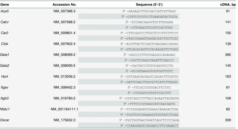
7900HT sequence detection system (Life Technologies). The housekeeping geneHprtwas used as endogenous control. cDNA segments of indicated genes (Table 1) were amplified from OC or SC cDNAs with a standard PCR protocol and cloned using the pGEM1-T easy vector system (Promega, Madison, WI, USA) as stated in the user’s manual. Absolute gene expression was calculated from standard curves of serial dilutions (107–102copies) of cloned plasmid DNAs. Relative fold changes were normalized to values of respective SCs. The calculation of Ct-values and absolute gene expression data was done with the SDS 2.4 software (Life Technologies). Nucleo-tide sequences of primers used for semi-quantitative and quantitative RT-PCR depicted in Table 1were either described previously [30–33] or designed with the Primer3 software. RT-PCR analyses were performed by touchdown PCR with cDNA samples. Plasmid DNAs (20 ng) of respective cloned gene fragments were used as positive control. H2O without template
DNA served as negative control.
Statistical analyses
Results
Time course and efficiency of osteoclastogenesis from ER-Hoxb8 SCs
As the central aim of this study was to establish OC differentiation from ER-Hoxb8-immorta-lized myeloid progenitor cells, we first generated ER-Hoxb8 cells from a BALB/c WT mouse as previously described [23]. For OC differentiation, ER-Hoxb8 SCs (24,000 cells per cm2) were treated with M-CSF (30 ng/ml) and sRANKL (50 ng/ml). Time course of osteoclastogenesis from these cells was monitored by measuring TRAP activity in culture supernatants between d1 and d7 after cell seeding (Fig 1A). TRAP activity was not detectable in supernatants until d4. Enzyme activity appeared at d5 and reached maximum at differentiation d6 (Fig 1A). OC differentiation for longer time periods, as shown for d7, again resulted in significantly reduced TRAP activity. This indicates a narrow time frame for optimal OC differentiation between d5 and d6 after cell seeding, characterizing ER-Hoxb8-derived OCs, similar to OCs derived from primary BMMs, as relatively short-lived cells.
Since a critical negative correlation between seeded cell number and OC differentiation effi-ciency has been reported [34], in a next step, progenitor cells were inoculated for OC differenti-ation in 96-well plates with cell densities ranging from 2,000 up to 320,000 cells per cm2. As can be seen inFig 1B, TRAP activities of cell supernatants at d5 of differentiation showed strik-ing differences dependstrik-ing on initially seeded cell numbers. Highest TRAP activity was observed at intermediary cell densities whereas higher cell densities resulted in a substantial reduction in TRAP activity as an indicator of less efficient OC differentiation (Fig 1B).Fig 1C and 1Dshow representative examples of OC cell morphology and number of nuclei per cell observed at intermediary cell densities (Fig 1C) compared to high density (Fig 1D) approaches. Thus, by Table 1. Primers used for standard, semi-quantitative and quantitative RT-PCR.
Gene Accession No. Sequence (5’-3’) cDNA, bp
Acp5 NM_007388.3 5’-AAGAACTTGCGACCATTGTTAGC 91
5’-CGTTCTCGTCCTGAAGATACTGCA
Calcr NM_007588.2 5’-TCCAACAAGGTGCTTGGGAA 141
5’-CTTGAACTGCGTCCACTGGC
Car2 NM_009801.4 5’-CTTCGATCCTTGCTCCCTTCTTCCT 150
5’-GTACGGAAATGAGACATCTGCTCGC
Ctsk NM_007802.4 5’-ACGTTACTCCAGTCAAGAACCAGGG 139
5’-GTCACACAGTCCACAAGATTCTGGG
Gata1 NM_008089.2 5’-GGCCCCTTGTGAGGCCAGAGAG 385
5’-CGCTCCAGCCAGATTCGACCC
Gata2 NM_008090.5 5’-CACTACCTGTGCAATGCCTG 145
5’-GCCATAAGGTGGTGGTTGTC
Hprt NM_013556.2 5’-GTTGAATACAGGCCAGACTTTGTTG 163
5’-GATTCAACTTGCGCTCATCTTAGGC
Itgav NM_008402.3 5’-TTCGCCGTGGACTTCTTC 81
5’-CTGGGTCGTGTTCGCTTT
Itgb3 NM_016780.2 5’-CGTCAGCCTTTACCAGAATTATAGTG 109
5’-TTTCCCGTAAGCATCAACAATG
Nfatc1 NM_001164111.1 5’-TCTGGGAGATGGAAGCAAAGACTGA 82
5’-TGGTTGCGGAAAGGTGGTATCTCAA
Oscar NM_175632.3 5’-TGCTGGTAACGGATCAGCTCCCCAGA 309
5’-CCAAGGAGCCAGAACCTTCGAAACT
combination of data from TRAP activity assays of supernatants and fixed cells, we were able to depict an optimal seeding density for successful OC differentiation within the range of 16,000– 32,000 ER-Hoxb8 SCs per cm2.
Comparison of OC differentiation from ER-Hoxb8 SCs with conventional
OC progenitor cell sources
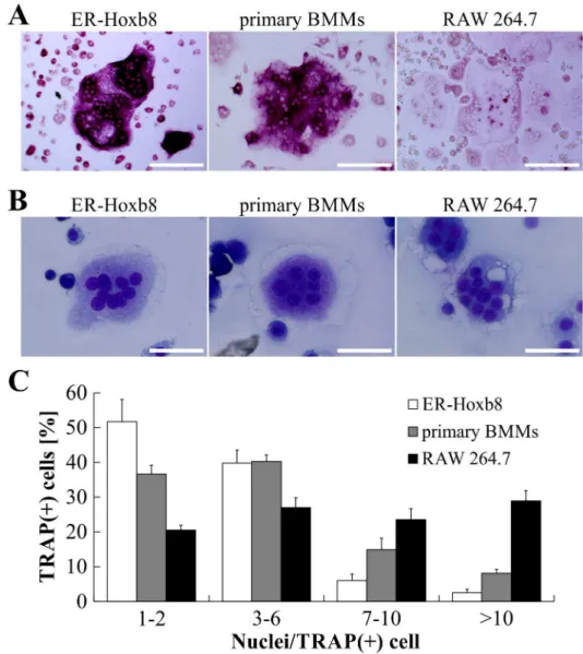
In a next step, we compared osteoclastogenesis from ER-Hoxb8 cells with conventional OC dif-ferentiation methods using primary BMMs and the macrophage cell line RAW 264.7 as OC progenitors. TRAP-stained cells inFig 2Aillustrate that no remarkable morphological differ-ences between RAW 264.7-, ER-Hoxb8- and primary BMM-derived OCs were detectable. However, whereas ER-Hoxb8- and primary BMM-derived OCs showed a comparable extent of TRAP staining, intensities of TRAP signals in RAW 264.7-derived OCs were less pronounced, indicating differences between RAW 264.7- and the two BM-derived OC cell cultures.
DiffQuik1staining after Cytospin1centrifugation of mature OCs did not show any strik-ing morphological discrepancies between ER-Hoxb8 generated OCs and conventional OCs (Fig 2B). In order to calculate differentiation efficiencies of the different methods, the number of nuclei per TRAP-positive cells was determined and grouped into several categories (1–2, 3–6, 7–10,>10 nuclei per cell). ER-Hoxb8 cultures displayed a higher percentage of
TRAP-Fig 1. Time course and efficiency of osteoclastogenesis from BALB/c-derived ER-Hoxb8 SCs.(A) The time course of OC differentiation from ER-Hoxb8 SCs (24,000 cells per cm2) is illustrated by quantification of TRAP activity in supernatants isolated between d1 and d7. No signs of TRAP activity are detectable in supernatants until d4. Enzyme activity starts to develop at d5 of differentiation and reaches its maximum at d6. Data are shown as the mean±SD,n= 3. (B) SCs were inoculated with indicated cell counts. TRAP activity of supernatants was measured at d5 of M-CSF/sRANKL-induced OC differentiation. Data are displayed as the mean±SD,n= 4. (C, D) Representative bright-field microscopy images of TRAP-stained ER-Hoxb8-derived OCs at d5 of cultivation in the presence of M-CSF and sRANKL. Remarkable differences in cell morphology and multi-nucleation can be seen between wells of intermediary (C) and high cell density (D) inoculations. Scale bars = 100μm.
positive committed OC progenitors with 1–2 nuclei, whereas freshly isolated BMMs used as OC progenitor resulted in a slightly higher percentage of TRAP-positive cells with 7–10 or>10
nuclei compared to ER-Hoxb8-derived OCs (Fig 2C). Statistical analyses of multi-nucleation in OCs from ER-Hoxb8 and primary BMM progenitors revealed statistically significant differ-ences in categories 1–2, 7–10 and>10, however biological significance is questionable since
the percentage of physiological OCs with 3–6 nuclei is indiscriminative. Surface area of OCs derived from different progenitor sources, as exemplified inFig 2B, were compared but did not exhibit obvious differences in diameter or size of OCs with comparable multi-nucleation status (data not shown). RAW 264.7-derived OCs showed an almost equal distribution of cells in the Fig 2. Comparison of OC differentiation from ER-Hoxb8 SCs with conventional OC progenitor sources.(A) Representative microscopy images of OCs derived from ER-Hoxb8 SCs (BALB/c WT), primary BMMs (BALB/c WT) or RAW 264.7 macrophages after formalin fixation and TRAP staining. Scale
bars = 150μm. (B) Examples of bright-field microscopy images of mature OCs after Cytospin1centrifugation
and modified Wright-Giemsa staining (DiffQuik1
). Scale bars = 50μm. (C) Graphic illustration of percentage of TRAP-positive cells with indicated number of nuclei per cell. OCs derived from RAW 264.7 cells show higher percentage of TRAP-positive cells with 7 or more nuclei. Data are displayed as the mean±SD,n= 4 (4 different 24-wells).
four different groups and thus a significantly higher percentage of OCs with 7 or more nuclei than both ER-Hoxb8- and BMM-derived OCs.
Comparison of OC differentiation using ER-Hoxb8 SCs from different
mouse strains
We next tested whether the osteclastogenesis from ER-Hoxb8 cells is dependent on the genetic background of the SC donor mouse. ER-Hoxb8 cells, generated from the three most widely used WT mouse strains C57BL/6, BALB/c and C3H/HeJ, were each plated at a density of maximal 16,000 cells per cm2(5,000 cells per 96-well; 25,000 cells per 24-well) for OC differentiation. TRAP activity of supernatants was tested at d5 of differentiation. As illustrated inFig 3A, effi-ciency of OC differentiation was remarkably different between SCs from analyzed inbred mice. While osteoclastogenesis with C3H/HeJ-derived ER-Hoxb8 cells was more efficient compared to BALB/c cells, supernatants of C57BL/6-derived cells showed comparatively little TRAP activ-ity. These findings were confirmed by microscopic analyses of TRAP-stained cells (Fig 3B). OCs, indicated by TRAP staining and multi-nucleation, were observed in cells of all three mouse strains, however OC yields and intensities of TRAP staining showed differences. Due to these genotype-specific OC differentiation efficiencies, we performed independent ER-Hoxb8 transduction attempts using BM cells from two additional C57BL/6 animals. However, OC dif-ferentiation did not show any obvious differences between these three independent C57BL/ 6-derived cell lines (data not shown) arguing for robust and reproducible mouse strain-specific differences. Thus, in order to use C57BL/6 mouse strains for generation of ER-Hoxb8 stem cells, and to achieve optimal OC differentiation efficiencies for the respective cell line, different parameters such as concentrations of sRANKL and M-CSF, differentiation kinetics and cell den-sities of OC progenitors will have to be further improved in future differentiation experiments.
Modulation of OC differentiation and activity using cytokines
It is well known that OCs, which share similarities with macrophage-like foreign-body giant cells [35], and could be considered to be specialized giant macrophages [12], are sensitive to factors released from immune cells and directly interact with cells of the immune system [12]. To investigate the responsiveness of ER-Hoxb8-derived OCs to different OC-inhibitory cyto-kines (GM-CSF, IL-4, INF-γ) as wells as OC-stimulatory cytocyto-kines (IL-6, IL-15, TNF-α), both were tested during OC differentiation.Fig 4illustrates TRAP activity of supernatants (Fig 4A) and cells (Fig 4B) derived from either primary BMMs (BALB/c WT) or BALB/c WT and IL-4R KO ER-Hoxb8 SCs at d4 (primary cells) or d5 (ER-Hoxb8 cells) of OC or macrophage
enhanced OC differentiation of freshly isolated BMMs (Fig 4A and 4B), whereas unexpectedly this effect was not visible in TRAP-stained ER-Hoxb8-derived OCs. We further detected a markedly different level of TRAP activity in supernatants of TNF-α-treated IL-4R KO and BALB/c WT ER-Hoxb8-derived OC differentiation conditions. These unexpected differential effects of TNF-αtreatment on ER-Hoxb8 cells are currently under in-depth investigation. Fig 3. Comparison of osteoclastogenesis using ER-Hoxb8 cells from different WT mouse strains.(A) ER-Hoxb8 OC progenitors (16,000 cells per cm2) obtained from BM cells of C57BL/6J, BALB/c and C3H/HeJ WT mice were subjected to osteoclastogenesis with M-CSF and sRANKL. TRAP activity of supernatants was measured at d5 of differentiation. Data are displayed as the mean±SD,n= 3 (three differentiation
experiments from identical batches of ER-Hoxb8 SCs). Significant differences in average TRAP activity compared to C57/BL6J-derived OC values are indicated by asterisks (*p<0.05,**p<0.01, and
***p<0.001, Student’st-test). (B) Representative microscopy images of formalin-fixed and TRAP-stained ER-Hoxb8-derived OCs of indicated WT origin. Representative multi-nucleated and TRAP-positive OCs are shown at lower (left column; scale bars = 200μm) and higher magnification (right column; scale
bars = 100μm).
Functional characterization of ER-Hoxb8-derived OCs
In order to clarify whether ER-Hoxb8-derived OCs may be designated as a new source of bona fide OCs, we examined the functionality of these cells in comparison to OCs derived from con-ventional OC progenitor sources. Mature and functional OCs are known to develop an F-actin Fig 4. Effect of inhibitory and stimulatory cytokines on OC differentiation of primary BMs or ER-Hoxb8 SCs.(A) BALB/c BMMs or ER-Hoxb8 OC precursor cells of BALB/c and IL-4R KO origin (23,500 cells per cm2) were cultured with 1) M-CSF alone (
“MФ”), 2) M-CSF and sRANKL (“OC”), or 3) the additional supplementation of indicated cytokines (GM-CSF: 10 ng/ml, remaining cytokines: 20 ng/ml). TRAP activity of supernatants was measured and compared to control differentiation (“OC”). Data are presented as the mean±SD,n= 4. Significant differences of average TRAP activity compared to respective control values (“OC”) are indicated by asterisks (*p<0.05,**p<0.01, and***p<0.001, Student’st-test). (B) Representative microscopy images illustrating morphology and TRAP staining of formalin-fixed cells after treatment with inhibitory and stimulatory modulators during OC differentiation. M-CSF-treated BMMs and Hoxb8 SCs as well as IL-4-M-CSF-treated BALB/c ER-Hoxb8 and BMMs do not show signs of TRAP staining. IL-4R KO cells are not sensitive to IL-4 and thus show normal OC differentiation potential. Scale bars = 150μm.
ring, which is important for the formation of a sealing zone that is associated with OC bone resorption activity [11]. Prior to the arrangement of a mature F-actin ring, actin structures of OCs pass through different intermediate developmental stages called podosome cluster, podo-some ring and podopodo-some belt [11,37]. To investigate these structures in ER-Hoxb8-derived OCs, cells were subjected to FITC-phalloidin staining at d5 of OC differentiation.Fig 5 illus-trates podosome cluster, podosome ring and podosome belt structures in multi-nucleated cells (C3H/HeJ WT) differentiated on glass coverslips. These structures, which were also visible in BALB/c WT and p62 KO ER-Hoxb8-derived OCs (data now shown), were detectable in com-parable quantities and qualities when cells were first differentiated and subsequently plated on biomimetic CaP-coated coverslips (data not shown). It is known that F-actin ring structures, which indicate actively resorbing OCs, are optimally visible on dentin or biomimetic bone sub-strates, for example on CaP-coated cell culture plates or glass coverslips, and thus fully devel-oped F-actin rings are not likely to be detectable on non-coated plates or coverslips. In order to overcome these limitations, mature OCs, initially differentiated from ER-Hoxb8 progenitors in standard cell culture dishes, were isolated and subsequently plated on CaP-coated glass cover-slips. Following incubation for 48 h, cells were stained for F-actin with FITC-phalloidin.Fig 5 (lowest panel) shows a representative multi-nucleated OC containing a heavily stained F-actin ring structure on the cell periphery. Cell movement and signs of OC-associated bone resorp-tion activity at the interface of the F-actin ring are depicted inS1 Fig. The overview of merged bright-field, FICT-phalloidin and DAPI images from ER-Hoxb8-derived OCs (C3H/HeJ WT, p62 KO) points to a directional CaP resorption activity, which is clearly linked to and depen-dent on its F-actin ring formation on the cell periphery.
For further functional characterization of ER-Hoxb8-derived OCs, we analyzed bone resorption, another hallmark of mature OCs. As a starting point, ER-Hoxb8 OC progenitors were directly differentiated in biomimetic CaP-coated cell culture plates for 5 d as previously shown. Subsequently, adherent cells on CaP substrate were stained to determine their TRAP activity (Fig 6A), or alternatively removed in order to visualize remaining CaP deposits via AgNO3treatment. Although OCs showed substantial extra- and intracellular TRAP deposits
(Fig 6A), almost no white spots, as an indicator of active resorption, were visible after AgNO3
treatment of CaP-coated cell culture plates (data not shown). Thus, we next tried to improve resorption activity of ER-Hoxb8-derived OCs by performing a first differentiation step of ER-Hoxb8 OC precursors in cell culture dishes, followed by the subsequent re-plating of mature OCs in CaP-coated cell culture plates.Fig 6Bshows representative TRAP-positive multi-nucleated OCs surrounded by areas clear of CaP coating, as an indicator of resorbing cells. In parallel, we stained for CaP deposits after previous removal of OCs from culture plates and detected multiple white holes of varying size on the CaP coatings (Fig 6C) indicative of resorption activity of multi-nucleated OCs. Interestingly, substantial resorption activity on CaP-coated plates was also detected with OCs from ER-Hoxb8 SCs generated from p62-defi-cient mice (S2 Fig), facilitating, for example, the investigation of the influence of p62-deficiency on the development and progress of the OC-associated PDB phenotypes.
ER-Hoxb8 BALB/c-derived OCs showed a slightly lower resorption activity (5.5 x 105μm2). OCs differentiated from RAW 264.7 macrophages displayed the highest CaP resorption values of all cell lines that were analyzed in our experiments (33.5 x 105μm2). In summary, CaP resorption of ER-Hoxb8-derived OCs is comparable to conventional OC sources and previ-ously published results [28].
Fig 5. C3H/HeJ ER-Hoxb8-derived OCs show different stages of F-actin ring formation.Visualization of F-actin ring and podosome structures in permeabilized (Triton™X-100) OCs was enabled by interaction of FITC-conjugated phalloidin (green) with F-actin. Nuclei were counterstained with DAPI (blue). Representative fluorescence microscopy images of F-actin structures observed in ER-Hoxb8-derived OCs show examples of podosome cluster, podosome ring and podosome belt in cells differentiated on uncoated glass coverslips. A representative example of fluorescence microscopy images of mature F-actin ring structures in OCs which were differentiated on 100 mm dishes (300,000 cells) and subsequently plated on CaP-coated glass coverslips is shown in the lowest panel. Scale bars = 50μm.
Fig 6. Resorption activity of C3H/HeJ ER-Hoxb8-derived OCs on CaP-coated cell culture plates.(A) Representative bright-field microscopy images of TRAP-stained OCs (seeding density of SCs: 20,000 cells per cm2) after 5 d of direct differentiation on CaP-coated plates show intracellular as well as longitudinal tracks of extracellular TRAP enzyme activity. Scale bars = 100μm. (B) Representative images of TRAP-stained OCs on AgNO3-colored CaP-coated cell culture plates surrounded by areas without CaP indicating actively resorbing OCs. OCs were differentiated on unCaP-coated dishes for 5 d, plated on CaP-coated 24-wells and incubated for a further 48 h. Scale bars = 100μm. (C) Examples of microscopy images at 4x (left column; scale bars = 200μm) and 20x magnification (right column; scale bars = 100μm) showing resorption activity of ER-Hoxb8-derived OCs on CaP-coated cell culture plates after AgNO3 and UV treatment. Resorption is visualized as white spots lacking CaP and remaining unstained despite application of AgNO3.
doi:10.1371/journal.pone.0142211.g006
Fig 7. Resorption activity of ER-Hoxb8-derived OCs compared to conventional OCs.(A) Representative microscopy images of pit formation assays performed with dentin discs and mature OCs from indicated sources. Resorption pits were visualized after removal of cells and toluidine blue staining. RAW 264.7 macrophages (“MΦ”) were used as negative control. Scale bars = 400μm. (B) Microscopy images of mature OCs re-plated in CaP-coated cell culture plates after formalin-fixation and TRAP plus AgNO3staining. Scale bars = 100μm. (C) Representative examples of merged and inverted overviews of 24-well cell culture plates obtained from 7x7 individual microscopic images at 20x magnification. Resorption areas are visible as black spots. Scale bars = 1 mm.
In order to rule out nonspecific matrix loss or CaP resorption, e.g. as a consequence of acidi-fication of the culture medium, several differentiation experiments with ER-Hoxb8 myeloid progenitor cells were performed as resorption controls. After removal of SCF and estradiol, ER-Hoxb8 cells were treated 1) with M-CSF (macrophage differentiation), 2) with M-CSF and sRANKL in combination (OC differentiation) or 3) with M-CSF, sRANKL and IL-4 (20 ng/ml, inhibition of normal OC differentiation). Differentiated and adherent cells were isolated at d6 and seeded on CaP-coated cell culture plates for a further 48 h. Medium alone (data not shown) and ER-Hoxb8 SCs with no known bone resorption activity were used as negative con-trols. Multi-nucleated and TRAP-positive cells with active resorption were present only in wells with OC differentiation conditions (S3 Fig). Furthermore, the development of resorption pits characterized by the absence of brown CaP deposits after AgNO3staining was completely
inhibited by IL-4 (S3 Fig).
Collectively, our results show that multi-nucleated and TRAP-positive ER-Hoxb8-derived cells exhibit typical characteristics of functional bona fide OCs, e.g. F-actin ring formation and bone resorption activity on dentin discs and CaP-coated cell culture plates.
Analysis of gene expression profiles during OC differentiation
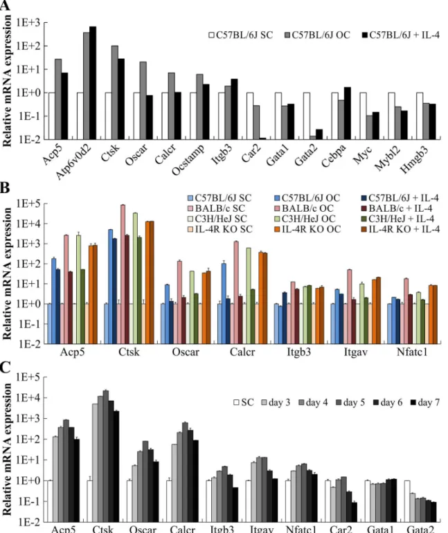
We next aimed to analyze OC differentiation of ER-Hoxb8 myeloid progenitor cells at the level of gene expression. Since p62 mice, which will be used in our future experiments to generate and further characterize PDB-related ER-Hoxb8-derived OCs, are on a C57BL/6 background [25], we decided to initiate gene expression experiments with C57BL/6-derived cells. Thus, expression profiles from p62 (data not shown) and C57BL/6 ER-Hoxb8 SCs as well as from the respective ER-Hoxb8-derived OCs were compared by applying Mouse Gene 2.0 ST Array from Affimetrix. Relative mRNA expression levels of a selection of OC (Acp5[TRAP],Atp6v0d2, Ctsk,Oscar,Calcr,Ocstamp,Itgb3,Car2) and SC marker genes (Gata1,Gata2,Cebpa,Myc, Mybl2,Hmgb3) in cells of C57BL/6 WT origin are summarized inFig 8A, respectively. In OCs, we detected a strong upregulation of almost all analyzed OC markers up to 366-fold
(Atp6v0d2) compared to untreated ER-Hoxb8 myeloid progenitors (Fig 8A, white and gray bars). Absolute gene expression level (data not shown) ofCar2, encoding the OC-associated carbonic anhydrase 2, was already high in ER-Hoxb8 SCs and not further elevated in OCs. However, administration of OC-inhibitory cytokine IL-4 completely abolishedCar2expression pointing to its importance for OC functionality (Fig 8A). IL-4 also reduced (Acp5,Ctsk, Ocstamp) or abolished (Oscar,Calcr) expression of most other analyzed OC-specific genes, which is in line with the strong inhibitory effect of IL-4 on OC differentiation (Fig 8A, black bars). In contrast, IL-4 further enhancedAtp6v0d2 and Itgb3expression compared to
ER-Hoxb8 SCs or ER-Hoxb8-derived OCs (Fig 8A). Besides the observed upregulation of OC-specific genes, OCs showed substantial downregulation of SC marker genes, especiallyGata2 andMyc(Fig 8A), which further indicated the genetic reprogramming during OC differentia-tion. Of interest, however, was that these effects on SC marker genes, except forCebpa, were not reversed by IL4 treatment (Fig 8A).
In order to validate the gene array data presented inFig 8A, we subsequently used SYBR1
Fig 8. Gene expression profiles of ER-Hoxb8-derived OCs and ER-Hoxb8 SCs.(A) Relative mRNA expression of selected OC or SC marker genes probed with cDNA originating from C57BL/6J ER-Hoxb8 SCs compared to cDNAs of ER-Hoxb8-derived OCs and IL-4-treated OC differentiations. Expression was determined by Mouse Gene 2.0 ST Array from Affimetrix. (B) qRT-PCR analyses of indicated OC and SC marker genes illustrate OC differentiation. RNAs were isolated from ER-Hoxb8 cells of indicated WT or IL-4R KO origin after 5 d under OC differentiation conditions (20,000 cells per cm2). Gene expression of respective SC cultures served as reference control. Data are mean±SD of technical duplicates. Each sample was pooled together from three 24-wells. Housekeeping geneHprtwas used for normalization of samples. (C) qRT-PCR analyses of indicated OC and SC marker genes were performed with samples isolated from BALB/c cells after 3–7 d under OC differentiation conditions. Gene expression of respective SC culture served as reference control. Data are mean±SD of technical duplicates. Samples were pooled together from three 24-wells. Expression ofHprtwas used for normalization. Note log scale on y axis (A-C).
expression of OC marker genes to an extent between OC and SC values, IL-4 had, as expected, no inhibitory effect on OC differentiation in IL-4R KO-derived OCs (Fig 8B). Expression of SC markerGata2strongly decreased during OC differentiation and the addition of IL-4 caused only marginal effects onGata2expression (S4 Fig). Additional experiments were performed to analyze the time course of gene expression during OC differentiation from d3 to d7 using BALB/c WT ER-Hoxb8 cells (Fig 8C). Expression of OC marker genesAcp5,Ctsk,Oscar, Calcr,Itgb3,ItgavandNfatc1strongly increased during OC differentiation and reached a maxi-mum level at d5 (Fig 8C). In contrast,Car2andGata2gene expression levels continuously decreased during OC differentiation andGata1-mRNA levels varied only marginally (Fig 8C).
Semi-quantitative RT-PCR analyses using a selection of samples from the qRT-PCR men-tioned above, further confirmed the qRT-PCR results (Fig 9). We observed a strong increase in OC marker gene expression, with maximum signals at d5, and a decrease in ER-Hoxb8 SC-spe-cific gene expression during osteoclastogenesis. Furthermore, the inhibitory effect of IL-4 on BALB/c WT-, but not on IL-4R KO-derived OC progenitors, can be estimated by comparing the intensities of the respective DNA bands after completed PCR reactions. In summary, by using quantitative as well as semi-quantitative RT-PCR analyses, we were able to prove the OC differentiation potential of ER-Hoxb8 SCs on the level of gene expression.
Discussion
Previous OC-associated research focused preferentially on osteoclast-osteoblast co-culture sys-tems or thein vitrodifferentiation of freshly isolated spleen and BM-derived OC precursors [14,15,16,18,19,20]. Alternatively, murine RAW 264.7 cells, which can be differentiated into OCs by simple incubation with sRANKL, have been used in basic approaches [21]. Further progress has also been made recently as protocols became available for the cryopreservation of OC precursors [15], for the isolation and cultivation of OCs from mouse BM [38], and for the OC differentiation from ESC and iPSC sources [17]. However, despite their benefits for basic OC research, these methods have numerous drawbacks. Directex vivoisolation of OCs is lim-ited by very low yields and cells cannot be cultivated for longer time periods. Repeated experi-mental attempts with freshly isolated progenitor cells and their resulting differentiated OCs are both highly time and animal consuming, with subsequent findings suffering from a relatively high variability. Cryopreservation of untreated OC precursors from mouse BM and spleen cells results in a limited half-life [15]. OC differentiation from RAW 264.7 OC progenitors is theo-retically possible in unlimited amounts, but it is also artificial and cells have to be genetically manipulated for targeted gene knockdown [39]. Different RAW 264.7 cell clones may further-more have extremely variable OC differentiation potential [40,41]. In addition, experiments with RAW 264.7-derived OCs may be influenced by the inherent genetic background (BAB/ 14), or the transforming molecular mechanisms operative in this Abelson leukemia virus-transformed mouse monocyte-macrophage cell line.
Redeckein which the authors were able to differentiate functional macrophages, neutrophil granulocytes and dendritic cells from ER-Hoxb8-immortalized SCs [23,24]. Thus, ER-Hoxb8 progenitor cells display a broad myeloid differentiation potential for different cell types of the respective lineages, including OCs, that are difficult to isolate or cultivate directly in high num-bersex vivoand without pre-activating them by using a conventional isolation procedure.
In the present study, differentiation efficiencies and OC yields using ER-Hoxb8 SCs were slightly lower but still comparable to both primary BMM preparations and RAW 264.7 cells. However, osteoclastogenesis was found to depend on the genetic background of the SC donor mice. Since LPS is known to inhibit osteoclastogenesis [42], our finding that C3H/HeJ-derived cells, with natural mutation in the TLR-4 receptor [43], showed the highest OC differentiation capacity, which may be due to a lack of TLR-4-mediated signals. Our future experiments will test this hypothesis.
When functionality of thein vitrogenerated OCs was tested, bone resorption capacity and cytokine responsiveness, among other functions, were found to display typical OC features. However, some cytokine effects were only moderate and not as pronounced as expected or as previously described. This moderate impact on OCs could be due to indirect or artificial effects of cytokines, which may not be fully visible in ourin vitrosystem composed of only OCs and no other contaminating cell types. For example, IL-6 is known to inhibit OCs by direct Fig 9. RT-PCR analyses of gene expression in ER-Hoxb8-derived OCs and ER-Hoxb8 SCs.Agarose gels of RT-PCR analyses showing expression of selected OC and SC marker genes in BALB/c- or IL-4R KO-derived SCs, OCs or IL-4-treated OC differentiation. Time course of gene expression between d3 and d7 of OC differentiation is illustrated in BALB/c-derived cells. Gene expression of OC marker genes is maximal at d5 of differentiation and decreases again at d6 and d7. UniformHprtexpression is shown as housekeeping control. Plasmid DNAs served as positive controls (+); water was used as negative control (-).
treatment, but stimulate them in cooperation with osteoblasts [44]. Moreover, some cytokines may only be effective at supra-physiological concentrations. Beyond this, we detected a signifi-cantly different TRAP activity level in supernatants of TNF-α-treated IL-4R KO and BALB/c WT ER-Hoxb8-derived OCs compared to conventional OCs differentiated from primary BMMs. These highly reproducible and very interesting observations concerning the effects of TNF-αon OC precursor cells under these specific OC differentiation conditions were defined as a starting point for further ongoing experiments with IL-4R KO and BALB/c WT
ER-Hoxb8-derived OCs.
The present study describes a reproducible and simple method for the quantitative genera-tion of murinein vitroOCs, which, since they share important characteristics, may substitute for the use of repeated batches of fresh BM preparations, thus reducing the time, cost and num-ber of animals required. On the basis of our results,in vitrogenerated ER-Hoxb8-derived OCs are available in theoretically unlimited amounts. Due to this simple availability, these cells can be used for different applications, e.g. large drug screenings to detect substances which influ-ence the differentiation and resorption activity of OCs. Since genetic reprograming of
ER-Hoxb8 SCs is already completed at d5 of OC differentiation (Figs8and9), we assume that even approaches targeting the effects of estrogens on mature ER-Hoxb8-derived OCs may be feasible. However, since we currently cannot completely rule out any non-physiological effects of estrogens on mature ER-Hoxb8 OCs, this point will have to be addressed in future investiga-tions for final clarification.
As a further advantage of ER-Hoxb8-derived OCs, signaling events induced by various pharmaceuticals or mediators, e.g. cytokines, may be analyzed more easily by phosphoprotein western blot or proteome analyses requiring relatively high amounts of cellular protein. In addition, the investigation of the effects on OC biology caused by mutations or gene deficien-cies may be improved by the use of ER-Hoxb8-derived OCs. For example, our results clearly show the feasibility of this new approach for examining the OC differentiation and CaP resorp-tion capacity of p62-deficient ER-Hoxb8-derived OCs, whereas previously only an incomplete siRNA-mediated knockdown of p62 in RAW 264.7 cells yielded the opposite results [39]. Thus,in vivoeffects, e.g. mutations or deletions in p62, which are associated with PDB [4], and which are known to ultimately enhance OC resorption activity, may be detected more readily with the ER-Hoxb8-dependent OC differentiation model presented here.
Nonetheless, some limitations of the ER-Hoxb8 technique should also be mentioned. Since statistically significant effects between different mouse lines can be detected most accurately by the use of larger cohorts inin vivoexperiments, ER-Hoxb8-derived OCs should not replace but initiate and supportin vivoexperiments. In this context, the use of ER-Hoxb8-derived OCs may be a good starting point for initial experiments with variable test parameters and condi-tions. Promisingin vitroresults may then be applied to largerin vivoexperiments in order to replicate and expand upon previously established data from ER-Hoxb8-derived OCs.
In summary, we established a novel and feasible method for differentiating OCs from ER-Hoxb8-immortalized myeloid progenitor cells. These OCs were further characterized on a functional and genetic level. We compared ER-Hoxb8-derived OCs with conventionally pro-ducedin vitroOCs and showed that they share typical standard OC characteristics. To the best of our knowledge, this is the first study using ER-Hoxb8 cells for OC differentiation and we propose that suchin vitrogenerated ER-Hoxb8-derived murine OCs represent a new signifi-cant and quantitative source of bona fide OCs and a valid tool for future OC-associated research.
Conclusions
The present work establishes a novelin vitromethod for the quantitative generation of func-tional murine OCs from ER-Hoxb8-immortalized myeloid progenitor cells. These new bona fide OCs offer an inexpensive and valid alternative to conventional OCs differentiated from primary BMC preparations, which are both time and animal consuming, or from RAW 264.7 cell lines, which are artificial and may have variable OC differentiation efficiencies. Thus, ER-Hoxb8-derived OCs have the potential to improve and facilitate future OC-associated research projects.
Supporting Information
S1 Fig. Resorption activity of ER-Hoxb8-derived OCs from different mouse lines.Images of merged bright-field, FICT-phalloidin and DAPI channels show the resorption activity of ER-Hoxb8-derived OCs from C3H/HeJ (upper panel) or p62 KO mice (lower panel) on CaP-coated cell culture plates. Areas lacking CaP as a consequence of OC resorption are schemati-cally visualized by black borders in microscopy images taken in bright-field channel mode (first column). Direction of resorption and motility of OCs can be estimated from FITC-phal-loidin-stained F-actin ring structures (green) concentrated on the cell periphery. Nuclei were counterstained with DAPI (blue). Scale bars = 50μm.
(TIF)
S2 Fig. Resorption activity of ER-Hoxb8-derived OCs of p62 KO origin.(A) Representative images of large (left) and small (right) multi-nucleated TRAP-stained p62 KO OCs on AgNO3
-colored CaP-coated cell culture plates. Scale bars = 100μm. (B) Examples of microscopic images at lower (left column; scale bars = 200μm) and higher magnification (right column; scale bars = 100μm) showing resorption of p62 KO ER-Hoxb8-derived OCs on CaP-coated cell culture plates after removal of cells, addition of AgNO3, and treatment with UV light. (C)
Representative examples of merged and inverted overviews of 24-well cell culture plates obtained from 7x7 individual microscopic images at 20x magnification. Resorption areas are visible as black spots. Scale bars = 1 mm.
(TIF)
S3 Fig. Resorption activity of ER-Hoxb8 SCs, ER-Hoxb8 macrophages, ER-Hoxb8 OCs and IL-4-treated OC differentiations.Mature cells that had subsequently been cultured on CaP substrate for 48 h are visualized by TRAP staining in combination with AgNO3and UV
treat-ment (upper panel). After removal of cells and AgNO3staining, resorption areas show up as
white spots (lower panel). CaP resorption is exclusively present in ER-Hoxb8-derived OCs. Scale bars = 100μm.
(TIF)
ER-Hoxb8 cells of indicated WT or IL-4R KO origin after 5 d under OC differentiation condi-tions (20,000 cells per cm2). Gene expression of respective SC cultures served as reference con-trol. Data are mean ± SD of technical duplicates. Each sample was pooled together from three 24-wells. Housekeeping geneHprtwas used for normalization of samples.
(TIF)
Acknowledgments
We thank St. Jude Children’s Research Hospital, Memphis, TN, USA and Hans and Georg Haecker for providing ecotropic packaging and 3HA-ERHBH-HoxB8-Neo plasmids as well as Nicole Bezold and Lisa Zeller for their excellent experimental support.
Author Contributions
Conceived and designed the experiments: FZ AG. Performed the experiments: FZ AM. Ana-lyzed the data: FZ AM AG. Contributed reagents/materials/analysis tools: AG. Wrote the paper: FZ AG.
References
1. Sims NA, Martin TJ. Coupling the activities of bone formation and resorption: a multitude of signals within the basic multicellular unit. Bonekey Rep. 2014; 3:481. doi:10.1038/bonekey.2013.215PMID: 24466412
2. Teti A. Mechanisms of osteoclast-dependent bone formation. Bonekey Rep. 2013; 2:449. doi:10.1038/ bonekey.2013.183PMID:24422142
3. Walsh MC, Kim N, Kadono Y, Rho J, Lee SY, Lorenzo J, et al. Osteoimmunology: interplay between the immune system and bone metabolism. Annu Rev Immunol. 2006; 24:33–63. PMID:16551243
4. Galson DL, Roodman GD. Pathobiology of Paget's Disease of Bone. J Bone Metab. 2014; 21(2):85– 98. doi:10.11005/jbm.2014.21.2.85PMID:25025000
5. Rodan GA, Martin TJ. Therapeutic approaches to bone diseases. Science. 2000; 289(5484):1508–14. PMID:10968781
6. Jones D, Glimcher LH, Aliprantis AO. Osteoimmunology at the nexus of arthritis, osteoporosis, cancer, and infection. J Clin Invest. 2011; 121(7):2534–42. doi:10.1172/JCI46262PMID:21737885
7. Park SJ, Bae HS, Cho YS, Lim SR, Kang SA, Park JC. Apoptosis of the reduced enamel epithelium and its implications for bone resorption during tooth eruption. J Mol Histol. 2013; 44(1):65–73. doi:10. 1007/s10735-012-9465-4PMID:23124894
8. Roodman GD. Biology of osteoclast activation in cancer. J Clin Oncol. 2001; 19(15):3562–71. PMID: 11481364
9. Yoneda T. Cellular and molecular mechanisms of breast and prostate cancer metastasis to bone. Eur J Cancer. 1998; 34(2):240–5. PMID:9741327
10. Boyle WJ, Simonet WS, Lacey DL. Osteoclast differentiation and activation. Nature. 2003; 423 (6937):337–42. PMID:12748652
11. Saltel F, Chabadel A, Bonnelye E, Jurdic P. Actin cytoskeletal organisation in osteoclasts: a model to decipher transmigration and matrix degradation. Eur J Cell Biol. 2008; 87(8–9):459–68. doi:10.1016/j. ejcb.2008.01.001PMID:18294724
12. Cappariello A, Maurizi A, Veeriah V, Teti A. Reprint of: The Great Beauty of the osteoclast. Arch Bio-chem Biophys. 2014; 561:13–21. doi:10.1016/j.abb.2014.08.009PMID:25282390
13. Charles JF, Aliprantis AO. Osteoclasts: more than 'bone eaters'. Trends Mol Med. 2014; 20(8):449–59. doi:10.1016/j.molmed.2014.06.001PMID:25008556
14. Chen H, Gilbert LC, Lu X, Liu Z, You S, Weitzmann MN, et al. A new regulator of osteoclastogenesis: estrogen response element-binding protein in bone. J Bone Miner Res. 2011(10: ):2537–47. doi:10. 1002/jbmr.456PMID:21773989
16. Li X, Okada Y, Pilbeam CC, Lorenzo JA, Kennedy CR, Breyer RM, et al. Knockout of the murine prosta-glandin EP2 receptor impairs osteoclastogenesis in vitro. Endocrinology. 2000; 141(6):2054–61. PMID: 10830290
17. Nishikawa K, Iwamoto Y, Ishii M. Development of an in vitro culture method for stepwise differentiation of mouse embryonic stem cells and induced pluripotent stem cells into mature osteoclasts. J Bone Miner Metab. 2014; 32(3):331–6. doi:10.1007/s00774-013-0547-5PMID:24366621
18. Takahashi N, Yamana H, Yoshiki S, Roodman GD, Mundy GR, Jones SJ, et al. Osteoclast-like cell for-mation and its regulation by osteotropic hormones in mouse bone marrow cultures. Endocrinology. 1988; 122(4):1373–82. PMID:3345718
19. Takahashi N, Akatsu T, Udagawa N, Sasaki T, Yamaguchi A, Moseley JM, et al. Osteoblastic cells are involved in osteoclast formation. Endocrinology. 1988; 123(5):2600–2. PMID:2844518
20. Yasuda H, Shima N, Nakagawa N, Yamaguchi K, Kinosaki M, Mochizuki S, et al. Osteoclast differentia-tion factor is a ligand for osteoprotegerin/osteoclastogenesis-inhibitory factor and is identical to TRANCE/RANKL. Proc Natl Acad Sci U S A. 1998; 95(7):3597–602. PMID:9520411
21. Hsu H, Lacey DL, Dunstan CR, Solovyev I, Colombero A, Timms E, et al. Tumor necrosis factor recep-tor family member RANK mediates osteoclast differentiation and activation induced by osteoprotegerin ligand. Proc Natl Acad Sci U S A. 1999; 96(7):3540–5. PMID:10097072
22. Krishnaraju K, Hoffman B, Liebermann DA. Lineage-specific regulation of hematopoiesis by HOX-B8 (HOX-2.4): inhibition of granulocytic differentiation and potentiation of monocytic differentiation. Blood. 1997; 90(5):1840–9. PMID:9292516
23. Wang GG, Calvo KR, Pasillas MP, Sykes DB, Häcker H, Kamps MP. Quantitative production of macro-phages or neutrophils ex vivo using conditional Hoxb8. Nat Methods. 2006; 3(4):287–93. PMID: 16554834
24. Redecke V, Wu R, Zhou J, Finkelstein D, Chaturvedi V, High AA, et al. Hematopoietic progenitor cell lines with myeloid and lymphoid potential. Nat Methods. 2013; 10(8):795–803. doi:10.1038/nmeth. 2510PMID:23749299
25. Komatsu M, Waguri S, Koike M, Sou YS, Ueno T, Hara T, et al. Homeostatic levels of p62 control cyto-plasmic inclusion body formation in autophagy-deficient mice. Cell. 2007; 131(6):1149–63. PMID: 18083104
26. Mohrs M, Ledermann B, Köhler G, Dorfmüller A, Gessner A, Brombacher F. Differences between IL-4-and IL-4 receptor alpha-deficient mice in chronic leishmaniasis reveal a protective role for IL-13 recep-tor signaling. J Immunol. 1999; 162(12):7302–8. PMID:10358179
27. Pear WS, Nolan GP, Scott ML, Baltimore D. Production of high-titer helper-free retroviruses by transient transfection. Proc Natl Acad Sci U S A. 1993; 90(18):8392–6. PMID:7690960
28. Maria SM, Prukner C, Sheikh Z, Mueller F, Barralet JE, Komarova SV. Reproducible quantification of osteoclastic activity: characterization of a biomimetic calcium phosphate assay. J Biomed Mater Res B Appl Biomater. 2014; 102(5):903–12. doi:10.1002/jbm.b.33071PMID:24259122
29. Patntirapong S, Habibovic P, Hauschka PV. Effects of soluble cobalt and cobalt incorporated into cal-cium phosphate layers on osteoclast differentiation and activation. Biomaterials. 2009; 30(4):548–55. doi:10.1016/j.biomaterials.2008.09.062PMID:18996589
30. Baek JM, Kim JY, Cheon YH, Park SH, Ahn SJ, Yoon KH, et al. Aconitum pseudo-laeve var. erectum inhibits receptor activator of nuclear factor kappa-B ligand-induced osteoclastogenesis via the c-Fos/ nuclear factor of activated T-cells, cytoplasmic 1 signaling pathway and prevents lipopolysaccharide-induced bone loss in mice. Molecules. 2014; 19(8):11628–44. doi:10.3390/molecules190811628 PMID:25100255
31. Bohuslavova R, Kolar F, Sedmera D, Skvorova L, Papousek F, Neckar J, et al. Partial deficiency of HIF-1αstimulates pathological cardiac changes in streptozotocin-induced diabetic mice. BMC Endocr Disord. 2014; 14:11. doi:10.1186/1472-6823-14-11PMID:24502509
32. Aziz MM, Ishihara S, Mishima Y, Oshima N, Moriyama I, Yuki T, et al. MFG-E8 attenuates intestinal inflammation in murine experimental colitis by modulating osteopontin-dependent alphavbeta3 integrin signaling. J Immunol. 2009; 182(11):7222–32. doi:10.4049/jimmunol.0803711PMID:19454719
33. Youn BU, Kim K, Kim JH, Lee J, Moon JB, Kim I, et al. SLAT negatively regulates RANKL-induced oste-oclast differentiation.
34. Ishida N, Hayashi K, Hoshijima M, Ogawa T, Koga S, Miyatake Y, et al. Large scale gene expression analysis of osteoclastogenesis in vitro and elucidation of NFAT2 as a key regulator. J Biol Chem. 2002; 277(43):41147–56. PMID:12171919
36. Yamada A, Takami M, Kawawa T, Yasuhara R, Zhao B, Mochizuki A, et al. Interleukin-4 inhibition of osteoclast differentiation is stronger than that of interleukin-13 and they are equivalent for induction of osteoprotegerin production from osteoblasts. Immunology. 2007; 120(4):573–9. PMID:17343616
37. Destaing O, Saltel F, Géminard JC, Jurdic P, Bard F. Podosomes display actin turnover and dynamic self-organization in osteoclasts expressing actin-green fluorescent protein. Mol Biol Cell. 2003; 14 (2):407–16. PMID:12589043
38. Tevlin R, McArdle A, Chan CK, Pluvinage J, Walmsley GG, Wearda T, et al. Osteoclast derivation from mouse bone marrow. J Vis Exp. 2014;(93: ):e52056. doi:10.3791/52056PMID:25407120
39. Li RF, Chen G, Ren JG, Zhang W, Wu ZX, Liu B, et al. The adaptor protein p62 is involved in RANKL-induced autophagy and osteoclastogenesis. J Histochem Cytochem. 2014; 62(12):879–88. doi:10. 1369/0022155414551367PMID:25163928
40. Watanabe T, Kukita T, Kukita A, Wada N, Toh K, Nagata K, et al. Direct stimulation of osteoclastogen-esis by MIP-1alpha: evidence obtained from studies using RAW264 cell clone highly responsive to RANKL. J Endocrinol. 2004; 180(1):193–201. PMID:14709158
41. Cuetara BL, Crotti TN, O'Donoghue AJ, McHugh KP. Cloning and characterization of osteoclast precur-sors from the RAW264.7 cell line. In Vitro Cell Dev Biol Anim. 2006; 42(7):182–8. PMID:16948499
42. Takami M, Kim N, Rho J, Choi Y. Stimulation by toll-like receptors inhibits osteoclast differentiation. J Immunol. 2002; 169(3):1516–23. PMID:12133979
43. Poltorak A, He X, Smirnova I, Liu MY, Van Huffel C, Du X, et al. Defective LPS signaling in C3H/HeJ and C57BL/10ScCr mice: mutations in Tlr4 gene. Science. 1998; 282(5396):2085–8. PMID:9851930
44. Yoshitake F, Itoh S, Narita H, Ishihara K, Ebisu S. Interleukin-6 directly inhibits osteoclast differentiation by suppressing receptor activator of NF-kappaB signaling pathways. J Biol Chem. 2008; 283