Hhex
Is Necessary for the Hepatic
Differentiation of Mouse ES Cells and Acts via
Vegf
Signaling
Adam S. Arterbery*, Clifford W. Bogue
Department of Pediatrics, Section of Critical Care Medicine, Yale School of Medicine, Yale University, New Haven, Connecticut 06520, United States of America
Abstract
Elucidating the molecular mechanisms involved in the differentiation of stem cells to hepatic cells is critical for both understanding normal developmental processes as well as for opti-mizing the generation of functional hepatic cells for therapy. We performedin vitro differenti-ation of mouse embryonic stem cells (mESCs) with a null mutdifferenti-ation in the homeobox gene
Hhexand show thatHhex-/-mESCs fail to differentiate from definitive endoderm (Sox17+/ Foxa2+) to hepatic endoderm (Alb+/Dlk+). In addition, hepatic culture elicited a>7-fold increase inVegfamRNA expression inHhex-/-cells compared toHhex+/+cells. Further-more, we identified VEGFR2+/ALB+/CD34-in earlyHhex+/+hepatic cultures. These cells were absent inHhex-/-cultures. Finally, through manipulation ofHhexandVegfa expres-sion, gain and loss of expression experiments revealed thatHhexshares an inverse rela-tionship with the activity of theVegfsignaling pathway in supporting hepatic differentiation. In summary, our results suggest thatHhexrepressesVegfsignaling during hepatic differen-tiation of mouse ESCs allowing for cell-type autonomous regulation ofVegfr2activity inde-pendent of endothelial cells.
Highlights
• Hhex-/-ESCs fail to differentiate from definitive endoderm to hepatic endoderm
• This defect involves perturbation of VEGF signaling pathway
• Differentiation involving this pathway produces VEGFR2+ hepatic progenitor cells
• VEGF regulation of hepatic specification is independent of endothelial cells
a11111
OPEN ACCESS
Citation:Arterbery AS, Bogue CW (2016)HhexIs Necessary for the Hepatic Differentiation of Mouse ES Cells and Acts viaVegfSignaling. PLoS ONE 11(1): e0146806. doi:10.1371/journal.pone.0146806
Editor:Johnson Rajasingh, University of Kansas Medical Center, UNITED STATES
Received:July 10, 2015
Accepted:December 22, 2015
Published:January 19, 2016
Copyright:© 2016 Arterbery, Bogue. This is an open access article distributed under the terms of the Creative Commons Attribution License, which permits unrestricted use, distribution, and reproduction in any medium, provided the original author and source are credited.
Data Availability Statement:All relevant data are within the paper and its Supporting Information files.
Funding:This study was supported by financial assistance from NIH Grants DK061146-06A2 to CWB and 5T32HL07272-32 to CWB/ASA.
Introduction
The liver originates from the foregut definitive endoderm (DE), which forms from the mesen-doderm of the anterior region of the primitive streak [1]. These endodermal precursors give rise to cells for both the liver and pancreas. DE movement is accompanied by epithelial-mesen-chymal transition and the hepatic endoderm (HE) is specified and begins to bud from DE around embryonic day (E) 8.5–9.5 in the mouse [2]. Throughout development, liver growth is maintained by a population of progenitor cells called hepatoblasts [3]. These progenitor cells are thought to give rise to the two main cell types in the liver, hepatocytes and biliary cells. Interestingly, a growing body of evidence indicates that the adult liver has functional stem cells. These adult hepatic progenitor cells can differentiate, trans-differentiate, and trans-determine between multiple terminal cell fates of DE origin, including pancreas and intestine [4,5]. More strikingly, the genetic mechanisms behind fetal and adult liver homeostasis are very similar [6]. Therefore, characterizing the genetic components of the liver’s ability for continued self-regen-eration through multiple developmental stages is fundamental to understanding the biology of liver growth and regeneration. In addition, studies focused on progenitor cells rather than ter-minally-differentiated cells can offer unique insight into the genetic mechanisms underlying organogenesis [7]. In vitro ESC-derived HE cells offer great potential for the treatment of many liver diseases, can provide insight into processes involved in drug metabolism, and can provide important insight into congenital liver diseases. One of the main factors hindering progress in realizing the therapeutic potential of stem cell-derived liver progenitor cells is a core understanding of the molecular mechanisms involved in the early stages of hepatic commitment.
Hhex, also known asPrh, has been shown to have roles in many biological processes includ-ing cell cycle regulation, organ development, and cell differentiation via both transcriptional activation and repression [8]. During liver development,Hhexis first expressed broadly in the DE at E7.0 and then becomes restricted to the foregut endoderm one day later [9]. Around the time of liver budding (E8.5–9.0),Hhexexpression in the foregut is primarily restricted to the ventral medial foregut, where the liver bud forms [10]. Currently, little is known about the genes and/or signaling pathways acting downstream ofHhexduring hepatic specification and liver bud formation. However,Hhexhas been shown to be involved in events prior to and just after specification. InHhex-/-mice, no liver forms and it has been reported that in these mice foregut development is normal and initial hepatic specification occurs, yet liver precursor cells fail to form a liver bud lined by a pseudostratified epithelium and to subsequently migrate into the adjacent septum transversum mesenchyme [7,9,11]. In another mouse model, targeted deletion ofHhexexpression in the foregut and hepatic diverticulum at E8.5—E9.5 resulted in severe hepatic defects, including hypoplasia of the liver, absence of extra-hepatic and intrahe-patic bile ducts, and evidence of an hepatoblast differentiation defect [12]. In addition, studies suggest thatHhexhas transcriptional targets in ventral DE progenitor cells that influence their proliferation and that reduction ofHhexresults in the loss of both liver and pancreatic gene expression [8,13].
Vegfr2expression [17]. The authors concluded that the defect was due to a loss of endothelial cells during the early stages of liver organogenesis, leading to disrupted endodermal-endothe-lial communication and a failure of cell migration and liver bud formation. Additionally, a Vegfr2+early hepatic progenitor cell was recently identified in both mice and humans that is capable of terminal differentiation into mature endodermal liver cell types (hepatocytes and biliary epithelial cells) [18]. The transcriptional mechanisms supporting Vegfr2-mediated hepatic progenitor differentiation were found to be cell autonomous.
HowHhexregulates hepatic differentiation, and if Vegf signaling is downstream ofHhexin this process, are both unknown. Thus, to address these gaps in our knowledge, we differenti-ated DE and HE progenitor cells from wild type andHhex-/-mouse ESCs and compared the molecular signatures that accompanied the transition of DE progenitor cells to cells of the hepatic lineage. We show that the absence ofHhexexpression blocks HE differentiation, in part via a transcriptional pathway that involves Vegf signaling.
Materials and Methods
Materials
SeeS1–S4Tables for tissue culture, antibodies, and qPCR materials.
ESC Cultures
All animal work and sample collection in this study was done in accordance with protocols that were approved by the Yale University Institutional Animal Care and Use Committee. Cell culture was performed at 37°C with 5% CO2.Hhex-/-Jet-BL6 ESCs were derived via homolo-gous recombination, isolated, and obtained from the Yale Animal Genomics Service as previ-ously reported [19]. DE and HE cells were derived as previously reported with some
Quantitative Real Time PCR (qPCR)
cDNA was prepared from DNase treated RNA using QScript cDNa supermix (Quanta Biosci-ences, Gaithersburg, MD). Reactions were run in duplicate and analyzed using a 7900HT Fast Real Time PCR System (Applied Biosystems Foster City, CA) under the following cycling con-ditions: 50°C for 2 minutes; 95°C for 10 minutes; then 45 cycles of 95°C for 5 seconds followed by 30 seconds at 60°C (annealing temperature). Each QPCR reaction contained the following: 10μL Perfecta SYBR GREEN Fast Mix with Rox (Quanta Biosciences, Gaithersburg, MD), 1μl
of forward and reverse primers, 2μL cDNA, and 6μL nuclease-free water. SeeS3 Tablefor a
list of genes analyzed and primers used. qPCR was performed on both EB-derived cultures and sorted cells. Raw values were converted to absolute copy number using a standard curve cover-ing a linear range of 5x10⁶to 10 copies. Absolute copy numbers were then normalized toActb
andGapdhand averaged. These normalized values were used for comparisons across all
sam-ples. We performed absolute quantification of mRNA expression using real-time PCR on the HE cultures with the gene expression data presented asHhex+/+HE cultures relative toHhex
-/-HE cultures (normalized expression divided by normalized expression). SeeS3 Tablefor primer details. RNA was harvested from whole culture, not FACS isolated, cells for DE mRNA analysis.
For quantitative gene expression analysis of mouse embryonic livers, DNase treated RNA was isolated from E11.5 and E13.5 livers using Qiagen RNeasy kit (Qiagen, Valencia, CA) according to manufacturers protocol. The production of cDNA and the real-time PCR protocol were preformed as above.
Immunofluorescence
Following optimization experiments, cells were washed in PBS, fixed in 4% paraformaldehyde for 15 minutes, permeabilized with PBS-0.05% Tween 20 (PBST) for 10 minutes, and blocked in PBST-1% BSA for 30 minutes. Primary antibodies (or IGG control antibodies) were diluted 1:100 in PBST-1% BSA and applied to coverslips overnight at 4C. The next day, the cells were washed in PBS and incubated with secondary antibody for 1 hour in the dark (Alexa Fluor 488 and 594—Invitrogen, Grand Island, NY). Following secondary incubation, the cells were washed in PBS, counterstained with Hoechst 33342 (Molecular Probes, Eugene, OR) at 0.5μg/
mL in the dark, and mounted to slides using Prolong Gold anti-fade reagent (Invitrogen, Grand Island, NY). Slides were dried overnight and visualized the next day on an Inverted upright Zeiss Axioscope, then stored at -4°C. Proteins used for antibody staining were as fol-lows: DE staining—Foxa2 and Sox17; HE staining—Aat and Alb. SeeS2 Tablefor antibody information.
Fluorescent Activated Cell Sorting (FACS)
Following optimization experiments, cells were trypsinized and blocked for one hour in PBS-1% BSA containing Fc Block (1ug/10^6 cells—BD Biosciences, San Jose, CA) on ice. Primary antibody [DE: Sox17+/Gsc+; HE: Alb+/Dlk+; hepatic progenitors: Vegfr2+/Alb+—seeS2 Table
Treatments
At the end of DE and HE differentiation, respectively, cells were treated with recombinant pro-tein or inhibitors for 72 hours and harvested for RNA as previously described.Hhex-/-HE cells were re-plated, allowed to become confluent, and were then treated with either 30 ng/uL Vegf protein or vehicle.Hhex-/-DE cells were re-plated in HE medium and grown to confluency and were then treated with either 10 uM Vegf inhibitor CBO-P11 or vehicle for control. To inhibit
Hhexexpression inHhex+/+HE cells, we used a combination of three predesigned siRNA oli-gos (IDTdna) in conjunction with the siRNA transfection reagent INTERFERin (Polyplus Transfection Inc.) according to the manufacturers instructions. Media was changed daily in all treatments. SeeS1 Tablefor protein and inhibitor details.
Statistics
All statistical analysis was done using JMP software and Wilcoxon/Mann-Whitney analysis with Dunn analysis for joint ranks. Data were log transformed and subjected to Kruskal-Wallis post-hoc analysis when necessary.
Results
Hhex
is not necessary for differentiation of mESCs to DE
In the present report, we used cell culture methods employing embryoid body formation to dif-ferentiate ESCs to DE and DE to HE as previously reported, with some modification [20–22,
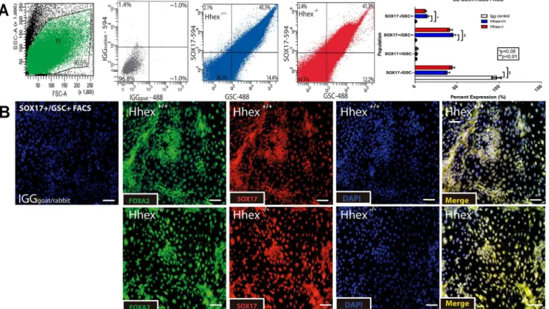
28]. Culture of cells produced from embryoid bodies in DE medium resulted in a highly differ-entiated population of DE cells that were SOX17+/FOXA2+ in both wild type andHhex-/- cul-tures (Fig 1–protein expression; andFig 2–mRNA expression). SOX17 and FOXA2 are two proteins with high nuclear expression in definitive endodermal cells and are the most efficient markers for isolating definitive endoderrmal cell types [29,30]. Thus,Hhexis not necessary for the differentiation of ESCs to DE. We also performed FACS for SOX17+/GSC+cells on both
Hhex+/+andHhex-/-EB cell cultures at the end of the 5-day DE differentiation protocol. SOX17 and GSC are two proteins previously used to identify DE for cell sorting [26]. Similar to the use of the nuclear protein FOXP3 for lineage segregation of Tcells using FACS [31], the nuclear pro-tein SOX17 has been used for lineage segregation of endoderm using FACS [32]. InHhex+/+DE cultures, 45.3% of the total population was SOX17+/GSC+(Fig 1A) and inHhex-/-DE cultures, 41.3% of the total population was SOX17+/GSC+(Fig 1A). Both of these percentages are consis-tent with previous reports on ESC-derived DE cells that used similar methods [33]. No dramatic differences were observed in the expression of mRNA genes betweenHhex+/+andHhex-/-DE cultures (Fig 2).
Hhex
is necessary for initiation of hepatic gene expression and
repression of
Vegf
signaling in differentiation of DE to HE
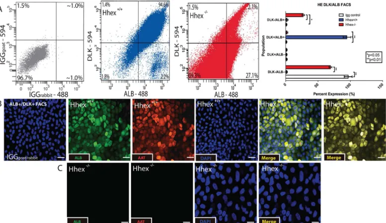
Previous studies have investigated the differentiation of mouse ESCs toward HE using various methods [20–22].Hhexhas been implicated in the process as evidenced by low expression lev-els ofAlbandAfpinHhex-/-ESCs differentiated towards the hepatic lineage [28]. After differ-entiation of DE cells from bothHhex+/+andHhex-/-ESCs, we re-plated the DE cells in HE media and cultured for an additional 7–10 days to obtain differentiated hepatic progenitor cells. After 7–10 days in HE medium, cells were subjected to FACS using ALB and DLK, a com-bination of proteins previously used to identify and sort HE progenitor cells [34]. Culture of
cells in HE medium for 10 days did not result in the differentiation of a large cell population that was double positive for ALB and DLK (Fig 3A). In addition, immunofluorescence analysis revealed that the 10 dayHhex+/+cultures exhibited a highly differentiated population of cells positive for both ALB and AAT, two proteins commonly used to identify hepatic progenitor cells [35] (Fig 3B). As expected,Hhex-/-ESCs did not produce any observable population of ALB+/AAT+cells (Fig 3C). Furthermore,Hhex+/+HE cultures showed significantly higher mRNA levels of genes whose expression is known to increase during HE differentiation, including: 87.8-fold higher expression ofHhex(inHhex+/+cells when HE cultures are com-pared to DE cultures), 13.9-fold higher expression ofAlb, 25.7-fold higher expression ofAat, 7.7-fold higher expression ofCebpα, 14.2-fold higher expression ofHnf4α, and 64.2-fold higher
expression ofTbx3, and 13.3-fold higher expression ofHnf6(Fig 2Efor genotype comparison within each differentiation stage, andS1 Figfor differentiation stage comparison within geno-type). We also analyzed mRNA quantity forVegfaand observed thatHhex-/-sorted HE cells maintained significantly higher levels of expression forVegfa(7.4-fold) and its receptors
Vegfr2(2.9-fold) andVegfr1(4.8-fold) (Fig 2Cfor genotype comparison within each differenti-ation stage, andS1 Figfor differentiation stage comparison within genotype). Finally, in addi-tion to being absent for ALB and AAT (S1 Fig),Hhex-/-HE cultures exhibited increased expression of DE genes and increased expression of pluripotency genes (Fig 2C and 2Dfor genotype comparison within each differentiation stage, andS1 Figfor differentiation stage
Fig 1. Differentiation of Definitive Endoderm. Fig 1:Hhex-/-ESCs show no defects when differentiated toward definitive endoderm.A)Analysis of FACS for SOX17 and GSC revealed that bothHhex+/+(blue) andHhex-/-(red) cultures produced similar percentages of differentiated DE cells. Light Scatter plot indicates the cells gated for sorting (green) and IGG plot (grey) confirms antigen specificity.B)Single channel and merged immunofluorescence staining of DE cultures after the 5-day culture. BothHhex+/+andHhex-/-cultures showed significantly more cells double positive for the nuclear expression of SOX17
and FOXA2/HNF3βcompared to IGG controls. (Scale bars = 100μM,*p<0.05,**p<0.01)
comparison within genotype). Thus, upon differentiation ofHhex-/-mESCs from DE to HE, the expression of Vegfa is increased, while the expression of liver-enriched genes is markedly attenuated compared to wild type cells. These results provide evidence for concurrent regula-tion ofHhex, HE genes, andVegfaduring the initial stages of HE differentiation in vitro.
In summary, comparison of mRNA expression between DE and HE stages forHhex+/+and
Hhex-/-cells indicated that the failure of HE differentiation from DE progenitor cells inHhex
-/-ESCs. This was characterized by a failure to increase HE gene expression (Hhex,Alb,Aat,
Cebpα,Hnf4α,Tbx3, andHnf6), maintenance of high levels of DE gene expression, and
dra-matically increased expression of the pluripotency marker geneOct4,Nanog, andAlkaline phosphatase). It should be noted that Alkaline phospahatse was chosen as a marker for pluripo-tency, as a high expression of SOX2 has been reported in cells driven toward the endodermal lineage using Activin A (SOX17+ cells) [36]. Additionally, this is accompanied by a significant increase inVegfa,Vegfr1, andVegfr2expression.
Fig 2. Genotype Comparison using QPCR at each Differentiation Stage.Hhex-/-HE cells did not show mRNA expression consistent with hepatic differentiation.A-E)Comparison of fold-change in normalized mRNA gene expression using differentiation-stage specific markers. Comparison of
pluripotency gene markers revealHhex-/-(red) cells showed increased pluripotency relative toHhex+/+(blue) at each differentiation stage, particularly during
HE differentiation(A, B, and E). Comparison of definitive endodermal gene markers revealHhex-/-cells fail to exhibit significant decreases in definitive
endodermal gene expression characteristic of HE differentiation(C), and as seen inHhex+/+HE cells.Hhex+/+HE cells showed dramatic increase in hepatic gene expression(D), whileHhex-/-cells showed a heavily attenuated expression. Comparison ofVegfsignaling gene markers showed thatHhex-/-cells exhibit increased levels of ligands (Vegf-a) and receptor (Vegfr1andVegfr2) gene expression at each differentiation stage, but particularly during HE differentiation(A, B, and C). (Note raw data is presented inS1 Fig) (*p<0.05,**p<0.01, and***p<0.001).
ESC-derived hepatic precursor cells express VEGFR2
Our data suggest thatVegfamRNA levels are inversely correlated with HE differentiation and are increased in the absence ofHhex(seeresultsabove). Based on these data, we looked for the presence of VEGFR2+murine hepatic progenitor cells inHhex+/+andHhex-/-HE cell cultures. As recently reported, VEGFR2+hepatic progenitors are amongst the earliest cells to differenti-ate from the DE stage toward the HE stage [18]. In addition, Vegfr2 is known to stimulate the majority of transcriptional activity in response to changes in Vegfa expression [37]. After one day of HE culture,Hhex+/+cells exhibited expression of VEGFR2 and ALB protein as assessed by immunofluorescnece (Fig 4B). This indicates that HE cells express VEGFR2 very early dur-ing in vitro differentiation, and are thus capable of utilizdur-ing Vegf signaldur-ing. CD34 is a marker for hematopoietic/endothelial cells, and was not observed to be co-expressed on ALB+cells (Fig 4C). These results suggest thatHhex+/+HE cells are not likely of endothelial origin and that there is no contamination of endothelial cells in our differentiated populations. While we do not see an indication of differentiation ofHhex-/-cells toward endothelial lineage under HE culture conditions, it is possible thatHhex-/-cells might more easily form endothelial cells under proper culture conditions. However, due to the reported role ofHhexin endothelial dif-ferentiation [38], endothelial maturation might be defective as well. We did not observe a sig-nificant amount of endothelial activity as a result of VEGF expression under HE conditions, however, the media conditions may preclude the differentiation toward the endothelial lineage
Fig 3. Differentiation of Hepatic Endoderm.Hhex-/-definitive endodermal cells did not differentiate toward hepatic endoderm.A)Analysis of FACS for ALB
and DLK revealed that onlyHhex+/+(blue) cultures produced significant populations of differentiated HE cells. IGG plot (grey) confirms antigen specificity.B)
Single channel and merged immunofluorescence staining of HE cultures. OnlyHhex+/+cells showed a uniform and highly differentiated population of HE cells that were double positive for ALB and AAT compared to IGG controls. (Scale bars = 50μM,*p<0.05,**p<0.01).
Fig 4. Differentiation of VEGFR2+ Hepatic Endoderm.Hhex-/-DE cells did not differentiate into VEGFR2+early hepatic progenitor cells.A)Analysis of FACS for ALB and VEGFR2 revealed that onlyHhex+/+(blue) cultures produced significant populations of HE progenitor cells and the majority of ALB+cells were also VEGFR2+inHhex+/+cultures. IGG plot confirms antigen specificity.B and C)Single channel and merged immunofluorescence staining ofHhex+/+
despite a potential predisposition to do so. At the end of HE culture, these same VEGFR2+
Hhex+/+hepatic cultures exhibited robust expression of ALB and AAT protein as assessed by immunofluorescence (Fig 4D). In addition,Hhex-/-HE cultures did not show any observable ALB or AAT expression (Fig 4E). Upon FACS analysis ofHhex+/+HE cells for both VEGFR2 and ALB, we observed many double-positive cells. In fact, 90.4% ofHhex+/+HE cells were ALB+/VEGFR2+while ALB+/VEGFR2-cells represented only 3.8% of the total population (Fig 4A). We then compared gene expression of bothHhex-/-andHhex+/+ALB+/ VEGFR2+sorted cells to their respective DE cell population and found, as expected, thatHhex-/-cells had very attenuated increases in hepatic gene expression with no change inVegfaexpression (Fig 4F). On the other hand,Hhex+/+cells had large increases in hepatic gene expression accompanied by a large decrease inVegfaexpression. In summary, the vast majority ofHhex+/+ESC-derived hepatic precursor cells express VEGFR2. The absence ofHheximpairs the differentiation of DE cells to HE and this is accompanied by dramatic perturbations inVegfaand Vegfr2 expression.
Hhex
expression is critical for the regulation of
Vegf
in HE differentiation
and maintenance of HE gene expression
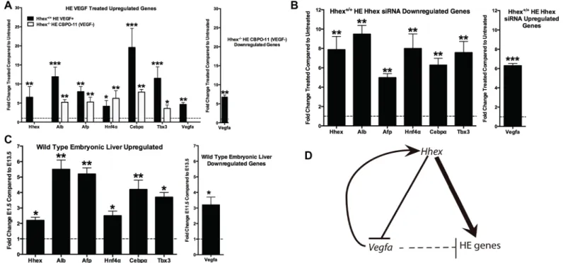
To determine the role ofHhexin regulating Vegf signaling during HE differentiation, we treated day-one HE cultures with 1) CBO-P11 to decrease Vegf signaling inHhex-/-cells, and 2) exogenous VEGFA to increase Vegf signaling inHhex+/+cells (Fig 5A). CBO-P11 is a 17-amino acid peptide derived from the region of the Vegf peptide that mediates binding to its receptors and blocks the binding of VEGFA to its cognate receptors VEGFR1 and VEGFR2 [39]. Inhibition and activation of the Vegf signaling pathway, such as with CBO-P11, has been previously shown to modulate the induction of VEGF secretion [40,41]. Accordingly, inhibi-tion of Vegf signaling with CBO-P11 is expected to decrease VEGFA/Vegfa secreinhibi-tion/expres- secretion/expres-sion in the current model and allow for the examination of the effect of reduced cellular stimulation from VEGF. When we inhibited Vegf signaling inHhex-/-HE cells using CBO-P11 (10uM for 72 hours resulting in a significant down-regulation ofVegfa6.75-fold), we observed significant increases in HE gene expression: 5.21-fold increase inAlb, 5.27-fold increase inAat, 6.22-fold increase inCebpα, 7.87-fold increase inHnf4α, and 3.58-fold increase inTbx3
expres-sion (Fig 5A). Unexpectedly, when VEGFA was added toHhex+/+HE cells at 30 ng/uL for 72 hours (resulting in a significant up-regulation ofVegfaexpression 4.84-fold),Hhexexpression increased 6.52-fold and was accompanied by significant increases in the expression of other HE genes: 11.91-fold increase inAlb, 7.99-fold increase inAat, 4.16-fold increase inCebpα,
19.59-fold increase inHnf4α, and an 11.50-fold increase inTbx3expression (Fig 5A). Thus, in
Hhex-/-cells, a decrease in HE gene expression is accompanied by elevatedVegfalevels due to the loss ofVegfarepression byHhex. When Vegf signaling is blocked inHhex-/-cells by CBO-P11 treatment, HE gene expression is rescued, demonstrating an important inhibitory role for Vegf signaling in HE gene expression. Interestingly, the expected decrease in HE gene expression upon treatment with exogenous VEGFA is blocked whenHhexis present, indicat-ing that the positive effect ofHhexon HE gene expression can overcome any inhibitory influ-ence on this process byVegfa.
ofHhex+/+andHhex-/-ALB+
/VEGFR2+sorted cells that were replated for 7 days in HE media. Despite rapid expansion of theHhex-/-sorted cells, only
Hhex+/+sorted cells showed a co-expression of ALB and AAT that is indicative of further/continued hepatic differentiation. (Scale bars = 50μM.)F)
Normalized mRNA expression of hepatic andVegfsignaling gene markers in ALB+/VEGFR2+sorted cells from bothHhex+/+andHhex-/-cultures.Hhex
-/-sorted cells show heavily attenuated mRNA expression for hepatic genes and no significant reduction inVegfaexpression when compared to cells from the previous DE differentiation stage. (*p<0.05,**p<0.01,***p<0.001).
To explore ifHhexregulates hepatic gene expression and Vegf signaling in ESCs that have already been differentiated to HE, we treated 10 day-oldHhex+/+cultures with a combination of threeHhexsiRNA oligos to knockdownHhexexpression (Fig 5B). Upon treatment of
Hhex+/+HE cells with a combination of three siRNA oligos targeted toHhexat 1uM each for 72 hours, we observed a significant decrease in the expression of HE genes: 7.87-fold decrease inHhex, 9.48-fold decrease inAlb, 4.99-fold decrease inAat, 7.99-fold decrease inCebpα,
6.28-fold decrease inHnf4α, and a 7.57-fold decrease inTbx3(Fig 5B). In addition, as expected,
we saw a 6.29-fold increase inVegfaexpression (Fig 5B). These data are consistent with the gene expression patterns we observed inHhex-/-HE cells and support aHhex-dependent mechanism in the post-specification maintenance of HE gene expression via Vegf signaling. These results also support the regulation of hepatic differentiation by bothHhexandVegfaand suggest thatVegfaprovides feedback input to increaseHhexexpression (suggesting the pres-ence of an inverse regulatory relationship—seeFig 5D). TreatingHhex+/+HE cells with exoge-nous VEGFA resulted in increased HE gene expression that was accompanied by an increase in
Hhexexpression. In summary, we found that in the presence ofHhex,Vegfaacts as a stimula-tor of HE gene expression by increasingHhexexpression. However, in the absence ofHhex,
Vegfaacts as a repressor of HE gene expression. Thus, while bothHhexandVegfaeffect HE gene expression, the positive effect ofHhexon hepatic differentiation is dominant over the inhibitory effect ofVegfa(Fig 5D).
Finally, to confirm that the gene expression pattern observed in the currentin vitroreport is relevant toin vivomechanisms, we subjected liver from embryonic mice (embryonic days E11.5 and E13.5) to qPCR assessment for hepatic markers. In the mouse, the liver is mainly a
Fig 5. ProposedHhex-VegfHepatic Regulatory Pathway.HhexandVegfsignaling modulate the commitment of DE cells toward the HE lineage.A)The addition of VEGFA protein toHhex+/+HE cells facilitated HE differentiation as indicated by increased hepatic gene marker expression (black bars). Similar
results were seen with the reduction of VEGF signaling inHhex-/-DE cells cultured in HE media (white bars).B)The reduction ofHhexusing siRNA inHhex+/ +HE cells resulted in decreased expression of hepatic gene markers and increasedVegfaexpression.C)Comparative qPCR analysis for the expression of
hepatic markers during the stages of mouse embryonic hepatic expansion, E11.5-E13.5. Hepatic markers change similarlyin vivowhen compared to the results obtained from thein vivodifferentiation from ESC in the current report.D)We propose anHhex-regulated model of HE specification wherebyHhexis necessary for the expression of hepatic genes, in part, via reducing/regulatingVegfsignaling. (*= p<0.05,**p<0.01,***p<0.001).
hematopoietic organ prior to embryonic day E12.5 [42], thus comparison of gene expression prior to and shortly after E12.5 will reflect the early stages of hepatic (metabolic) function of the organ. As expected, the results for the in vivo analysis reflect gene expression events occur-ring in the in vitro model here (namely increased hepatic gene expression–Hhex,Alb,Aat,
Hnf4a,Cebpa, andTbx3, and decreasedVegfexpression) (Fig 5C). These results parallel the gene expression pattern seen inHhex+/+HE cultures (Figs2and5, andS1 Fig), but is absent in
Hhex-/-HE cultures.
Discussion
The current results indicate thatHhexrepressesVegfsignaling during the differentiation of HE progenitor cells and that in the absence ofHhex, ESC-derived DE cells do not differentiate toward HE cells. Furthermore, we show thatHhexis necessary for the initial increase in hepatic gene expression during early HE differentiation in vitro and this is accompanied by a decrease inVegfaexpression, suggesting thatVegfaexpression must decrease in order for hepatic differ-entiation to proceed. We confirmed this finding by showing that inhibition of Vegf signaling in
Hhex-/-cells, in whichVegfalevels are markedly increased, rescued the molecular signature of HE differentiation. When we treatedHhex+/+HE cells with VEGFA to increase Vegf signaling, we saw an increase in HE gene expression accompanied by an increaseHhexexpression. This indicates that the inhibitory effect ofVegfaon HE differentiation is blocked in the presence of elevatedHhexlevels. Taken together, these results indicate that the repression of Vegf signaling viaHhex, which occurs in hematopoietic cells [14,38,43,44], supports the commitment of DE cells toward the HE lineage. However, Vegf-mediated regulation of HE gene expression may rely on bothHhex-dependent andHhex-independent mechanisms.
Hhexhas previously been linked to hepatic development both in vivo [7,9,11,45] and in vitro [28].Hhex-/-embryos fail to develop a liver bud [11,45] and it has been suggested that DE cells specify toward HE but fail to migrate and proliferate [7,9]. However, these conclu-sions are based on the expression of genes not essential, nor absolutely specific for hepatic dif-ferentiation (such asAlb,Afp,Foxa2, andProx1). For instance, it has been previously suggested that neither the expression ofFoxa2[46] norProx1is specific for hepatic differentiation [47]. Also, previous studies have shown thatAfpis expressed in other foregut derivatives such as gas-trointestinal and pancreatic tissues [48,49]. In addition, usingAlbexpression alone to define hepatic specification events has recently been questioned [50] and it has been shown that gut cells are capable of producingAlbtranscripts in response to stimulation byGata4[51]. How-ever, when these genes are used in combination with other genes whose expression is more spe-cific for the hepatic lineage (such asAat,Hnf4α,Cebpα, andTbx3as used in this study), a more
accurate assessment of hepatic differentiation events.
It has been suggested thatHhexexpression is correlated with hepatic specification events both in vivo [52–54] and in vitro [28]. Furthemore, it has been reported that hepatic cells from
Hhex-/-embryos show morphological defects related to liver organogenesis [7,9]. In support of conserved function forHhexregulation of HE specification, the use ofHhexmorpholinos in zebra fish results in similar hepatic defects as those seen in mouse [55]. Overexpression of
Hhexin DE cells has been suggested to induce HE gene expression while prolongedHhex
deterioration of the commitment toward a hepatic cell fate, as evidenced by a large decrease in the expression HE genes. Taken together, these results illustrate the complex nature ofHhex
regulation of HE specification and differentiation. ConcerningHhexand its potential relation-ship to pluripotency, it has been well documented that increases in lineage determinant genes (such asHhex) are paralleled by decreases in pluripotency genes (such asOct4,Nanog, and
Alkaline Phosphatasewhich are required for germ line transmission in mouse ESCs) [56]. However, it still remains to be determined what the mechanistic relationship between lineage determinants and pluripotency markers on the same cell. A growing body of evidence suggest that increased expression of pluripotency genes after lineage commitment may be representa-tive of the labile nature for multi-lineage potential of pluripotent cells [57]. It should be noted that we observed increased expression of pluripotency markers at all stages in theHhex
-/-mESCs when compared toHhex+/+mESCs from the same stage, although most notably in the HE stage (Fig 2). Thus, while our results suggest theHhex-/-mESCs acquire typical DE gene expression as seen inHhex+/+mESCs, pluripotency genes remain elevated representing a possi-ble disconnect between DE gene expression, differentiation, and/or pluripotency genes. How-ever, it has been previously reported that treatment withHhexshRNA does not impair the progressive down-regulation of Sox2 and Oct4 during the induction of primitive and precar-diac mesoderm [58]. In addition, we now report thatHhexhas an important role in the induc-tion of Vegf-regulated mechanisms that facilitate the specificainduc-tion, differentiainduc-tion, and continued hepatic commitment of HE cells from DE progenitors.
Vegfais the main ligand for stimulating theVegfsignaling pathway and mRNA levels for multipleVegfsignaling components (Vegfa,Vegfr1,Vegfr2) were higher inHhex-/-HE cells than inHhex+/+HE cells. Our results support a reciprocal relationship betweenHhexand
Vegfain HE cells in which increasedHhexexpression represses Vegfa and increased Vegfa increasesHhexexpression [8,14]. A similar relationship is known to exist in endothelial cells [14,15], and therefore may represent a conservedHhex-Vegfamolecular pathway that is shared between hepatic and endothelial cells. Activation ofVegfsignaling can occur as the result of stimulation from multiple transcriptional regulators, includingHhex, which has been shown to act as both a direct [14,43] and indirect [59] repressor ofVegfsignaling activity.
Hhexcan induce transcriptional activation or repression via binding to a core consensus bind-ing site motif (5’-C/tA/tATTAAA/g-3’) [8,60,61]. Furthermore,Hhexhas also been shown to bind non-consensus sites within target genes includingVegf,Vegfr1, andVegfr2[14,
43]. In addition to these direct transcriptional repression mechanisms,Hhexhas also been implicated via indirect repression where it has been shown to prevent transcriptional activation of theVegfr2gene by inhibiting the binding ofGata2to its promoter regions [59].
As discussed earlier, most of the evidence for transcriptional interaction ofHhexwithVegf
signaling comes from the hematopoietic literature (hematopoietic differentiation and leuke-mia). The predominate effect ofHhexin hematopoietic and leukemic cells is to regulate cellular growth [14,38,44], as high levels ofHhexexpression lead to cell death and decreasedVegf sig-naling [14,62]. Similarly, decreasingHhexexpression promotes cell growth via interruption of
Vegfsignaling-dependent apoptosis [14]. Reduction or absence ofHhexexpression may sensi-tize cells to changes inVegfsignaling sinceHhexis not present to modulateVegfsignaling. Therefore, the growth of cells with decreased or absentHhexexpression is likely to be shifted from anHhex-dominant mechanism to aVegf-dominant mechanism. Under this scenario, HE gene expression could be facilitated by both an increase inHhexexpression and a reduction in
HhexandVegfmay also apply to cell types outside of the hematopoietic compartment, includ-ing thyroid and mammary epithelial cells [14,65,66].
While a role forHhexin hepatic differentiation has been established by a number of investi-gators, it has been unclear, until recently, what role theVegfpathway plays in hepatocyte speci-fication/maturation, ifHhexandVegfinteract in the process, and if this interaction occurs between endothelial and endodermal cells or in endodermal cells alone. In 2009, Matsumoto et al. showed that the liver bud failed to form inVegfr2-/-embryos due either to the lack of sig-naling from, or absence of, endothelial cells [17]. They showed that at the time ofHhex expres-sion and function,Vegfr2is required for the outgrowth and expansion of hepatic endodermal explants. However, this defect could be the absence ofVegfr2expression on endodermal cells as well as endothelial cells. Importantly, the analysis from that study showed that in early hepatic endoderm tissue explants, VEGFR2 and ALB expression overlaps. Thus,Vegfsignaling may be intrinsic to both endothelial and hepatic cell types.
Interestingly, a recent report identified an in vivo population of non-endothelial, Vegfr2-po-sitive cells (VegfR2+/CD31-), that were isolated from murine liver using lineage tracing [18]. These cells were shown to be hepatic progenitor cells that support the commitment/maturation of other hepatic progenitors (e.g.,Vegfr2-/CD31-) by increasing the expression of HE genes. This maturational effect seems to occur cell autonomously within the VEGFR2+murine hepatic progenitor population. An endothelial cell population (VegfR2+/CD31+) was also iso-lated but did not support hepatic commitment/maturation. These VegfR2+/CD31-hepatic pro-genitors are true propro-genitors from which functional hepatic cells (both hepatocytes and biliary epithelial cells), from both in vivo and in vitro sources, can be derived. The authors note that these cells marked for HE specification may differentiate into functional and terminal liver cell types via a progressive down-regulation of VEGFR2 (potentially via anHhexmechanism). This recent report clearly establishes thatVegfr2, which has traditionally been defined as a mesodermal and hematopoietic/endothelial/vascular marker, is also expressed on, and plays a role in, HE progenitor cells that function to instruct early liver development via both non cell-autonomous (paracrine) and cell-cell-autonomous (autocrine) signaling.
Our results are in agreement with the study by Goldman et al (identification of VEGFR2+/ ALB+/CD34-murine hepatic progenitor cells) and provides strong support for the notion that
Vegfsignaling is intrinsic to the HE cell and independent of the presence of endothelial cells [18]. Additionally,Vegfsignaling in foregut epithelial cells has previously been proposed to be independent of an endothelial cell function [67–69]. While Vegf/Vegfr2 expression/activation has traditionally restricted to mesodermal and ectodermal derivatives, we now offer further support Vegf signaling as a marker for endodermal derivatives as well. Therefore we propose thatHhexfacilitates HE specification and differentiation (continued commitment) from DE progenitors in part via an intrinsicVegfa-Vegfr2mechanism.
Conclusions
In summary, our investigations suggest a novel transcriptional relationship betweenHhexand
Vegfsignaling in HE differentiation. We propose thatHhexis required for the onset of HE dif-ferentiation and thatHhexstimulates the expression of many hepatic genes. Its absence per-turbs the hepatic competence of differentiating DE cells in part by failing to reduceVegf
signaling activity. In addition, we observed thatVegfaaffects HE differentiation independent of
This complex role ofVegfsignaling in HE differentiation potentially involves the balanced acti-vation ofVegfr1/Vegfr2transcriptional mechanisms via modulation ofVegfa. There is known interplay and trade-offs between VEGFR2 and VEGFR1 stimulation and it is interesting to wonder how such a dynamic could influence hepatic specification. It is clear thatVegfahas a direct influence onHhexexpression and theVegfa-induced increase inHhexlimits the inhibi-tory effect that increasedVegfsignaling has on differentiation of DE to HE in vitro. Therefore, this reciprocal relationship betweenHhexandVegfsignaling (increasedHhexexpression with reducedVegf) may support a feedback mechanism that calibrates the onset and continued expression of hepatic genes resulting in a fine-tuned HE differentiation/maturation program. Future directions should aim to identify howHhexandVegfsignaling interact to exert their independent and combined effects on the hepatic lineage. In addition, during HE differentia-tion the reladifferentia-tionship betweenHhexandVegfasupports increased HE gene expression and differentiation of VEGFR2+hepatic progenitors from endodermal precursors via aHhex -domi-nant mechanism. Finally, in the absence ofHhex, aVegfa-dominant mechanism represses HE gene expression and impairs the ability of endodermal cells to differentiate toward the hepatic lineage.
Supporting Information
S1 Fig. Differentiation Stage QPCR.Hhex-/-HE cells did not show mRNA expression consis-tent with hepatic differentiation.A)Normalized mRNA expression of hepatic gene markers.
Hhex+/+(blue) HE cells showed dramatic increase in hepatic gene expression, whileHhex
-/-(red) cells showed a heavily attenuated expression.B)Normalized mRNA expression of defini-tive endodermal gene markers.Hhex-/-cells fail to show a decrease in definitive endodermal gene expression characteristic of HE differentiation, as seen inHhex+/+cells.C)Normalized mRNA expression of pluripotency gene markers.Hhex-/-cells showed increased pluripotency relative toHhex+/+at each differentiation stage (but particurlarly during HE differentiation) that further indicates a differentiation defect.D)Normalized mRNA expression of Vegf signal-ing gene markers.Hhex-/-cells showed increased levels of ligands (Vegf-a) and receptor (Vegfr1
andVegfr2) gene expression at each differentiation stage (but particularly during HE differenti-ation) that indicates aberrant cellular signaling.
(TIF)
S1 Table. Cell Culture. A)Products are listed with their manufacturer.B)Cell Culture recipes are listed in order of use.
(TIF)
S2 Table. IF/FACS Antibodies.Antibodies are listed along with company, species, catalogue number, concentration and method used.
(TIF)
S3 Table. qPCR primers.Primers are listed according to grouping of analysis (associations). Reverse primers are listed in their sense (not anti-sense) direction.
(TIF)
S4 Table. Antibody Optimization.Antibodies were optimized specifically for their use in the method(s) listed. Optimal signal was chosen based on concentration and time, as well as com-parison to unstained controls, Igg controls, and controls stained with secondary antibody only (background).
Acknowledgments
We would like to thank the Yale Center for Analytical Sciences for help with statistics. We would also like to thank two reviewers for their helpful comments on the manuscript.
Author Contributions
Conceived and designed the experiments: ASA. Performed the experiments: ASA. Analyzed the data: ASA. Contributed reagents/materials/analysis tools: ASA CWB. Wrote the paper: ASA CWB.
References
1. Livigni A, Villegas SN, Oikonomopoulou I, Rahman A, Morrison GM, Brickman JM. Differentiation of embryonic stem cells into anterior definitive endoderm. Curr Protoc Stem Cell Biol. 2009; Chapter 1: Unit 1G 3. Epub 2009/07/09. doi:10.1002/9780470151808.sc01g03s10PMID:19585461.
2. Houssaint E. Differentiation of the mouse hepatic primordium. I. An analysis of tissue interactions in hepatocyte differentiation. Cell Differ. 1980; 9(5):269–79. Epub 1980/10/01. PMID:7438211.
3. Lemaigre FP. Development of the biliary tract. Mech Dev. 2003; 120(1):81–7. Epub 2002/12/20. PMID: 12490298.
4. Delisle JC, Martignat L, Bach JM, Bosch S, Louzier V. Bipotential mouse embryonic liver (BMEL) cells spontaneously express Pdx1 and Ngn3 but do not undergo further pancreatic differentiation upon Hes1 down-regulation. BMC Res Notes. 2008; 1:136. Epub 2008/12/26. doi:10.1186/1756-0500-1-136
PMID:19108739; PubMed Central PMCID: PMC2649931.
5. Yechoor V, Liu V, Espiritu C, Paul A, Oka K, Kojima H, et al. Neurogenin3 is sufficient for transdetermi-nation of hepatic progenitor cells into neo-islets in vivo but not transdifferentiation of hepatocytes. Dev Cell. 2009; 16(3):358–73. Epub 2009/03/18. doi:10.1016/j.devcel.2009.01.012PMID:19289082; PubMed Central PMCID: PMC2676438.
6. Fausto N. Liver regeneration and repair: hepatocytes, progenitor cells, and stem cells. Hepatology. 2004; 39(6):1477–87. Epub 2004/06/09. doi:10.1002/hep.20214PMID:15185286.
7. Bort R, Signore M, Tremblay K, Martinez Barbera JP, Zaret KS. Hex homeobox gene controls the tran-sition of the endoderm to a pseudostratified, cell emergent epithelium for liver bud development. Dev Biol. 2006; 290(1):44–56. Epub 2005/12/21. doi:10.1016/j.ydbio.2005.11.006PMID:16364283.
8. Soufi A, Jayaraman PS. PRH/Hex: an oligomeric transcription factor and multifunctional regulator of cell fate. Biochem J. 2008; 412(3):399–413. Epub 2008/05/24. doi:10.1042/BJ20080035PMID: 18498250; PubMed Central PMCID: PMC2570084.
9. Bort R, Martinez-Barbera JP, Beddington RS, Zaret KS. Hex homeobox gene-dependent tissue posi-tioning is required for organogenesis of the ventral pancreas. Development. 2004; 131(4):797–806. Epub 2004/01/23. doi:10.1242/dev.00965PMID:14736744.
10. Ghosh B, Ganea GR, Denson LA, Iannucci R, Jacobs HC, Bogue CW. Immunocytochemical character-ization of murine Hex, a homeobox-containing protein. Pediatr Res. 2000; 48(5):634–8. Epub 2000/10/ 25. doi:10.1203/00006450-200011000-00014PMID:11044484.
11. Martinez Barbera JP, Clements M, Thomas P, Rodriguez T, Meloy D, Kioussis D, et al. The
homeobox gene Hex is required in definitive endodermal tissues for normal forebrain, liver and thyroid formation. Development. 2000; 127(11):2433–45. Epub 2000/05/11. PMID:10804184.
12. Hunter MP, Wilson CM, Jiang X, Cong R, Vasavada H, Kaestner KH, et al. The homeobox gene Hhex is essential for proper hepatoblast differentiation and bile duct morphogenesis. Dev Biol. 2007; 308 (2):355–67. Epub 2007/06/21. doi:10.1016/j.ydbio.2007.05.028PMID:17580084; PubMed Central PMCID: PMC2045067.
13. Zaret KS. Genetic programming of liver and pancreas progenitors: lessons for stem-cell differentiation. Nat Rev Genet. 2008; 9(5):329–40. Epub 2008/04/10. doi:10.1038/nrg2318PMID:18398419.
14. Noy P, Williams H, Sawasdichai A, Gaston K, Jayaraman PS. PRH/Hhex controls cell survival through coordinate transcriptional regulation of vascular endothelial growth factor signaling. Mol Cell Biol. 2010; 30(9):2120–34. Epub 2010/02/24. doi:10.1128/MCB.01511-09PMID:20176809; PubMed Central PMCID: PMC2863580.
16. Hallaq H, Pinter E, Enciso J, McGrath J, Zeiss C, Brueckner M, et al. A null mutation of Hhex results in abnormal cardiac development, defective vasculogenesis and elevated Vegfa levels. Development. 2004; 131(20):5197–209. Epub 2004/10/02. doi:10.1242/dev.01393PMID:15459110.
17. Matsumoto K, Yoshitomi H, Rossant J, Zaret KS. Liver organogenesis promoted by endothelial cells prior to vascular function. Science. 2001; 294(5542):559–63. Epub 2001/09/29. doi:10.1126/science. 1063889PMID:11577199.
18. Goldman O, Han S, Sourrisseau M, Dziedzic N, Hamou W, Corneo B, et al. KDR Identifies a Conserved Human and Murine Hepatic Progenitor and Instructs Early Liver Development. Cell Stem Cell. 2013; 12 (6):748–60. Epub 2013/06/12. doi:10.1016/j.stem.2013.04.026PMID:23746980.
19. Bogue CW, Zhang PX, McGrath J, Jacobs HC, Fuleihan RL. Impaired B cell development and function in mice with a targeted disruption of the homeobox gene Hex. Proc Natl Acad Sci U S A. 2003; 100 (2):556–61. doi:10.1073/pnas.0236979100PMID:12522149; PubMed Central PMCID: PMC141034.
20. Chinzei R, Tanaka Y, Shimizu-Saito K, Hara Y, Kakinuma S, Watanabe M, et al. Embryoid-body cells derived from a mouse embryonic stem cell line show differentiation into functional hepatocytes. Hepa-tology. 2002; 36(1):22–9. Epub 2002/06/27. doi:10.1053/jhep.2002.34136PMID:12085345.
21. Gouon-Evans V, Boussemart L, Gadue P, Nierhoff D, Koehler CI, Kubo A, et al. BMP-4 is required for hepatic specification of mouse embryonic stem cell-derived definitive endoderm. Nat Biotechnol. 2006; 24(11):1402–11. Epub 2006/11/07. doi:10.1038/nbt1258PMID:17086172.
22. Jones EA, Tosh D, Wilson DI, Lindsay S, Forrester LM. Hepatic differentiation of murine embryonic stem cells. Exp Cell Res. 2002; 272(1):15–22. Epub 2001/12/13. doi:10.1006/excr.2001.5396PMID: 11740861.
23. Schroeder IS, Wiese C, Truong TT, Rolletschek A, Wobus AM. Differentiation analysis of pluripotent mouse embryonic stem (ES) cells in vitro. Methods in molecular biology. 2009; 530:219–50. doi:10. 1007/978-1-59745-471-1_12PMID:19266345.
24. Gouon-Evans V, Boussemart L, Gadue P, Nierhoff D, Koehler CI, Kubo A, et al. BMP-4 is required for hepatic specification of mouse embryonic stem cell-derived definitive endoderm. Nat Biotechnol. 2006; 24(11):1402–11. doi:10.1038/nbt1258PMID:17086172.
25. Morrison GM, Oikonomopoulou I, Migueles RP, Soneji S, Livigni A, Enver T, et al. Anterior definitive endoderm from ESCs reveals a role for FGF signaling. Cell Stem Cell. 2008; 3(4):402–15. doi:10. 1016/j.stem.2008.07.021PMID:18940732.
26. Hansson M, Olesen DR, Peterslund JM, Engberg N, Kahn M, Winzi M, et al. A late requirement for Wnt and FGF signaling during activin-induced formation of foregut endoderm from mouse embryonic stem cells. Dev Biol. 2009; 330(2):286–304. Epub 2009/04/11. doi:10.1016/j.ydbio.2009.03.026PMID: 19358838; PubMed Central PMCID: PMC2730582.
27. Kim PT, Ong CJ. Differentiation of definitive endoderm from mouse embryonic stem cells. Results and problems in cell differentiation. 2012; 55:303–19. doi:10.1007/978-3-642-30406-4_17PMID: 22918814.
28. Kubo A, Kim YH, Irion S, Kasuda S, Takeuchi M, Ohashi K, et al. The homeobox gene Hex regulates hepatocyte differentiation from embryonic stem cell-derived endoderm. Hepatology. 2010; 51(2):633– 41. Epub 2010/01/12. doi:10.1002/hep.23293PMID:20063280.
29. Yasunaga M, Tada S, Torikai-Nishikawa S, Nakano Y, Okada M, Jakt LM, et al. Induction and monitor-ing of definitive and visceral endoderm differentiation of mouse ES cells. Nat Biotechnol. 2005; 23 (12):1542–50. Epub 2005/11/29. doi:10.1038/nbt1167PMID:16311587.
30. Snykers S, De Kock J, Rogiers V, Vanhaecke T. In vitro differentiation of embryonic and adult stem cells into hepatocytes: state of the art. Stem Cells. 2009; 27(3):577–605. Epub 2008/12/06. doi:10. 1634/stemcells.2008-0963PMID:19056906; PubMed Central PMCID: PMC2729674.
31. Oh U, Grant C, Griffith C, Fugo K, Takenouchi N, Jacobson S. Reduced Foxp3 protein expression is associated with inflammatory disease during human t lymphotropic virus type 1 Infection. The Journal of infectious diseases. 2006; 193(11):1557–66. doi:10.1086/503874PMID:16652285.
32. Pan Y, Ouyang Z, Wong WH, Baker JC. A new FACS approach isolates hESC derived endoderm using transcription factors. PloS one. 2011; 6(3):e17536. PMID:21408072; PubMed Central PMCID: PMC3052315.
33. Shiraki N, Yamazoe T, Qin Z, Ohgomori K, Mochitate K, Kume K, et al. Efficient differentiation of embry-onic stem cells into hepatic cells in vitro using a feeder-free basement membrane substratum. PLoS One. 2011; 6(8):e24228. Epub 2011/09/03. PMID:21887386; PubMed Central PMCID: PMC3162614.
35. Zhao R, Duncan SA. Embryonic development of the liver. Hepatology. 2005; 41(5):956–67. Epub 2005/04/21. doi:10.1002/hep.20691PMID:15841465.
36. Kallas A, Pook M, Trei A, Maimets T. SOX2 Is Regulated Differently from NANOG and OCT4 in Human Embryonic Stem Cells during Early Differentiation Initiated with Sodium Butyrate. Stem cells interna-tional. 2014; 2014:298163. doi:10.1155/2014/298163PMID:24707296; PubMed Central PMCID: PMC3951062.
37. Waltenberger J, Claesson-Welsh L, Siegbahn A, Shibuya M, Heldin CH. Different signal transduction properties of KDR and Flt1, two receptors for vascular endothelial growth factor. J Biol Chem. 1994; 269(43):26988–95. Epub 1994/10/28. PMID:7929439.
38. Kubo A, Chen V, Kennedy M, Zahradka E, Daley GQ, Keller G. The homeobox gene HEX regulates proliferation and differentiation of hemangioblasts and endothelial cells during ES cell differentiation. Blood. 2005; 105(12):4590–7. doi:10.1182/blood-2004-10-4137PMID:15728128; PubMed Central PMCID: PMC1895005.
39. Zilberberg L, Shinkaruk S, Lequin O, Rousseau B, Hagedorn M, Costa F, et al. Structure and inhibitory effects on angiogenesis and tumor development of a new vascular endothelial growth inhibitor. J Biol Chem. 2003; 278(37):35564–73. Epub 2003/07/03. doi:10.1074/jbc.M304435200PMID:12837752.
40. Chatterjee S, Heukamp LC, Siobal M, Schottle J, Wieczorek C, Peifer M, et al. Tumor VEGF:VEGFR2 autocrine feed-forward loop triggers angiogenesis in lung cancer. The Journal of clinical investigation. 2013; 123(4):1732–40. doi:10.1172/JCI65385PMID:23454747; PubMed Central PMCID:
PMC3613914.
41. Zilberberg L, Shinkaruk S, Lequin O, Rousseau B, Hagedorn M, Costa F, et al. Structure and inhibitory effects on angiogenesis and tumor development of a new vascular endothelial growth inhibitor. The Journal of biological chemistry. 2003; 278(37):35564–73. doi:10.1074/jbc.M304435200PMID: 12837752.
42. Crawford LW, Foley JF, Elmore SA. Histology atlas of the developing mouse hepatobiliary system with emphasis on embryonic days 9.5–18.5. Toxicologic pathology. 2010; 38(6):872–906. doi:10.1177/ 0192623310374329PMID:20805319; PubMed Central PMCID: PMC3490618.
43. Noy P, Sawasdichai A, Jayaraman PS, Gaston K. Protein kinase CK2 inactivates PRH/Hhex using mul-tiple mechanisms to de-repress VEGF-signalling genes and promote cell survival. Nucleic Acids Res. 2012; 40(18):9008–20. Epub 2012/07/31. doi:10.1093/nar/gks687PMID:22844093; PubMed Central PMCID: PMC3467080.
44. Topisirovic I, Culjkovic B, Cohen N, Perez JM, Skrabanek L, Borden KL. The proline-rich homeodomain protein, PRH, is a tissue-specific inhibitor of eIF4E-dependent cyclin D1 mRNA transport and growth. Embo J. 2003; 22(3):689–703. Epub 2003/01/30. doi:10.1093/emboj/cdg069PMID:12554669; PubMed Central PMCID: PMC140753.
45. Keng VW, Yagi H, Ikawa M, Nagano T, Myint Z, Yamada K, et al. Homeobox gene Hex is essential for onset of mouse embryonic liver development and differentiation of the monocyte lineage. Biochem Bio-phys Res Commun. 2000; 276(3):1155–61. Epub 2000/10/12. doi:10.1006/bbrc.2000.3548PMID: 11027604.
46. Dufort D, Schwartz L, Harpal K, Rossant J. The transcription factor HNF3beta is required in visceral endoderm for normal primitive streak morphogenesis. Development. 1998; 125(16):3015–25. Epub 1998/07/22. PMID:9671576.
47. Sosa-Pineda B, Wigle JT, Oliver G. Hepatocyte migration during liver development requires Prox1. Nat Genet. 2000; 25(3):254–5. Epub 2000/07/11. doi:10.1038/76996PMID:10888866.
48. Kwon GS, Fraser ST, Eakin GS, Mangano M, Isern J, Sahr KE, et al. Tg(Afp-GFP) expression marks primitive and definitive endoderm lineages during mouse development. Dev Dyn. 2006; 235(9):2549– 58. Epub 2006/05/19. doi:10.1002/dvdy.20843PMID:16708394; PubMed Central PMCID:
PMC1850385.
49. Jones EA, Clement-Jones M, James OF, Wilson DI. Differences between human and mouse alpha-fetoprotein expression during early development. J Anat. 2001; 198(Pt 5):555–9. Epub 2001/06/30. PMID:11430694; PubMed Central PMCID: PMC1468244.
50. Stanger BZ, Podolsky DK. Development of the gastrointestinal system. In: Yamada T, Alpers D.H., Kal-loo A.N., Kaplowitz N., Owyang C., Powell D.W., editor. Textbook of Gastroenterology. 5th ed: Wiley-Blackwell Publishing; 2009. p. 567–602.
51. Bossard P, Zaret KS. Repressive and restrictive mesodermal interactions with gut endoderm: possible relation to Meckel's Diverticulum. Development. 2000; 127(22):4915–23. Epub 2000/10/25. PMID: 11044405.
53. Tanaka T, Inazu T, Yamada K, Myint Z, Keng VW, Inoue Y, et al. cDNA cloning and expression of rat homeobox gene, Hex, and functional characterization of the protein. Biochem J. 1999; 339 (Pt 1):111– 7. Epub 1999/03/23. PMID:10085234; PubMed Central PMCID: PMC1220134.
54. Thomas PQ, Brown A, Beddington RS. Hex: a homeobox gene revealing peri-implantation asymmetry in the mouse embryo and an early transient marker of endothelial cell precursors. Development. 1998; 125(1):85–94. Epub 1997/12/09. PMID:9389666.
55. Wallace KN, Yusuff S, Sonntag JM, Chin AJ, Pack M. Zebrafish hhex regulates liver development and digestive organ chirality. Genesis. 2001; 30(3):141–3. Epub 2001/07/31. PMID:11477693.
56. Palmqvist L, Glover CH, Hsu L, Lu M, Bossen B, Piret JM, et al. Correlation of murine embryonic stem cell gene expression profiles with functional measures of pluripotency. Stem cells. 2005; 23(5):663–80. doi:10.1634/stemcells.2004-0157PMID:15849174.
57. Loh KM, Lim B. A precarious balance: pluripotency factors as lineage specifiers. Cell stem cell. 2011; 8 (4):363–9. doi:10.1016/j.stem.2011.03.013PMID:21474100.
58. Liu Y, Kaneda R, Leja TW, Subkhankulova T, Tolmachov O, Minchiotti G, et al. Hhex and Cer1 mediate the Sox17 pathway for cardiac mesoderm formation in embryonic stem cells. Stem cells. 2014; 32 (6):1515–26. doi:10.1002/stem.1695PMID:24585688; PubMed Central PMCID: PMC4260090.
59. Minami T, Murakami T, Horiuchi K, Miura M, Noguchi T, Miyazaki J, et al. Interaction between hex and GATA transcription factors in vascular endothelial cells inhibits flk-1/KDR-mediated vascular endothe-lial growth factor signaling. J Biol Chem. 2004; 279(20):20626–35. Epub 2004/03/16. doi:10.1074/jbc. M308730200PMID:15016828.
60. Crompton MR, Bartlett TJ, MacGregor AD, Manfioletti G, Buratti E, Giancotti V, et al. Identification of a novel vertebrate homeobox gene expressed in haematopoietic cells. Nucleic Acids Res. 1992; 20 (21):5661–7. Epub 1992/11/11. PMID:1360645; PubMed Central PMCID: PMC334400.
61. Williams H, Jayaraman PS, Gaston K. DNA wrapping and distortion by an oligomeric homeodomain protein. J Mol Biol. 2008; 383(1):10–23. Epub 2008/08/30. doi:10.1016/j.jmb.2008.08.004PMID: 18755198.
62. Nakagawa T, Abe M, Yamazaki T, Miyashita H, Niwa H, Kokubun S, et al. HEX acts as a negative regu-lator of angiogenesis by modulating the expression of angiogenesis-related gene in endothelial cells in vitro. Arterioscler Thromb Vasc Biol. 2003; 23(2):231–7. Epub 2003/02/18. PMID:12588764.
63. Jankovic D, Gorello P, Liu T, Ehret S, La Starza R, Desjobert C, et al. Leukemogenic mechanisms and targets of a NUP98/HHEX fusion in acute myeloid leukemia. Blood. 2008; 111(12):5672–82. doi:10. 1182/blood-2007-09-108175PMID:18388181.
64. Topisirovic I, Guzman ML, McConnell MJ, Licht JD, Culjkovic B, Neering SJ, et al. Aberrant eukaryotic translation initiation factor 4E-dependent mRNA transport impedes hematopoietic differentiation and contributes to leukemogenesis. Mol Cell Biol. 2003; 23(24):8992–9002. PMID:14645512; PubMed Central PMCID: PMC309660.
65. D'Elia AV, Tell G, Russo D, Arturi F, Puglisi F, Manfioletti G, et al. Expression and localization of the homeodomain-containing protein HEX in human thyroid tumors. J Clin Endocrinol Metab. 2002; 87 (3):1376–83. Epub 2002/03/13. PMID:11889211.
66. Puppin C, Puglisi F, Pellizzari L, Manfioletti G, Pestrin M, Pandolfi M, et al. HEX expression and locali-zation in normal mammary gland and breast carcinoma. BMC Cancer. 2006; 6:192. Epub 2006/07/21. doi:10.1186/1471-2407-6-192PMID:16854221; PubMed Central PMCID: PMC1550255.
67. Gouysse G, Couvelard A, Frachon S, Bouvier R, Nejjari M, Dauge MC, et al. Relationship between vas-cular development and vasvas-cular differentiation during liver organogenesis in humans. J Hepatol. 2002; 37(6):730–40. Epub 2002/11/26. PMID:12445412.
68. Kamiya A, Kinoshita T, Ito Y, Matsui T, Morikawa Y, Senba E, et al. Fetal liver development requires a paracrine action of oncostatin M through the gp130 signal transducer. Embo J. 1999; 18(8):2127–36. Epub 1999/04/16. doi:10.1093/emboj/18.8.2127PMID:10205167; PubMed Central PMCID: PMC1171297.