Adr1 and Cat8 Mediate Coactivator Recruitment and
Chromatin Remodeling at Glucose-Regulated Genes
Rhiannon K. Biddick, G. Lynn Law, Elton T. Young*
Department of Biochemistry, University of Washington, Seattle, Washington, United States of America
Background.Adr1 and Cat8 co-regulate numerous glucose-repressed genes inS. cerevisiae, presenting a unique opportunity to explore their individual roles in coactivator recruitment, chromatin remodeling, and transcription.Methodology/Principal Findings.We determined the individual contributions of Cat8 and Adr1 on the expression of a cohort of glucose-repressed genes and found three broad categories: genes that need both activators for full derepression, genes that rely mostly on Cat8 and genes that require only Adr1. Through combined expression and recruitment data, along with analysis of chromatin remodeling at two of these genes,ADH2andFBP1, we clarified how these activators achieve this wide range of co-regulation. We find that Adr1 and Cat8 are not intrinsically different in their abilities to recruit coactivators but rather, promoter context appears to dictate which activator is responsible for recruitment to specific genes. These promoter-specific contributions are also apparent in the chromatin remodeling that accompanies derepression:ADH2requires both Adr1 and Cat8, whereas, at FBP1, significant remodeling occurs with Cat8 alone. Although over-expression of Adr1 can compensate for loss of Cat8 at many genes in terms of both activation and chromatin remodeling, this over-expression cannot complement all of thecat8D phenotypes. Conclusions/Significance. Thus, at many of the glucose-repressed genes, Cat8 and Adr1 appear to have interchangeable roles and promoter architecture may dictate the roles of these activators.
Citation: Biddick RK, Law GL, Young ET (2008) Adr1 and Cat8 Mediate Coactivator Recruitment and Chromatin Remodeling at Glucose-Regulated Genes. PLoS ONE 3(1): e1436. doi:10.1371/journal.pone.0001436
INTRODUCTION
Alteration in gene expression patterns in response to variations in environmental signals is a tightly regulated process. In the yeastS.
cerevisiae large changes in transcription accompany the diauxic
shift, as organisms switch from using glucose as the main carbon source to non-fermentable carbon sources [1]. Many of the genes that are activated upon glucose exhaustion are under the control of the upstream kinase, Snf1, a homolog of the mammalian AMP-activated kinase [2]. Within the Snf1 network there is a wide range of finer control which includes two gene-specific activators, Adr1 and Cat8, that are necessary for the expression of about two hundred of these genes [3]. Broadly, Adr1 regulates genes involved in peroxisomal proliferation, b-oxidation and utilization of non-fermentable carbon sources (pathways involved in metabolism of ethanol, glycerol and lactate) [3] whereas Cat8 regulates gluconeogenic genes and most of the enzymes in the glyoxylate cycle [4]. However both Adr1 and Cat8 are known to work in tandem to obtain complete derepression of some genes, for exampleADH2andACS1[5].
This overlapping regulation created by this non-redundant pair of activators is a unique system for controlling gene expression. Other pairs of transcription factors in yeast exist, but they do not mimic the situation observed with Adr1 and Cat8. For example, Ace2 and Swi5 are similar because they co-regulate some genes, while acting independently at others, but Ace2 and Swi5 are highly homologous and bind to the same DNA target sequence, unlike Adr1 and Cat8 [4]. Oaf1 and Pip2 form another set of activators that, although there is some Pip2-independent regula-tion by Oaf1, are structurally very similar and form a heterodimer at most promoters [6]. Msn2 and Msn4 comprise yet another common pair of activators, but these function almost entirely as a unit, and even their own expression is intimately connected (MSN4 expression depends on Msn2) [7]. Even Cat8 itself shares its binding site (Carbon Source Response Element or CSRE) with Sip4, another zinc cluster transcription factor, although evidence suggests that Cat8 is more important for regulation of CSRE-containing genes than Sip4 [8]. Unlike the pairs mentioned above,
which arose by genome duplication, Adr1 and Cat8 are structurally different, independently expressed transcription factors with distinct binding sites. They often bind to the same promoter under activating conditions, and yet their individual contributions to expression of the gene vary widely.
One mechanism for co-regulation is cooperative binding of transcription factors. This seems to occur at a few promoters regulated by both Adr1 and Cat8. At theADH2promoter, Cat8-binding is enhanced in the presence of Adr1, but the reverse is not true [9]. The same situation is observed at a few other promoters but for the most part, their binding seems to be independent.
Another potential avenue for differential contributions to activation is in a transcription factor’s ability to recruit coactivators to the promoter. The paradigm for eukaryotic activator-dependent transcription relies on such recruitment: DNA-binding transcrip-tion factors provide specificity by binding preferentially to unique sequences, and then recruit a number of coactivators, such as complexes involved in chromatin remodeling and adaptors between activators and polymerase, as well as the actual transcription machinery itself. A number of these coactivators are non-essential and are only recruited to some promoters [10], a
Academic Editor:Geraldine Butler, University College Dublin, Ireland
ReceivedJuly 27, 2007;AcceptedDecember 17, 2007;PublishedJanuary 16, 2008
Copyright:ß2008 Biddick et al. This is an open-access article distributed under the terms of the Creative Commons Attribution License, which permits unrestricted use, distribution, and reproduction in any medium, provided the original author and source are credited.
Funding:R. Biddick was supported in part by PHS NRSA T32 GM07270 from NIGMS. The research was supported by a grant from the National Institutes of Health GM26079 to ETY.
Competing Interests:The authors have declared that no competing interests exist.
possible explanation for the range of dependence on Adr1 and Cat8. The HO promoter in yeast is activated by Swi5 and the heterodimer Swi4/Swi6 (SBF). Swi5 binds first and recruits the coactivator Swi/Snf, followed by Swi4/Swi6 binding [11]. Swi5 also recruits Mediator, a multi-subunit complex thought to act as a bridge between gene-specific activators and the holoenzyme, but does not recruit polymerase itself [12]. A variation on this mechanism is seen with Gcn4, which has two activation domains, each with their own functionality [13]. We asked whether or not Adr1 and Cat8 could also operate in a ‘‘division of labor’’ mechanism to co-regulate many genes, and if so, at the genes dependent only on one activator, if that activator then does the work of two recruiters, or if some coactivators are dispensable.
Starting from previous microarray studies [3], we confirmed the dependencies on Adr1 and Cat8 for derepression of a number of glucose-repressed genes. Based on these results, we further refined the subset of genes at which to study coactivator recruitment
(ADH2, ADY2, FBP1 and JEN1). We found that although Adr1
and Cat8 share a common ability to recruit coactivators, this inherent property may be mediated through promoter architec-ture, which dictates the factor that will be the dominant recruiter. We also looked at chromatin remodeling of two promoters,ADH2
and FBP1. The effects on chromatin remodeling at these
promoters upon derepression caused by deletion of Adr1 and/or Cat8 correlated with expression data. When Adr1 was over-expressed however, the role of Cat8 in remodeling was abolished at theADH2promoter, and this again was mirrored in expression levels of this high copy Adr1cat8Dstrain. These data support the conclusion that for coactivator recruitment and chromatin remodeling, Adr1 and Cat8 are not intrinsically unique, and that the observed differences between them may arise from differences in promoter context.
RESULTS
A subset of glucose-regulated genes exhibit a range
of dependence on the two transcription factors Adr1
and Cat8 for expression
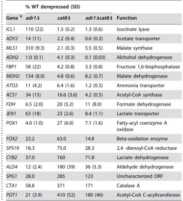
Microarray analysis under glucose-limiting conditions in strains deleted for either Adr1 and Cat8 singly or in combination identified over a hundred Adr1-dependent genes and nearly twice that many Cat8-dependent genes, with 30 genes overlapping these sets [3]. We confirmed the dependence on these transcription factors at a number of genes using real-time quantitative PCR (QPCR) from mRNA isolated from a wild-type strain, aadr1Dstrain, acat8Dstrain and a adr1Dcat8Dstrain (Table 1).ADH2, the canonical Adr1-dependent gene, but which is also known to be both bound and regulated by Cat8 [9], showed a strong dependence on both Adr1 and Cat8 for expression. Some genes showed a strong requirement for just Cat8
(ICL1, MLS1 and MDH2), whereas others (SPG1, CYB2, CTA1,
POT1 and ALD4) were only dependent on Adr1. Many genes, however, showed a dependence on both Adr1 and Cat8 that varied between these extremes. We note that at these extremes, however, loss of the unnecessary factor (i.e. loss of Adr1 atMLS1or loss of Cat8 at SPG1) resulted in significantly higher than wild-type levels of expression. This may indicate a metabolic requirement for elevated transcription of some genes to compensate for the loss of other gene products resultant in the absence of activator. Of the strongly Adr1-dependent genes, onlySPG1contains a consensus CSRE, making it unlikely that Cat8 is acting as a direct repressor. All genes tested, even those that are strongly Cat8-dependent, contain Adr1 binding sites, and furthermore, binding of Adr1 has been observed at a number of these promoters ([9] and references therein).
Expression data is accurately reflected in the
observed occupancy of Pol II and TFIIB
An important function of activators is to recruit and stabilize coactivators and the transcription machinery. Recruitment, therefore, was a candidate for differential roles of Adr1 and Cat8. Using chromatin immunoprecipitation (ChIP) we examined this possibility. We narrowed our recruitment studies to four genes,
ADH2, ADY2, FBP1andJEN1, all of which are dependent on both
factors but to differing degrees. In addition, ADH2, ADY2 and FBP1have well-characterized promoters.
We first assayed binding of RNA polymerase II (Pol II). A 5-fold increase on average over the amount of pol II detected at the promoter in repressing conditions was generally observed upon derepression. The amount of pol II at each of the glucose-regulated promoters in repressing conditions was not elevated compared to a region of the telomere not expected to be bound by pol II (additional negative control regions included the coding region of the Pol I structural gene and theADH2ORF yielded the same results). This was true for all of the coactivators we studied (with the exception of TFIID, see below) so data was shown only for derepressed conditions. For mutant strains, the results were expressed as the percent of the factor bound compared to 100% binding in the wild-type strain, both in derepressing conditions. Pol II binding, based on three biological samples, was generally consistent with expression data (compare Fig. 1A and Table 1). The notable exception was the greater than wildtype occupancy atFBP1inadr1Dstrain. Although Adr1 is not the dominant factor at this gene in terms of expression, it does bind to this promoter [9] and has a minor affect on expression. The increased levels of Pol II in this background may indicate that Adr1 does have a small role and that its deletion requires compensation by increased levels of coactivators.
Table 1.Expression in activator mutantsa
. . . .
% WT derepressed (SD)
Geneb adr1D cat8D adr1Dcat8D Function
ICL1 110 (22) 1.5 (0.2) 1.3 (0.6) Isocitrate lyase
ADY2 14 (11) 2.2 (0.4) 0.6 (0.3) Acetate transporter
MLS1 310 (9.3) 2.1 (0.3) 5.5 (0.5) Malate synthase
ADH2 1.0 (0.1) 4.1 (0.3) 0.1 (0.03) Alchohol dehydrogenase
FBP1 58 (22) 4.2 (0.8) 3.3 (0.8) Fructose 1,6-bisphosphatase
MDH2 154 (8.0) 4.8 (0.6) 8.2 (0.7) Malate dehydrogenase
ATO3 11 (4.2) 6.4 (1.6) 1.2 (0.3) Ammonia transporter
ACS1 34 (15) 16.6 (3.6) 4.2 (0.5) Acetyl-CoA synthase
FDH 6.5 (2.0) 20 (5.2) 11 (8.0) Formate dehydrogenase
JEN1 63 (18) 23 (2.6) 8.4 (1.1) Lactate transporter
POX1 4.0 (1.0) 27 (6.0) 7.1 (1.6) Fatty-acyl coenzyme A oxidase
FOX2 22.2 63.0 14.8 Beta-oxidation enzyme
SPS19 18.3 75.0 28.3 2,4 -dienoyl-CoA reductase
CYB2 37.0 160 71.8 Lactate dehydrogenase
ALD4 12 (2.4) 180 (39) 36 (5.3) Aldehyde dehydrogenase
SPG1 28.0 285 123 Uncharacterized ORF
CTA1 58.8 371 171 Catalase A
POT1 21 (3.9) 410 (52) 180 (46) Acetyl-CoA C-acyltransferase
a
Values are the average of 3 biological samples, each one quantitated in duplicate and normalized to ACT1 values. The standard deviation is shown in parenthesis, except in cases where the samples were pooled prior to qPCR. b
Genes are arranged in descending order of Cat8-dependence doi:10.1371/journal.pone.0001436.t001
....
...
...
....
...
...
....
...
...
....
...
...
....
...
...
....
...
...
....
...
...
....
...
...
....
...
...
....
...
To further confirm the presence of the Pol II machinery, occupancy at the promoter by TFIIB was determined (Fig. 1B). Again, TFIIB binding is consistent with expression data, although there is some activator-independent binding atFBP1andJEN1. It is unsurprising that the patterns of Pol II recruitment closely matched those of TFIIB, given their tight association. We did notice, however, that levels of TFIIB occupancy in activator mutants was generally higher than the occupancy of Pol II, which may be an artifact of the ChIP assay (i.e.TFIIB cross-links more efficiently), or may represent a more stable association at the promoter, even in the absence of activator. We did not observe either Pol II or TFIIB at any promoter under repressed conditions, as compared to a negative control locus (data not shown).
Adr1 and Cat8 recruit an array of coactivators upon
derepression
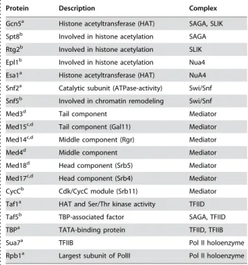
We extended our ChIP analysis to study the recruitment of a number of coactivators, in addition to components of the general transcription machinery. To assay occupancy at the promoter, we
used epitope tagged representative proteins from each complex (Table 2) to facilitate ChIP analysis in either a wildtype,adr1D, cat8D
or adr1Dcat8D strain. We looked at the recruitment of several chromatin remodeling complexes (SAGA, NuA4 and Swi/Snf), the Mediator complex, and TFIID (Fig 1C–H). In no case did we observe binding at any promoter tested under repressed conditions, and we present here data after four hours of derepression.
The requirement for SAGA is not universal at all yeast promoters, as there are reported examples of activated transcrip-tion in the absence of both the histone acetyl transferase (HAT) function of SAGA as well as its non-HAT function [14]. Gcn5, the HAT component of SAGA, which is specific for histone H3, was observed at the promoter regions of all four genes tested under derepressing conditions in a wildtype strain. At the ADY2 and FBP1 promoters, both Adr1 and Cat8 are required for recruitment, but at ADH2 and JEN1, Adr1 alone sufficed for more than 50% of the recruitment (Fig. 1C). Gcn5 is also a member of the SLIK (SAGA-like) complex [15]. In order to determine whether or not Gcn5 was being recruited as part of SAGA or SLIK, we tagged unique components of both of these Figure 1. Recruitment profiles under derepressed conditions. A–G:ChIP analysis for coactivators/general transcription machinery at the indicated promoter regions in a wildtype strain (black),adr1D(pink),cat8D(blue) andadr1Dcat8D(green). Binding is expressed as the percent of the wildtype derepressed value (set to 100%) after normalization to theTELnegative control locus. Error bars represent the standard deviation of biological replicates (two or more). Data is shown at 4 hours of derepression. H: ChIP analysis for Taf1 in repressing (grey bars) and derepressing (four hours, black bars) conditions. Error bars represent technical replicates. Inset: ChIP analysis by PCR at for Taf1 atACT1. The protein(s) assayed for in each case is listed in Table 2.
complexes and observed the identical pattern of recruitment only with the SAGA-specific protein (Spt8) and not with the SLIK-specific protein (Rtg2) (which was detected at the SLIK-dependent promoterCIT2) (data not shown). Esa1, the HAT component of NuA4, which mainly acetylates the H4 tail, bound to promoters upon derepression, however in contrast to SAGA, its recruitment was largely activator independent, suggesting it may play a global, rather than targeted role (Fig. 1D). Similar results were obtained with a second component of NuA4, Epl1 (data not shown).
Regulation ofADH2has been reported in the literature to be both dependent on the ATP-dependent remodeling complex Swi/ Snf, and independent of it [16], [17]. Our ChIP data for Snf2, the catalytic subunit of Swi/Snf, suggest an activating role for this complex at this subset of genes (Fig. 1E). The recruitment of Swi/ Snf was strongly dependent on Adr1 at all promoters tested, and also strongly dependent on Cat8 at ADH2 and FBP1. The contribution to its recruitment by Cat8 is reduced but still observable atADY2andJEN1. We also looked at the binding of Snf5 and saw a similar pattern of recruitment (data not shown).
Recent reports on the importance of Mediator reached contradictory conclusions, with some data suggesting Mediator is involved at only a small fraction of yeast genes, and then only when cells are under stress [18], and other data arguing that Mediator is as important as other general transcription factors [19]. It has also been reported that Mediator occupies not only promoter regions, but ORFs as well, and that this occupancy is not necessarily correlated with gene activation [20]. The importance of Mediator under respiratory conditions, however, has never been explored. We first determined the occupancy of Mediator (based on six components, two from each submodule) at both the promoters and ORFs of ADH2 and ADH1 (Table 3). ADH1 is highly expressed under glucose conditions, and down regulated
under derepressed conditions. We find that all components of Mediator follow the same pattern of binding, namely, when the gene is active, the complex is found at the promoter. We detected Mediator in the ORF of ADH1 under repressed (activating) conditions, but at a much lower amount than in the promoter. For the genes we assayed, Mediator occupancy was highest at the promoter under activating conditions.
The analysis of Mediator was extended to look at the involvement of activators using three components (Med15 (Gal11), Med14 (Rgr1) and Med17 (Srb4). The patterns of occupancy for these three components were the same and therefore results were averaged together (Fig. 1F). Activator-dependency was expected given the putative role of Mediator as a bridge between transcription factors and general transcription machinery, although at most promoters, one activator by itself was enough to achieve maximal or near-maximal recruitment. Binding was also observed for CycC atADH2 under derepressed conditions (data not shown). The presence of CycC/Cdk8, commonly thought of as a repressor [10], along with the rest of Mediator under activating conditions argues against any repressive role of Mediator at these genes.
This subset of genes contains TATA-boxes, so TBP occupancy is expected upon activation. ChIP for TBP reflects this requirement, as a small but significant increase in binding occurs upon derepression. It was difficult to assess the role of activators in this recruitment due to the small range of signal (Fig. 1G). It is clear, however, that Adr1 (and not Cat8) is important for proper recruitment of TBP atADH2andADY2. TBP was expressed from a 2mplasmid, and the resulting high levels of TBP may contribute to non-specific binding, accounting for the low specific signals.
ChIP for TFIID, using Taf1 (TafII145) (which was not identified as part of SAGA [21]), suggests that under normal conditions (e.g. in a wildtype strain), these four glucose-repressed genes are TAF-independent, as there is no observable increase in binding upon derepression of a wild-type strain (Fig. 1H). Deletion of activators did not effect the apparent level of Taf1 binding (data not shown). To confirm these results, we looked at the promoter of a gene previously reported to be TAF-dependent, ACT1 [22], and accordingly saw a large signal under repressed conditions when this gene is more active, and a weaker signal under derepressed conditions when activity decreases (Fig. 1H, inset). Previous reports suggesting that TFIID interacts with Adr1 and is important for ADH2 expression were based primarily on Taf5 (TAFII90) [23], which has since been shown to be a member of both SAGA and TFIID [21], leading to misinterpretation of the importance of TFIID. We detected Taf5 at all four of these promoters, in a similar pattern as Gcn5 (data not shown), further supporting the conclusion that SAGA is required for activation, and TFIID is not. Table 2.Coactivators used in ChIP Analysis
. . . .
Protein Description Complex
Gcn5a Histone acetyltransferase (HAT) SAGA, SLIK
Spt8b Involved in histone acetylation SAGA
Rtg2b Involved in histone acetylation SLIK
Epl1b Involved in histone acetylation Nua4
Esa1a Histone acetyltransferase (HAT) NuA4
Snf2a Catalytic subunit (ATPase-activity) Swi/Snf
Snf5b Involved in chromatin remodeling Swi/Snf
Med3d Tail component Mediator
Med15c,d Tail component (Gal11) Mediator
Med14c,d Middle component (Rgr) Mediator
Med4d Middle component Mediator
Med18d Head component (Srb5) Mediator
Med17c,d Head component (Srb4) Mediator
CycCb Cdk/CycC module (Srb11) Mediator
Taf1a HAT and Ser/Thr kinase activity TFIID
Taf5b TBP-associated factor SAGA, TFIID
TBPa TATA-binding protein TFIID, TFIIB
Sua7a TFIIB Pol II holoenzyme
Rpb1a Largest subunit of PolII Pol II holoenzyme
a
Results in Figure 1 b
Results not shown c
Results are averaged and shown in Figure 1f dResults shown in Table 3
doi:10.1371/journal.pone.0001436.t002
...
...
...
....
...
...
....
...
...
....
...
...
....
...
...
....
...
...
....
...
...
....
...
...
....
...
...
....
...
.
Table 3.ChIP Analysis of Mediator Componentsa
. . . .
ADH1 ADH2
Promoter ORF Promoter ORF
Component R D R D R D R D
Med3 6.5 1.5 2.5 0.83 1.6 3.7 0.56 1.1
Med15 7.1 2.1 2.0 0.88 1.3 4.7 0.60 1.5
Med4 9.4 2.4 2.6 1.5 1.1 4.5 0.78 2.3
Med14 11 1.5 2.8 1.1 1.5 3.8 1.0 1.3
Med17 22 3.0 8.6 1.4 0.84 2.1 0.98 0.40
Med18 12 1.5 7.1 2.1 1.6 4.1 1.5 1.9
aValues expressed as the %IP’ed divided by the %IP’ed at theTEL doi:10.1371/journal.pone.0001436.t003
....
...
....
...
...
....
...
...
....
...
...
....
...
...
Recruitment of these coactivators directly affects
expression of this subset of genes
Recruitment of coactivators to a promoter indicates that they play a direct role. However, due to the redundancy of coactivator function, it is important to assess their importance in transcription by gene expression studies. To assess this, we isolated mRNA from strains defective in SAGA, Swi/Snf, or Mediator and looked at expression levels over several hours of derepression, after normalization to ACT1 (Table 4). The loss of Gcn5 led to reductions in expression ofADH2andFBP1. Deletion of Ada1, an adaptor protein necessary for the structural integrity of SAGA, led to a more severe diminution of gene expression of ADH2 and FBP1, as well as reductions in expression of ADY2 and JEN1 suggesting that SAGA is important for appropriate gene expression both in its capacity as a HAT as well as another, non-HAT function.
Swi/Snf also plays an important role in regulating expression of these genes (Table 4), as shown by the strong reduction in transcription of either asnf2Dorsnf5Dstrain in the case ofADH2 andFBP1. The effect of the coactivator deletion was less severe at ADY2andJEN1in the case ofsnf2D, but all genes were affected by the loss of Snf5.
Using a temperature sensitive (ts) mutation in Med17, we were able to study the affect of inactivating Mediator. Expression of most of the genes assayed decreased at least 10-fold when Med17 was inactivated (Table 4).ADH2was an exception to the near total loss of activity in this strain, with levels still 19% of the wild-type expression. In summary, the expression studies support the conclusions reached from coactivator recruitment. Saga, Swi/ Snf1, and Mediator are recruited in a glucose-sensitive manner to the promoters of all four glucose-regulated genes, and all four of them play an important role in transcription.
Because genome-wide studies indicated that the defects caused by loss of these coactivators are widespread [10], we also measured the expression of the activators themselves throughout derepres-sion (Table 4). In large part, transcription levels of Adr1 and Cat8 are unaffected by loss of these coactivators. The slight decreases observed are likely due to a combination of global reduction in transcription and slow growth of these strains, in accordance with the severity of the mutation. Adr1 binding persists at wild-type levels in thegcn5D andmed17 ts strains, indicating that although protein levels may be diminished up to 50% compared to a wild-type strain, there is still enough transcription factor to fully occupy the promoter (data not shown).
Chromatin remodeling at the ADH2 and FBP1
promoters have different requirements for Adr1 and
Cat8
Differential recruitment of known coactivators seems unable to explain the dependence of gene expression on both Adr1 and Cat8. Specific recruitment of unknown coactivators may explain this apparent conundrum. Another possibility is that a step in gene activation requires the simultaneous participation of both Adr1 and Cat8. Previous studies in our lab and others show that specific changes in promoter architecture at bothADH2andFBP1occur upon derepression and these changes do not occur in aadr1cat8 double mutant [24], (Infante, unpublished). To determine whether or not the two activators have redundant roles in terms of chromatin structure, we used a nucleosome scanning assay, or NuSA, to measure both nucleosome occupancy and location within the promoter in different yeast strains [25]. Figure 2A shows chromatin remodeling at theADH2promoter in a wild-type strain upon derepression: there is a reduction in protection at all 3 nucleosome positions as predicted [25]. These changes depended on both Adr1 and Cat8, as loss of either one of these activators in derepressing conditions abolished this remodeling. These findings agree with the expression data, in that both activators are required for a substantial increase in expression upon derepression (Table 1). The NuSA at theFBP1promoter with a wild-type strain showed changes in chromatin structure upon derepression–a major reduction of protection at N-1 and N-2 and a shift in position of N-2 (Fig. 2B) ([25], Infante unpublished data). However, in this case, differences were seen between the two deletion strains. In the
Dadr1strain, a significant amount of remodeling occurred. There was a major reduction of occupancy at N-1, occupancy at N-2 was reduced to approximately 50% of repressed levels and the position of N-2 shifted downstream as in wildtype under derepressing conditions. Remodeling in the absence of Cat8, however, was reduced. Some reduction in the N-1 occupancy occurred but less than in theDadr1strain, and the occupancy of N-2 did not change, other than a partial shift in the position. Again the FBP1 chromatin remodeling agrees with the expression data, in that there is a role for both Adr1 and Cat8, albeit an unequal one.
Over expression of Adr1 compensates for loss of
Cat8 at most genes
The ChIP experiments revealed that both Adr1 and Cat8 are inherently capable of recruiting each coactivator individually (i.e. no coactivator is absolutely dependent on both Adr1 and Cat8 for recruitment) (Fig. 1, and discussion); however, both are necessary for complete chromatin remodeling. This led us to question whether or not they perform unique non-recruitment functions at the promoter, or if there is simply a requirement for a threshold level of activator, achieved at some promoters through a combination of Adr1 and Cat8 and at other promoters largely through only one activator. In order to investigate this possibility, we looked at the levels of activation in strains over-expressing Adr1 in the presence or absence of Cat8. This was done by creating strains chromosomally deleted for either Adr1 or Adr1 and Cat8 together, and then expressing Adr1 from a high-copy plasmid under control of theADH1 promoter. At most genes that were dependent on both Adr1 and Cat8, over-expression of Adr1 greatly reduced the requirement for Cat8 (Table 5). Even at the strongly Cat8-dependent genesICL1, MLS1andMDH2,there was a reduction in the dependence on Cat8, but not full compensation for its loss. Adr1 does bind to all three of these promoters, suggesting this is a direct effect, despite the fact that normally Adr1 Table 4.Gene Expression Levels in Coactivator Deletion
Strainsa
. . . .
strain
Gene gcn5D ada1D snf2D snf5D med17 ts
ADH2 65 3.6 19 1.8 19
ADY2 110 10 77 11 5.3
FBP1 36 1.7 23 0.27 1.30
JEN1 190 47 63 7.0 9.0
ADR1 45 61 160 95 110
CAT8 65 77 110 88 53
aValues expressed as % wildtype after 4 hours of derepression doi:10.1371/journal.pone.0001436.t004
...
....
...
...
....
...
...
....
...
...
....
...
...
....
is not required for activation [9] (Table 1).FBP1was the sole gene where excess Adr1 cannot alleviate the requirement for Cat8.
We used NuSA to ask whether or not the mechanism by which excess Adr1 efficiently activates ADH2 but not FBP1 can be explained in terms of chromatin remodeling. Figure 2C shows that the remodeling at theADH2promoter in strains over expressing Adr1 was nearly identical to the remodeling seen in the derepressed wildtype strain, regardless of Cat8, in that protection at N-1 and N-2 was very low compared to protection at CEN3. The same was seen at FBP1 (Fig. 2D), where protection in derepressing conditions was similar to the wild-type levels when Adr1 was over expressed, again regardless of Cat8. The lack of full expression ofFBP1in the absence of Cat8 cannot be attributed to chromatin remodeling and may be due to missing or reduced levels of coactivators that are normally recruited by this activator.
DISCUSSION
A consistent set of coactivators is recruited to and
regulates glucose-repressed genes
We have established a set of coactivators that are directly involved in the regulation of glucose-repressed genes based on ChIP and expression studies. In a wild-type strain, recruitment of coactiva-tors did not vary from promoter to promoter. SAGA, Swi/Snf, and Mediator complexes, but not NuA4 or TFIID, are all directly involved in transcription of these genes.
We confirmed the genetic evidence that SAGA plays a role in expression of these genes and extended the analysis to show that it is required in both a HAT and non-HAT capacity. Recruitment of the HAT component Gcn5 (Fig. 1C) demonstrates a direct role for it at the promoter, and the expression data in agcn5Dstrain supports this conclusion (Table 4). The increased severity of the defect in
activation seen in theada1Dstrain, however, signifies that SAGA is required beyond just its function as a HAT. Furthermore, the absence of TFIID (Fig. 1H) suggests that the additional role of SAGA is to recruit TBP, a known alternative to its recruitment as part of TFIID [26]. NuA4, the other HAT complex thought to be involved in regulation of these genes, showed weak derepression-dependent binding (,2 fold increase over background from repressed to derepressed), but it appeared to be largely independent of activators (Fig. 1D). We conclude that it does not have a targeted role. This is supported by other findings of NuA4 as a global regulator of acetylation levels (reviewed in [27]). Esa1, the HAT component, has been previously shown to affect expression of ADH2 and other glucose-regulated genes [28], in agreement with our ChIP data.
The ATP-dependent chromatin remodeling complex Swi/Snf is part of the set of coactivators required for appropriate expression of glucose-repressed genes. Based on ChIP evidence (Fig. 1E) and the expression data from two separate deletions (snf2Dandsnf5D, Table 2) performed in biological duplicate, we conclude Swi/Snf is involved in direct regulation ofADH2andFBP1. The variation in expression betweensnf2D and snf5D may reflect a less stringent requirement for this coactivator. Indeed, previous literature gives conflicting results as to the importance of this complex in regulation of ADH2, which may have arisen due to differences in derepressing media, time of derepression, and choice of subunit. In agreement with genome-wide localization studies [10] and recentin vitrotranscription assays [19], we found an important role for Mediator in activation. Strong binding under derepressed conditions was found for three Mediator components. Our results (Fig. 1F and Table 3) indicate that it is required, as a whole complex (i.e. all submodules act in concert) for expression of these genes. Further-more, we found no evidence of Mediator acting as a repressor.
A recent genome-wide study by Steinmetz et al reported that Pol II occupancy does not strictly correlate with gene activity [29], Figure 2. Differential roles for Adr1 and Cat8 in chromatin remodeling atADH2andFBP1.NuSA results are displayed as the amount of relative protection, after normalization to the well-positioned nucleosome at CEN3. The position of each amplicon (referenced to the middle of each amplicon) within the promoter is shown on the x-axis, with approximate location of nucleosomes shown. (A) and (C) are the results atADH2,(B) and (D) are atFBP1. (A) and (B) compare NuSA results ofDadr1 (pink) andDcat8(green) in derepressed conditions to a wildtype strain either in repressed (dark blue) or derepressed (light blue) conditions. (C) and (D) compare NuSA results between over-expression of Adr1 with (red) or without Cat8 (blue) in derepressed conditions.
opening up the question of whether pre-formed preinitiation complexes (PICs) may exist at promoters which are inactive, but poised for fast release from repression, in a similar fashion to what is observed at the heat shock promoters [30]. Despite the relative rapid kinetics of derepression (polymerase can be detected at the promoter as early as ten minutes after glucose depletion (data not shown)), we do not find any evidence for a pre-assembled PIC at these promoters, as neither Pol II nor TFIIB are detectable at promoter regions prior to derepression (Fig. 1A and B).
Adr1 and Cat8 are not intrinsically unique in their
functions
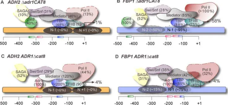
The possibility that these two activators made unique contributions via their ability to recruit different coactivators is ruled out through our extensive ChIP experiments, demonstrating that both Adr1 and Cat8 are capable of recruiting any of the coactivators we studied. Figure 3 summarizes the roles of Adr1 and Cat8 at two contrasting genes,ADH2(A and C) andFBP1(B and D). The fact that Mediator, for example, is recruited largely by Adr1 atADH2, but by Cat8 at FBP1argues that protein-protein interactions are not the governing principle, but rather that it is promoter context that dictates which transcription factor dominates these interactions (see below).
Another possible manner in which they may make distinct contributions to regulation is via chromatin remodeling. This is evidenced by the fact that Adr1 and Cat8 are differentially required for nucleosome remodeling atADH2andFBP1(Fig. 3). The non-recruitment function, be it solely through chromatin remodeling or through an unidentified action, may not be very significant, however, in light of our finding that excess Adr1 compensates for the loss of Cat8 in the activation of co-regulated genes (Table 5). At promoters where Cat8 is largely responsible for recruitment (i.e. FBP1), excess Adr1 reduces the need for Cat8, but does not abolish it.
Table 5.Expression incat8Dstrains with single copy or multi-copy Adr1
. . . .
%CAT8 derepressed (SD)
Gene single copya multi-copyb
ICL1 1.5 (0.2) 14 (7.4)
ADY2 2.2 (0.4) 75 (47)
MLS1 2.1 (0.3) 44 (56)
ADH2 4.1 (0.3) 104 (5.3)
FBP1 4.2 (0.8) 5.7 (7.2)
MDH2 4.8 (0.6) 35 (11)
ATO3 6.4 (1.6) 350 (47)
ACS1 16.6 (3.6) 160 (48)
FDH 20 (5.2) 2900 (670)
JEN1 23 (2.6) 1200 (180)
POX1 27 (6.0) 830 (180)
FOX2 63.0 130 (10)
SPS19 75.0 130 (49)
CYB2 160 150 (83)
ALD4 180 (39) 800 (110)
SPG1 285 310 (0)
CTA1 371 240 (39)
POT1 410 (52) 290 (54)
a
Data taken from Table 1 (% WT strain)
bValues are an average of biological duplicates after 4 hours of derepression, normalized toACT1and expressed as the percent of activation in a multi-copy Adr1CAT8strain, with the standard deviation shown in parenthesis. doi:10.1371/journal.pone.0001436.t005
....
....
...
...
....
...
...
....
...
...
....
...
...
....
...
...
....
...
...
....
...
...
....
...
...
....
...
...
....
...
Figure 3. Adr1 and Cat8 play different roles atADH2andFBP1.Comparison between theADH2(A and C, orange) andFBP1(B and D, blue) promoters in eitheradr1D (A and B) or acat8D(C and D). Combined expression, recruitment and chromatin remodeling data at 4 hours of derepression are shown. Values in parenthesis indicate the percent binding of the wild-type derepressed value, after normalization (an asterisk represents previously published data [9]). Gene activation is reported as the percent of the wild-type derepressed level. Binding sites are shown at their approximate locations along the promoter (Cat8 in green, Adr1 in pink, and TATA box in blue). Nucleosome positions under derepressed conditions are depicted as shaded ovals on the promoter, and the degree of chromatin remodeling estimated as a percent of the remodeling observed in a wild-type strain. Locations of coactivators are not intended to reflect interactions.
Through mass action, high levels of Adr1 may be able to recruit and stably retain more coactivators than it would at wild-type levels. At promoters where Adr1 primarily recruits coactivators (i.e. ADH2), Cat8 is dispensable when Adr1 is over expressed, despite what is otherwise a strong requirement for Cat8 in terms of expression. Since the binding sites recognized by Adr1 and Cat8 are very dissimilar, the possibility that stable PIC formation requires the DNA to be bound at two positions or by two separate factors at these promoters is ruled out. Additionally, there is no consistent arrangement of the Adr1 and Cat8 binding sites within promoters of co-regulated genes [9], supporting the conclusion that two separate activator binding events are not necessary for activation of these genes.
Interestingly, we observed that multi-copy Adr1 strains lacking Cat8 fail to grow on glycerol, ethanol, acetate and lactate, all carbon sources whose utilization normally require Cat8 (data not shown) (reviewed in [31] and references therein). These observa-tions suggest that the functional redundancy between these activators appears to be limited to a subset of their target genes, perhaps indicative that they have specialized roles during growth on specific non-fermentable carbon sources. Although we did not perform the converse experiment with over expressed Cat8, we predict that this would yield similar results, with compensation for loss of Adr1 at most genes in this subset, with the exception of those that do not have Cat8 binding sites (POT1,for example).
Promoter architecture may dictate the roles that
activators assume at each promoter
In wild-type cells, even at co-regulated genes, subtle differences exist between the roles of Adr1 and Cat8. While they may both share the function of recruiting and stabilizing coactivators, the fact that some promoters display a bias for Adr1 while others depend primarily on Cat8, indicates that under physiological conditions factors exist which dictate which activator dominates regulation (or if both activators will contribute equally). A candidate for such a factor is the promoter architecture, which includes such things as the number and position of nucleosomes, the arrangement of binding sites, and the overall spacing of elements throughout the promoter. For example, it has been demonstrated that the correct helical phasing of Adr1 and Cat8-binding sites is required for wild-type levels of expression of ADH2 [32]. Similar constraints may play a role in the choice of Adr1 versus Cat8 as the more prominent activator at other co-regulated genes.
Nucleosome positioning is another factor that may be important. For example, ADH2 has a repressive chromatin structure under high glucose conditions, with significant remod-eling of the nucleosomes upon activation (unpublished data), [28]. The preference for Adr1 at ADH2 may be a result of the fact that the UAS1 is in a nucleosome-free region, whereas at FBP1, the presumed Adr1 binding site is less accessible due to the position of the 21 nucleosome (Fig. 2), and so Cat8, which binds in a nucleosome-free region, becomes the dominant factor.
Based on our studies with these four genes, it appears that despite an overarching shared regulation, the finer workings of the mechanism of activation remain varied. Although there are some commonalities (a consistent set of coactivators), we conclude that the mechanism of activation must be examined on a promoter by promoter basis, even amongst highly related genes. Additionally, the fact that we see no inherent differences in the abilities of Adr1 and Cat8 to recruit coactivators and yet some genes are more heavily dependent on one than the other further supports the conclusion that promoter architecture is equally important for activated transcription.
MATERIALS AND METHODS
Yeast strains and growth of cultures
All strains used in this study were derived from W303. Deletions of activators and epitope tagging of all protein components were introduced according to previously published work [33], [34]. The
med17tsstrain was constructed by first transforming with asrb4-138–
containing URA3 plasmid, followed by deletion of chromosomal Med17 according to [33]. TBP was C-terminally 13-Myc tagged and expressed from a 2mplasmid. Cultures were grown in YPD (with the exception of strains carrying the TBP plasmid, which were grown in SM lacking leucine for plasmid selection) with either 5% glucose (repressing conditions) or 0.05% glucose (derepressing conditions) at 30u. For themed17 tsexperiments, cultures were grown overnight at room temperature and then shifted to 37u for 30 minutes before taking the repressed sample; cultures were then spun down in a pre-warmed centrifuge and resuspended in 37ulow glucose media and grown for 4 hours. Wild-type strains were grown in the same manner for comparison to eliminate indirect effects of the high temperature. Strains over-expressing Adr1 were created by introducing plasmid YEpNKA1(URA), based on pKD17, which expresses Adr1 from the ADH1promoter, with only a minor modification from its original form [35] in a strain chromosomally deleted for Adr1 or both Adr1 and Cat8. For comparison, the single copy Adr1 strain was created in the same strain background using pKD16, a centromeric plasmid carrying a single copy ofADR1with its endogenous promoter [35]. These strains were grown in synthetic media lacking uracil or tryptophan, respectively, for plasmid selection with either 5% glucose (repressed) or 0.05% glucose (derepressed).
ChIP and real-time PCR (QPCR)
ChIP was performed as previously described, using both dimethy-ladipimate (Pierce chemicals) and formaldehyde as crosslinking agents and using QPCR instead of standard PCR (Tachibana, 2005). We note that the use of an additional cross-linker did make a substantial difference in the observed binding for some proteins. Monoclonal antibodies against c-Myc (9E10)(Santa Cruz Biotech-nology sc-40) and HA (F-7)(Santa Cruz BiotechBiotech-nology sc-7293) epitopes were used for all immunoprecipitations except for Pol II, which was immunoprecipitated using the 8WG16 antibody (Abcam ab817). Primers (IDT) for gene-specific QPCR were designed to cover promoter regions. Sequences are available upon request. QPCR was performed on an MJ Research DNA Engine using Power-SYBR Green (Applied Biosystems). Samples were run in triplicate and relative amounts of DNA were calculated using a standard curve generated from serial dilutions of the input DNA. Standard curves were included for every primer set. Experiments were performed in biological duplicate or triplicate and all results were averaged together. Except where noted, values were calculated as the ratio of the percent immunoprecipitation of ChIP to input at the specific locus to the percent immunoprecipitation at the telomeric region. For repressed samples, this ratio was approximately one. Derepressed values were expressed as the percent of the wild-type value. The associated error results from the standard deviation of the biological replicates. Additional analysis using previously published methods [34], [36] all yielded similar patterns (data not shown).
mRNA isolation and QPCR
performing QPCR. Gene-specific QPCR was performed in duplicate using primers near the 39end of the ORF regions (sequences available upon request). Relative amounts were obtained by comparison to a standard curve and normalized to ACT1 levels. Samples were prepared from biological triplicates and quantitated in duplicate.
Nucleosome scanning assay
(NuSA)
Two hundred mL cell cultures in either repressing or derepressing conditions were processed using the procedures in the yeast culture, micrococcal nuclease digestion, protein degradation and DNA purification steps as outlined in [38]. Specifically, repressed cells were incubated with zymolyase for 15 min. at 30uC while derepressed cells were incubated for 45 min. After the RNAse A digestion, samples were analyzed on 2%-agarose gel to determine the extent of digestions and only samples that were highly enriched in mononucleosomal DNA were gel-extracted using a Qiagen gel extraction kit. DNA samples were diluted 1/300–1/500 and used in QPCR reactions to quantify the presence of a specific amplicon. The
protection value set for each amplicon corresponds to the fold-enrichment of that amplicon in the mononucleosomal DNA compared to the undigested-sample and normalized toCEN3values. The amplicon used forCEN3covered the region shown previously to have a well-positioned nucleosome [39]. QPCR primers were designed to cover the promoter ofADH2andFBP1with amplicons averaging 100 bp in size (sequences available upon request).
ACKNOWLEDGMENTS
We thank K. Khaw for creation ofada1D, gcn5Dandsnf2Dstrains and E. Arms for her contribution to the expression studies in activator mutants as well as in the strains over-expressing Adr1. We also thank C. Tachibana for her insightful discussions. The TBP plasmid was a gift from S. Hahn.
Author Contributions
Conceived and designed the experiments: EY GL RB. Performed the experiments: EY GL RB. Analyzed the data: EY GL RB. Contributed reagents/materials/analysis tools: GL RB. Wrote the paper: RB.
REFERENCES
1. DeRisi JL, Iyer VR, Brown PO (1997) Exploring the metabolic and genetic control of gene expression on a genomic scale. Science 278: 680–686. 2. Hardie DG, Carling D, Carlson M (1998) The AMP-activated/SNF1 protein
kinase subfamily: metabolic sensors of the eukaryotic cell? Annu Rev Biochem 67: 821–855.
3. Young ET, Dombek KM, Tachibana C, Ideker T (2003) Multiple pathways are co-regulated by the protein kinase Snf1 and the transcription factors Adr1 and Cat8. J Biol Chem 278: 26146–26158.
4. Haurie V, Perrot M, Mini T, Jeno P, Sagliocco F, et al. (2001) The transcriptional activator Cat8p provides a major contribution to the repro-gramming of carbon metabolism during the diauxic shift in Saccharomyces cerevisiae. J Biol Chem 276: 76–85.
5. Kratzer S, Schuller HJ (1997) Transcriptional control of the yeast acetyl-CoA synthetase gene, ACS1, by the positive regulators CAT8 and ADR1 and the pleiotropic repressor UME6. Mol Microbiol 26: 631–641.
6. Karpichev IV, Small GM (1998) Global regulatory functions of Oaf1p and Pip2p (Oaf2p), transcription factors that regulate genes encoding peroxisomal proteins in Saccharomyces cerevisiae. Mol Cell Biol 18: 6560–6570. 7. Estruch F, Carlson M (1993) Two homologous zinc finger genes identified by
multicopy suppression in a SNF1 protein kinase mutant of Saccharomyces cerevisiae. Mol Cell Biol 13: 3872–3881.
8. Roth S, Kumme J, Schuller HJ (2004) Transcriptional activators Cat8 and Sip4 discriminate between sequence variants of the carbon source-responsive promoter element in the yeast Saccharomyces cerevisiae. Curr Genet 45: 121–128. 9. Tachibana C, Yoo JY, Tagne JB, Kacherovsky N, Lee TI, et al. (2005)
Combined global localization analysis and transcriptome data identify genes that are directly coregulated by Adr1 and Cat8. Mol Cell Biol 25: 2138–2146. 10. Holstege FC, Jennings EG, Wyrick JJ, Lee TI, Hengartner CJ, et al. (1998)
Dissecting the regulatory circuitry of a eukaryotic genome. Cell 95: 717–728. 11. Cosma MP, Tanaka T, Nasmyth K (1999) Ordered recruitment of transcription
and chromatin remodeling factors to a cell cycle- and developmentally regulated promoter. Cell 97: 299–311.
12. Bhoite LT, Yu Y, Stillman DJ (2001) The Swi5 activator recruits the Mediator complex to the HO promoter without RNA polymerase II. Genes Dev 15: 2457–2469.
13. Drysdale CM, Duenas E, Jackson BM, Reusser U, Braus GH, et al. (1995) The transcriptional activator GCN4 contains multiple activation domains that are critically dependent on hydrophobic amino acids. Mol Cell Biol 15: 1220–1233. 14. Lee TI, Causton HC, Holstege FC, Shen WC, Hannett N, et al. (2000) Redundant roles for the TFIID and SAGA complexes in global transcription. Nature 405: 701–704.
15. Pray-Grant MG, Schieltz D, McMahon SJ, Wood JM, Kennedy EL, et al. (2002) The novel SLIK histone acetyltransferase complex functions in the yeast retrograde response pathway. Mol Cell Biol 22: 8774–8786.
16. Taguchi AK, Young ET (1987) The cloning and mapping of ADR6, a gene required for sporulation and for expression of the alcohol dehydrogenase II isozyme from Saccharomyces cerevisiae. Genetics 116: 531–540.
17. Di Mauro E, Kendrew SG, Caserta M (2000) Two distinct nucleosome alterations characterize chromatin remodeling at the Saccharomyces cerevisiae ADH2 promoter. J Biol Chem 275: 7612–7618.
18. Fan X, Chou DM, Struhl K (2006) Activator-specific recruitment of Mediator in vivo. Nat Struct Mol Biol 13: 117–120.
19. Takagi Y, Kornberg RD (2006) Mediator as a general transcription factor. J Biol Chem 281: 80–89.
20. Andrau JC, van de Pasch L, Lijnzaad P, Bijma T, Koerkamp MG, et al. (2006) Genome-wide location of the coactivator mediator: Binding without activation and transient Cdk8 interaction on DNA. Mol Cell 22: 179–192.
21. Grant PA, Schieltz D, Pray-Grant MG, Steger DJ, Reese JC, et al. (1998) A subset of TAF(II)s are integral components of the SAGA complex required for nucleosome acetylation and transcriptional stimulation. Cell 94: 45–53. 22. Li XY, Bhaumik SR, Green MR (2000) Distinct classes of yeast promoters
revealed by differential TAF recruitment. Science 288: 1242–1244.
23. Komarnitsky PB, Klebanow ER, Weil PA, Denis CL (1998) ADR1-mediated transcriptional activation requires the presence of an intact TFIID complex. Mol Cell Biol 18: 5861–5867.
24. Di Mauro E, Verdone L, Chiappini B, Caserta M (2002) In vivo changes of nucleosome positioning in the pretranscription state. J Biol Chem 277: 7002–7009. 25. Tachibana C, Biddick R, Law GL, Young ET (2007) A poised initiation
complex is activated by Snf1. J Biol Chem 282: 37308–37315.
26. Dudley AM, Rougeulle C, Winston F (1999) The Spt components of SAGA facilitate TBP binding to a promoter at a post-activator-binding step in vivo. Genes Dev 13: 2940–2945.
27. Doyon Y, Cote J (2004) The highly conserved and multifunctional NuA4 HAT complex. Curr Opin Genet Dev 14: 147–154.
28. Agricola E, Verdone L, Xella B, Di Mauro E, Caserta M (2004) Common chromatin architecture, common chromatin remodeling, and common tran-scription kinetics of Adr1-dependent genes in Saccharomyces cerevisiae. Biochemistry 43: 8878–8884.
29. Steinmetz EJ, Warren CL, Kuehner JN, Panbehi B, Ansari AZ, et al. (2006) Genome-wide distribution of yeast RNA polymerase II and its control by Sen1 helicase. Mol Cell 24: 735–746.
30. Zanton SJ, Pugh BF (2006) Full and partial genome-wide assembly and disassembly of the yeast transcription machinery in response to heat shock. Genes Dev 20: 2250–2265.
31. Schuller HJ (2003) Transcriptional control of nonfermentative metabolism in the yeast Saccharomyces cerevisiae. Curr Genet 43: 139–160.
32. Donoviel MS, Kacherovsky N, Young ET (1995) Synergistic activation of ADH2 expression is sensitive to upstream activation sequence 2 (UAS2) orientation, copy number and UAS1-UAS2 helical phasing. Mol Cell Biol 15: 3442–3449. 33. Guldener U, Heck S, Fielder T, Beinhauer J, Hegemann JH (1996) A new
efficient gene disruption cassette for repeated use in budding yeast. Nucleic Acids Res 24: 2519–2524.
34. Knop M, Siegers K, Pereira G, Zachariae W, Winsor B, et al. (1999) Epitope tagging of yeast genes using a PCR-based strategy: more tags and improved practical routines. Yeast 15: 963–972.
35. Dombek KM, Young ET (1997) Cyclic AMP-dependent protein kinase inhibits ADH2 expression in part by decreasing expression of the transcription factor gene ADR1. Mol Cell Biol 17: 1450–1458.
36. Agricola E, Verdone L, Di Mauro E, Caserta M (2006) H4 acetylation does not replace H3 acetylation in chromatin remodelling and transcription activation of Adr1-dependent genes. Mol Microbiol 62: 1433–1446.
37. Collart MAaO, S (2005) ‘‘Preparation of Yeast RNA by extraction with hot acidic phenol’’. In: Ausubel FM, Brent R, Kingston RE, Moore DD, Seidman JG, Smith JA, Struhl K, eds (2005) Current Protocols in Molecular Biology. New York: Greene/Wiley Interscience. pp 13.12.11–13.12.15. 38. Liu CL, Kaplan T, Kim M, Buratowski S, Schreiber SL, et al. (2005)