Normal expression of IFN-
γ
γ
γ
γ
γ
R in
four patients with uncommon
mycobacterial infection phenotypes
1Grupos de Inmunodeficiencias Primarias and 2Inmunovirologia,
Facultad de Medicina, Corporación Biogénesis, Universidad de Antioquia, Medellín, Colombia
M.T. Rugeles1,2, B. Rincón1, C. Rugeles1, C.J. Montoya1, M. Hernández1, C. Estrada1, M.M. Olivares1 and P.J. Patiño1
Abstract
Several primary immunodeficiency diseases affecting the interleukin 12/interferon gamma (IFN-γ) pathway have been identified, most of
them characterized by recurrent and protracted infections produced by intracellular microorganisms, particularly by several species of myco-bacteria. In the present study we analyzed the expression of IFN-γ
receptor (IFN-γR) and signal transducer and activator of transcription
1 (STAT-1) in 4 children with Mycobacterium tuberculosis infection of uncommon clinical presentation. These molecules were evaluated by flow cytometry and Western blotting in B cells transformed with Epstein-Barr virus and mutations were scanned by single-strand con-formational polymorphisms and DNA sequencing. The expression of IFN-γR1 was normal in all 4 patients. The genetic analysis of IFN-γR1 and IFN-γR2 coding sequences did not reveal any mutation. The
expression of the STAT-1 molecule was similar in patients and healthy controls; however, when the phosphorylation of this transcription factor in response to IFN-γ activation was evaluated by Western blot, a significant lower signal was evident in one patient. These data indicate that there are no alterations in the expression or function of the IFN-γR chains in these patients. However, the low level of STAT-1 phosphorylation found in one of these patients might be explained by a defect in one of the molecules involved in the signal transduction pathway after IFN-γ interacts with its receptor. In the other three
patients the inability to eliminate the mycobacteria may be due to a defect in another effector mechanism of the mononuclear phagocytes.
Correspondence
P.J. Patiño
Laboratorio de Inmunología Facultad de Medicina Universidad de Antioquia A.A. 1226, Medellín Colombia (S.A.) Fax: +57-4-510-6047
E-mail: [email protected] Research supported by Fundación para la Promoción de la Investigación y la Tecnología del Banco de la República, by Instituto Colombiano para el Desarrollo de la Ciencia y la Tecnología, and by Comité para Investigación de la Universidad de Antioquia.
Received August 27, 2003 Accepted May 3, 2004
Key words
•Mycobacterial disease •IFN-γ
•IFN-γ receptor
•STAT-1 •SSCP-PCR
Introduction
The protective immune response against intracellular microorganisms such as myco-bacteria, salmonella and leishmania depends on immune mechanisms mediated by differ-ent cells. Perhaps the most relevant mechan-ism is the activation of mononuclear phago-cytes by cytokines, particularly by interferon
gamma (IFN-γ). This cytokine is produced as
a consequence of antigenic activation by natural killer (NK) and T cells, especially TH1, and its production is increased by anti-gen-presenting cell-derived cytokines such as interleukin 12 (IL-12) and 18 (IL-18). IFN-γ has regulatory effects on the innate
and NK cells, increased expression of major histocompatibility complex I and II, inhibi-tion of TH2 cell differentiainhibi-tion, inducinhibi-tion of CD8+ lymphocyte maturation into cytotoxic cells, and promotion of the immunoglobulin isotype switch in B cells towards immuno-globulin G1 (IgG1) and IgG3 (1-5).
The IFN-γ receptor (IFN-γR) is present in
almost every human cell and is composed by two different integral membrane proteins:
IFN-γR1 (α chain or CD119w), a 90-kDa protein
that binds IFN-γ with high affinity, and IFN-γR2 (ß chain or accessory factor 1), a 62-kDa
molecule that has a less active role in binding IFN-γ but that is essential for the signal
trans-duction triggered by IFN-γ. These proteins are
encoded by genes located in chromosome 6 and chromosome 21, respectively. Before ac-tivation, IFN-γR1 and IFN-γR2 are separated in the plasma membrane; however, when the IFN-γ homodimer binds two IFN-γR1
mol-ecules, two IFN-γR2 molecules interact with the IFN-γ/IFN-γR1 complex (1,5-9).
Two tyrosine kinases called Janus kinase 1 and 2 (JAK1 and JAK2) are bound to resting IFN-γR. JAK1 is bound to a
four-amino acid motif (266LPKS269) located in the intracellular domain of IFN-γR1, whereas
JAK2 is bound to a proline-rich sequence (263PPSIPLQIEEYL274) also in the intracel-lular region of IFN-γR2. The formation of the IFN-γ/IFN-γR1-IFN-γR2 complex allows
the interaction between JAK1 and JAK2 and their reciprocal transphosphorylation, which induces the phosphorylation of the IFN-γR1
440 tyrosine. This phosphorylated domain (440YDKPH445) is a dock site for the SH2 domain of the signal transducer and activa-tor of transcription 1 (STAT-1) molecule; the binding of STAT 1 to this site promotes its own phosphorylation, homodimerization and later translocation to the nucleus. The interaction of activated STAT 1 with differ-ent DNA consensus sequences induces the transcription of a first set of IFN-γ-regulated genes including the IFN-γ-regulating factor
1. In turn IFN-γ-regulating factor 1 activates
the transcription of a second set of genes induced by IFN-γ, which play an important role in antiviral activity, apoptosis, antigen processing, expression of major histocom-patibility complex, and development of TH1 cells. Furthermore, in the murine model there is clear evidence that the inducible nitric oxide synthase gene is up-regulated by
IFN-γ. This enzyme is essential for the
microbi-cidal activity of macrophages; however, in humans the data are still controversial (1,5,7,10-19).
Recently, the first cases of IFN-γR
defi-ciency have been described in humans, caused by mutations in the IFN-γR1 or IFN-γR2 gene (Online Mendelian Inheritance in
Man, 209950). The phenotype of these ge-netic defects has a main characteristic, i.e., an abnormal susceptibility to infections caused by non-pathogenic mycobacteria such as bacillus Calmette Guerin (BCG) and non-tuberculosis mycobacteria. These patients exhibit systemic infections and inability to develop mature granulomas in response to mycobacteria and frequently die as a conse-quence of this infection. Microorganisms other than mycobacteria such as Salmonella and some viruses also produce severe infec-tions (20-26).
In the present study we report the func-tional and molecular characterization of
IFN-γR in 4 patients followed at the Primary
Immunodeficiency Diseases Clinic at the School of Medicine of the University of Antioquia, who presented uncommon mani-festations of mycobacterial infections sug-gesting a defect in the immune response against this type of intracellular microorgan-ism.
Material and Methods
Patients
A deficiency in the IL-12/IFN-γ/IFN-γR/
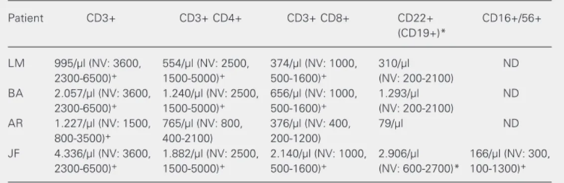
the criteria recommended by the Primary Immunodeficiency Committee of the World Health Organization, which indicate that this diagnosis must be considered in patients with severe infection by an intracellular mi-croorganism who present normal numbers of T, B and NK cells (Table 1) and normal levels of serum immunoglobulins. The study was approved by the Ethics Committee of the Research Medical Center of the Uni-versity of Antioquia and patients were sub-mitted to clinical evaluation and medical examination after giving written informed consent.
Case reports
Patient LM, a female, at 4 months of age presented severe abdominal symptoms that required surgical treatment. She also devel-oped respiratory manifestations and Myco-bacterium tuberculosis was identified in tra-cheal secretion. Diagnosis of miliary tuber-culosis was established and treatment was started. When she was two years old, tuber-culosis reactivation was confirmed by an intestinal biopsy positive for M. tuberculo-sis. The patient was treated by surgical re-moval of the affected region and by adminis-tration of ethambutol, ethionamide and iso-niazid, with an appropriate response. Cur-rently the patient is in good condition with
prophylactic treatment with trimethoprim-sulfamethoxazole (TMP-SMX). She had been vaccinated with BCG during the first week after birth.
Patient BA, a boy, at 2 months of age presented an abscess in the left thigh after application of anti-hepatitis B vaccine. In the following three years, four surgical pro-cedures were performed to remove the ab-scess. The biopsy demonstrated multiple granulomas, some of the solid and others of caseous consistency, and the Ziehl Nielsen stain was positive for mycobacteria. M. tu-berculosis was identified by culture. This patient was under anti-tuberculosis treatment and is currently in prophylaxis with TPM-SMX. He received the BCG vaccine after birth.
Patient AR, a 10-year-old boy, presented respiratory distress due to a granulomatous lesion in the oropharynx. The biopsy re-vealed a lesion characterized by reactive acanthosis, a necrotic center and a plasmo-cytoid infiltrate. One of the serial Koch’s bacillus analyses from this lesion was posi-tive; however, the mycobacterium species could not be identified. Despite the institu-tion of anti-tuberculosis treatment the pa-tient developed a severe deterioration of his general condition and died. This patient had not received the BCG vaccine.
Patient JF, a boy who presented jaundice,
Table 1. Lymphocyte populations in peripheral blood of patients quantified by flow cytometry.
Patient CD3+ CD3+ CD4+ CD3+ CD8+ CD22+ CD16+/56+
(CD19+)*
LM 995/µl (NV: 3600, 554/µl (NV: 2500, 374/µl (NV: 1000, 310/µl ND
2300-6500)+ 1500-5000)+ 500-1600)+ (NV: 200-2100)
BA 2.057/µl (NV: 3600, 1.240/µl (NV: 2500, 656/µl (NV: 1000, 1.293/µl ND
2300-6500)+ 1500-5000)+ 500-1600)+ (NV: 200-2100)
AR 1.227/µl (NV: 1500, 765/µl (NV: 800, 376/µl (NV: 400, 79/µl ND
800-3500)+ 400-2100) 200-1200)
JF 4.336/µl (NV: 3600, 1.882/µl (NV: 2500, 2.140/µl (NV: 1000, 2.906/µl 166/µl (NV: 300,
2300-6500)+ 1500-5000)+ 500-1600)+ (NV: 600-2700)* 100-1300)+
choluria and acholia at 2 months of age. Liver gammagraphy indicated bile tree atre-sia and a liver biopsy revealed a soft granu-loma. A chest X-ray showed enlarged lymph nodes and infiltration of the hilum. The tu-berculin test was positive (11 mm) and the biopsy of a cervical lymph node revealed chronic granulomatous inflammation com-patible with tuberculosis. When he was 5 months old he presented purulent secretion from axillaryand supraclavicular lymphad-enitis, and M. tuberculosis was identified by culture of this secretion. Functional tests of phagocytic cells were normal. The patient re-ceived anti-tuberculosis treatment followed by improvement and currently is in good con-dition. The patient was vaccinated with BCG.
Analysis of IFN-γγγγγR expression
The expression of IFN-γR1 was
deter-mined in Epstein-Barr virus transformed B (EBV-B) cells from patients and normal sub-jects. EBV-B cells were obtained after incu-bation of 107 blood mononuclear cells with B95-8 cell line supernatant (ATCC #CRL 1612) for 3 to 4 weeks. IFN-γR1 was
ana-lyzed in 5 x 10⁵ EBV-B cells by incubation for 20 min with 1 µg monoclonal antibody against IFN-γR1 labeled with rhodamine
(a gift of Dr. Steve Holland, NIAID, NIH). Cells were washed with cytometry buffer (PBS, 1% fetal calf serum (Gibco, Rock-ville, MD, USA) and 0.1% sodium azide) at 300 g for 7 min and fixed with fixation buffer (PBS, 1% paraformaldehyde and 1% sodium azide) and median fluorescence in-tensity was measured in an Epics XL flow cytometer (Coulter, Miami, FL, USA).
Analysis of STAT-1 expression by flow cytometry
The intracellular expression of STAT-1 was determined by flow cytometry in EBV-B cell lines obtained from our patients; 5 x 10⁵ EBV-B cells were permeabilized with
CITO Fix/PERM (Becton-Dickinson, San Jose, CA, USA) and incubated for 30 min at 4ºC in the dark with 1 µg anti-STAT-1 mono-clonal antibody (New England Biolabs, Bev-erly, MA, USA). Cells were washed with cytometry buffer (PBS, 1% fetal calf serum (Gibco) and 0.1% sodium azide) at 600 g for 7 min and 1 µg FITC-labeled anti-rabbit IgG (Santa Cruz Company,Santa Cruz, CA, USA) was added; then cells were fixed with fixa-tion buffer (PBS and 1% paraformaldehyde) and median fluorescence intensity was meas-ured with an Epics XL flow cytometer (Coulter).
Detection of STAT-1 and phosphorylated STAT-1 by Western blotting
To assess the function of the IFN-γ /IFN-γR activation pathway the phosphorylation of STAT-1 was analyzed in EBV-B cells from patients. Briefly, 107 cells were incu-bated with 100 ng/ml recombinant human IFN-γ (rhIFN-γ) for 15 min at 37ºC, the cell
phosphorylation was stopped by adding a 4ºC solution containing 2 mM EDTA and 400 mM sodium orthovanadate (O-NaVO4; Sigma, Saint Louis, MO, USA), and cells not incubated with rhIFN-γ were used as nega-tive control. Cells were centrifuged at 600 g for 10 min at room temperature and the cell pellets were lysed with 50 µl lysis buffer containing 0.5% Triton X-100 (Sigma), 50 mM HEPES, pH 7.2 (BioWhittaker, Walkersville, MD, USA), 150 mM NaCl, 5 mM EDTA (Sigma), 1 mM O-NaVO4, 10 mM NPGB (Sigma), 100 µg/ml aprotinin (Sigma), leupeptin (Sigma), 100 µg/ml PMSF (Sigma) and 100 µg/ml chymostatin (Boeh-ringer Mannheim, Indianapolis, IN, USA).
2 mercaptoethanol (Sigma), bromophenol blue (Sigma), and 5% SDS (Sigma), and finally boiled for 5 min. Protein samples were submitted to 8% SDS-PAGE and later transferred to PVDF membranes (Bio-Rad). For immunodetection, membranes were blocked with 5% BSA for 60 min, washed three times with PBS/Tween 20, 1 min each wash, incubated with 1 µg antibody to phos-phorylated STAT-1 (New England Biolabs) for 1 h, and washed again as before and 1 µg anti-rabbit IgG peroxidase-labeled antibody (Santa Cruz Company) was added. The reac-tion was developed by chemiluminescence using the ECL kit (Amersham Life Science, Buckinghamshire, UK) and exposed to X-Omat RX film (Eastman Kodak Co., Roch-ester, NY, USA). Non-phosphorylated STAT-1 was detected in non-stimulated cells using an antibody against non-phosphory-lated STAT-1 (New England Biolabs).
PCR-SSCP for IFN-γγγγγR genes
To find possible mutations in the
IFN-γR1 and R2 genes, PCR fragments were analyzed by single-strand conformational
polymorphisms (SSCP). Each exon of these two genes was amplified with the oligo-nucleotides listed in Table 2. The amplifica-tion was carried out with 100 ng genomic DNA, 0.5 µM of each primer, 200 µM dNTPs, 1.25 mM Mg2+, and 2U Taq DNA polymer-ase (Promega, Madison, WI, USA). The re-action was carried out for 25 cycles at 94ºC/ 30 s, 58ºC/45 s and 72ºC/40 s and a final incubation at 72ºC/5 min was performed using a 9600 PE thermocycler (Perkin Elmer, Foster City, CA, USA). To check for the proper amplification, PCR products were run on agarose gel and visualized by UV light after ethidium bromide staining. Next, 5 µl of each product was mixed with 1 µl of 6X loading buffer (formamide, xylene cyanol, and bromophenol blue), denatured at 80ºC/2 min and placed on ice. These fragments were then electrophoresed on 6% polyacrylamide gel under non-denaturing conditions at 35 W/4 h at 25ºC. To identify DNA bands, the gel was stained with silver (Bio-Rad).
DNA sequencing
All PCR products exhibiting an altered
Table 2. Synthetic oligonucleotides used for amplification of IFN-γR1 and IFN-γR2 genes from genomic DNA.
IFN-GR1 IFN-GR2
Name Sequence Name Sequence
1F 5'-CAC TCA AAT TCC TCC CAC AC-3' 1F 5'-GAG CCG AAT CCC CTC CAC CG-3'
1R 3'-GCA GCC CTG CCG CGA ACG AC-5' 1R 5'-CCA CCT GAT CTG AGC ACT CC F-3'
2F 5'-TAT CTG GGC AAT GTG GCA TC-3' 2F 5'-GCC TGT ACC AGT AGG GAC TC-3'
2R 3'-GGG AAT TTC CAA GGA CCT AA-5' 2R 5'-CAG CTG CAG CAG ATC CAA CAG-3'
3F 5'-CAC AGA CAG AAA TGG TTT GAC-3' 3F 5'-CTG CAG GAA TTC TGT GAA TTG-3' 3R 3'-CAG CAA CTG CTA ATA AAA GCA-5' 3R 5'-GAA GTC TAT ACT CAA GTT CTC-3' 4F 5'-TAT ACT TCC TCC TCC TCC TTC-3' 4F 5'-CTA TAA TAC ATA TGT GTA TGT GTG TGG-3' 4R 3'-CAA CTT TTG CTA GCT ACA CAA G-5' 4R 5'-CAT GGA GAC ACC CTG TTC TTG-3' 5F 5'-TTC TTC AGT TGT TTG AAC AGG A-3' 5F 5'-CAT TTA CAT GTG TGC TTG TGA TG-3' 5R 3'-AGA TCT TTT GAA ACT GCA AAT GA-5' 5R 5'-CAC TAT TGG AGG AGT ATT CTT TTC-3' 6F 5'-CTT AAT TGT AAC TTG TGA TTT C-3' 6F 5'-GTG CGT AGA AGA TCA TTC TG-3'
6R 3'-GTA GAC TGA CTG ATT GAT G-5' 6R 5'-CAC AGA GCA GCC CTG TCT C-3'
7AF 5'-ATC TTT AAT CAA TTT TTC TCC-3' 7F 5'-GGT CTG GTA TAC TGA ACT GGT AAA C-3' 7AR 3'-GAC CAC GTC AGG AAT ATT TTC-5' 7R 5'-GCT GAA ACT CTG CAG AAA ATA GGC-3' 7BF 5'-CAT GCA TCA CGA AGA CAA TCC AG-3'
electrophoretic pattern in the PCR-SSCP for IFN-γR genes were sequenced. Direct se-quencing of purified PCR products was per-formed using the ABI Big Dye Terminator Cycle Sequencing Ready Reaction DNA kit (Perkin Elmer) and the sequenced products were resolved on an ABI 310 automated sequencer (Perkin Elmer).
In patient AR the STAT1 cDNA was analyzed for a mutation in the region encod-ing the tail segment of the protein. Sense (5'- TCGACAGTCTTGGCACCTAACGTGC-3') and antisense (5'-TGCTATCAACAGGT TGCAGCG-3') primers were used to am-plify the complete cDNA. The STAT1 re-gion encoding L706 was amplified with sense (5'-TCGGTTGATGGAAAGCGTA-3') and antisense (5'-CTCTTCTGTGTTCACTTAC-3') primers. Genomic DNA from EBV-B cell lines was also amplified. The products were sequenced as previously described.
Results
Expression of IFN-γγγγγR1 in EBV-B cell lines from patients
The expression of IFN-γR1 in these
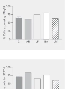
pa-tients was evaluated by flow cytometry in peripheral blood B cells transformed with EBV. This assay was repeated twice in each patient with cells transformed from two dif-ferent blood samples. As shown in Figure 1, the percentage of EBV-B cells expressing IFN-γR1 was similar for patients and control
subjects: 65.8 ± 5.2 for controls, 60.2 ± 8.1 for AR, 72 ± 11 for JF, 78.3 ± 16.1 for BA, and 62.8 ± 10.9 for LM.
Evaluation of the expression of STAT-1 and STAT-1 phosphorylation in EBV-B cell lines from patients
When the level of expression of STAT-1 was determined by flow cytometry using intracellular staining, no differences in the percentage of positive cells for this molecule were detected between EBV-B cells from patients and control subjects: 71.8 ± 18.8 for controls, 85.6 ± 13.5 for AR, 65.5 ± 10.7 for JF, 76.3 ± 9.6 for BA, and 61.4 ± 11.4 for LM (Figure 2); as for IFN-γR1 analysis, this as-say was repeated twice in each patient with cells transformed from two different blood samples. Similarly, when the expression of STAT-1 protein was analyzed by Western blotting no differences between cells from patients and healthy controls were demon-strated (Figure 3).
To evaluate the integrity of the IFN-γR
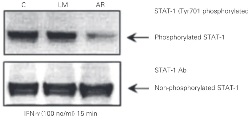
intracellular signaling pathway in the pa-tients of this study, the phosphorylation of STAT-1 protein in Tyr701 was determined after EBV-B cell stimulation with recombi-nant human IFN-γ. The assay revealed that
one of the patients (AR) had an evident reduction in the phosphorylation of STAT-1 when compared with other patients or with a healthy control after cell activation with
Figure 2. Analysis of intracellular signal transducer and activator of transcription 1 (STAT-1) ex-pression determined flow cytom-etry in EBV-B cell lines obtained from our patients. The control (C) value of percentage of cells ex-pressing intracellular STAT-1 was obtained from cell lines from five different healthy subjects while the patients were analyzed twice. The mean values are pre-sented. No statistically signifi-cant differences were observed (unpaired t-test).
Figure 1. Analysis of interferon-gamma receptor 1 (IFN-γR1) ex-pression by flow cytometry in EBV-B cells using a rhodamine-labeled monoclonal antibody against IFN-γR1. The control (C) value for the percentage of cells expressing the receptor was ob-tained from cell lines from five different healthy subjects; the patients were analyzed twice and their mean values are pre-sented. No statistically signifi-cant differences were observed (unpaired t-test).
1234 1234 1234 1234 1234 1234 1234 1234 1234 1234 1234
% Positive cells for STAT-1
100
75
50
25
0
C AR JF BA LM
100
75
50
25
0
C AR BA LM
1234 1234 1234 1234 1234 1234 1234 1234 1234 1234 1234 1234
% Cells expressing
IFN-γ
R1
rhIFN-γ (Figure 4). An even loading of
pro-tein extract indicated that there was less phosphorylation of the STAT-1 tyrosine resi-due in cells of the patient AR.
SSCP analysis and DNA sequencing of genomic DNA corresponding to the IFN-γγγγγR1 and IFN-γγγγγR2 genes
SSCP analysis of genomic DNA obtained from the present patients revealed an abnor-mal electrophoretic pattern in some of the PCR fragments spanning the coding region of the IFN-γR1 gene. One patient (AR)
ex-hibited an abnormal shift in the fragment corresponding to exon 2. When PCR frag-ments amplified from exon 7 were analyzed, patients AR and LM showed aberrant elec-trophoretic patterns (Figure 5). However, when these three PCR fragments were se-quenced in order to identify a possible muta-tion explaining the SSCP data, no sequence alterations with respect to the known se-quence of the IFN-γR1 gene were
demon-strable (data not shown). Furthermore, in PCR fragments amplified from genomic DNA corresponding to the coding region of the IFN-γR2 gene, SSCP analysis did not
demonstrate any abnormal pattern in PAGE carried out under non-denaturing conditions (data not shown).
As described above, patient AR exhib-ited a reduction in the phosphorylation of STAT-1 at Tyr701; therefore, in order to determine whether this patient had a muta-tion in the codon encoding this amino acid or in other near residues critical for STAT-1 function, as recently described (27), the DNA segment encoding the tail segment of STAT-1 protein was sequenced in this patient. Af-ter sequencing both strands of cDNA and genomic DNA no mutations were detected (data not shown).
Discussion
In this study we report the immunologic
Figure 5. Representative pic-tures of SSCP analyses of the PCR fragments corresponding to exons two and seven of the IFN-γR1 gene in different pa-tients. The arrows indicate the PCR products that presented an abnormal migration pattern after 6% native electrophoresis. These analyses were performed on two different occasions in or-der to confirm the findings. C = control.
and molecular characterization of the
IFN-γR/STAT-1 activation pathway in 4 patients suffering uncommon infections produced by mycobacteria. Our hypothesis was that these patients might present a genetic defect in one of the molecules involved in the IL-12/IFN-γ
Figure 4. Evaluation of signal transducer and activator of transcription 1 (STAT-1) phosphory-lation in response to interferon-gamma (IFN-γ) in EBV-B cells from two patients and one
healthy control (C). Cells were incubated with 100 ng/ml rhIFN-γ for 15 min/37ºC, then an antibody to P-Tyr701 was used to identify phosphorylated STAT-1 in the lysis extract after electrophoresis and blotting onto a PVDF membrane. In order to confirm an equal load of protein extract, the membrane was removed and incubated with the antibody against the non-phosphorylated form of STAT-1. This assay was repeated twice to confirm the result. Figure 3. Detection of non-phosphorylated signal transducer and activator of transcription 1 (STAT-1) by Western blotting in resting EBV-B cell lines from patients and two healthy controls (C) using an antibody against the non-phosphorylated form of STAT-1. This assay was repeated at least once for each patient.
STAT-1
C AR JF BA LM CN
STAT-1 (Tyr701 phosphorylated) Ab
Phosphorylated STAT-1
STAT-1 Ab
Non-phosphorylated STAT-1
IFN-γ (100 ng/ml) 15 min
C LM AR
C AR BA C C AR LM C
pathway based on their age and clinical pres-entation of these defects; furthermore, no other primary or acquired defects of the immune system such as severe combined immunodeficiency or HIV infection were detected. To determine the cell surface ex-pression of IFN-γR1, the presence of this protein in EBV-B cells was assessed by flow cytometry. The expression of IFN-γR1
ob-served in all patients was similar to that found in healthy controls. Since this result did not rule out the presence of a defect that could affect the function of IFN-γR1 but not its structure, the IFN-GR1 was characterized at the genomic level. The SSCP analysis of coding regions for this gene revealed some abnormal migration profiles in exons 2 and 7 in 2 patients; however, the DNA sequencing of these fragments did not show any nucleo-tide change.
Different possibilities might explain the inconsistency of these results. During PCR prior to SSCP analysis some DNA fragments exhibiting a different conformation during electrophoresis might have been generated. The altered electrophoretic mobility could also be a consequence of the thermal and physical conditions of electrophoresis (28). In other cases, the changes in DNA respon-sible for the altered mobility in the SSCP might be located in the sequence comple-mentary to the primers used during PCR, in regions very difficult to define by the se-quencing reaction used.
Based on these results, the next step was the molecular analysis of the IFN-GR2 gene; in this case the SSCP did not indicate any possible sequence alteration of the PCR frag-ments spanning the coding region of this gene.
Since alterations were not found in the IFN-GR1 or IFN-GR2 genes, we decided to evaluate the expression and phosphorylation of STAT-1, a molecule essential for the sig-nal transduction pathway in response to
IFN-γ. In the 4 patients the intracellular levels of
STAT-1, assessed by flow cytometry and
Western blotting, were similar to those found in healthy controls; however, the phospho-rylation of cellular STAT-1 in response to human IFN-γ was found to be substantially
reduced in one patient (AR). A recent report described two unrelated kindred affected by a heterozygous dominant STAT-1 mutation with a leucine substitution for a serine at amino acid position 706 (L706S), which is a loss-of-function mutation that severely im-pairs the phosphorylation of tyrosine 701 (27). In order to determine whether the alter-ation observed in our patient could be ex-plained by a mutation in one of the nucleo-tides encoding these critical tyrosines, the DNA segment encoding the tail segment of the STAT-1 protein was sequenced. After sequencing the DNA region spanning these critical residues, no mutations were detected. The protective immune response to intra-cellular pathogens such as mycobacteria es-sentially depends on adequate activation and function of mononuclear phagocytic cells. In order to achieve a full effector response, these cells need soluble signals from the microenvironment; of particular importance is the production of IFN-γ by NK and T
helper cells. IFN-γ secretion is induced by IL-12, a cytokine produced by macrophages and dendritic cells. IFN-γ activates different
effector mechanisms of phagocytic cells, which ensure appropriate control and elimi-nation of phagocytized microorganisms. The immune response against intracellular mi-croorganisms also involves lysis of infected cells by cytotoxic T and NK cells (2,5,6,20). The importance of the immune effector mechanisms related to IL-12 and IFN-γ has
been clearly demonstated by a growing group of patients with Mendelian susceptibility to mycobacterial infections, a primary immu-nodeficiency disease caused by genetic de-fects in the IL-12/IFN-γ activation pathway.
susceptibility to the development of dissemi-nated mycobacterial disease or local recur-rent infections by non-tuberculosis myco-bacteria and, in some cases, chronic infec-tions by other agents such as Salmonella species and certain viruses (20,22-27,29-36). Many of these characteristics are simi-lar to the clinical features observed in our patients (Table 3); therefore, a defect in one of the molecules involved in the
IL-12/IFN-γ/IFN-γR/STAT-1 activation pathway is a
possible explanation for their phenotype. Patients with defects in the IL-12/IFN-γ
pathway exhibit differences in susceptibility to mycobacterial disease. Complete abroga-tions of IFN-γR1 or IFN-γR2 are strongly
associated with an early onset of severe and often fatal infection with low virulence my-cobacterial species, such as non-tuberculo-sis mycobacteria or BCG. In these patients the infection continues despite the instaura-tion of an appropriate anti-tuberculosis treat-ment; furthermore, the lesions reveal poor granuloma formation and are rich in myco-bacteria. In contrast, partial deficiency of IFN-γR1 or IFN-γR2 is associated with a
milder clinical presentation and an impaired but not abolished response to IFN-γ. Most of the time these patients develop infections by more aggressive mycobacteria such as M. avium or M. tuberculosis; the granulomas found in the lesions of these patients are
mature, well formed and with few mycobac-teria; moreover, they respond very well to anti-tuberculosis treatment (2).
Patients who present mutations affecting the expression or function of 12p40 or IL-12Rß1 molecules generally develop dissemi-nated but curable mycobacterial infections with non-tuberculosis mycobacteria or BCG, a similar phenotype to that observed in pa-tients with partial deficiency of IFN-γR1 or
IFN-γR2. This less severe clinical picture
seems to be explained by a residual IL-12-independent IFN-γ secretion (25,32,37-39).
Several patients with mutations in STAT-1 gene have been recently described; how-ever, they show differences in their clinical presentation. As mentioned before, two kin-dred with the same heterozygous mutation affecting the dimerization of STAT-1 to form gamma-activated factor were susceptible to mycobacteria but resistant to viruses, with a phenotype similar to that of patients with partial IFN-γR (28). A more recent report
described two other children who developed disseminated BCG after vaccination, with remission occurring with antibiotic treatment. However, both patients died from dissemi-nated viral disease, produced in one of them by herpes simplex virus 1, while no virus was identified in the other (40). In these patients two different homozygous muta-tions, which produced a complete deficiency
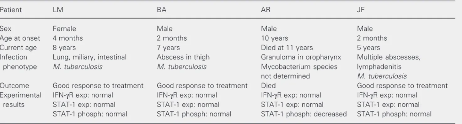
Table 3. Summary of the characteristics, clinical outcome and experimental findings for four patients with uncommon presentation of mycobacterial infections.
Patient LM BA AR JF
Sex Female Male Male Male
Age at onset 4 months 2 months 10 years 2 months
Current age 8 years 7 years Died at 11 years 5 years
Infection Lung, miliary, intestinal Abscess in thigh Granuloma in oropharynx Multiple abscesses,
phenotype M. tuberculosis M. tuberculosis Mycobacterium species lymphadenitis
not determined M. tuberculosis
Outcome Good response to treatment Good response to treatment Died Good response to treatment
Experimental IFN-γR exp: normal IFN-γR exp: normal IFN-γR exp: normal IFN-γR exp: normal
results STAT-1 exp: normal STAT-1 exp: normal STAT-1 exp: normal STAT-1 exp: normal
STAT-1 phosph: normal STAT-1 phosph: normal STAT-1 phosph: decreased STAT-1 phosph: normal
of STAT-1, were found, affecting both the formation of gamma-activated factor and the formation of the STAT-1/STAT-2/p48 tri-mer (transcription factor IFN-stimulated gene factor 3) that is essential for the cellular response to IFN-α/ß.
The present results indicate that mol-ecules that constitute the IFN-γR/STAT-1
pathway were intact and functioning in at least three of the 4 patients studied: normal surface expression of IFN-γR1, no
muta-tions found in the IFN-GR1 or IFN-GR2 genes, and normal expression and phospho-rylation of STAT-1 in 3 patients (Table 3). The phenotype of these 3 patients might be explained by a defect in IL-12/IL-12R that precludes an adequate activation of mono-nuclear phagocytes or by an alteration in a step downstream from STAT-1 activation; furthermore, an IFN-γ-independent
anti-my-cobacterial mechanism could be affected in any of these patients. In order to identify the genetic defect associated with the phenotype of these 3 patients it is necessary to continue studying the molecules involved in the
acti-vation of mononuclear phagocytes, particu-larly IL-12/IL-12R.
On the other hand, in patient AR the reduced phosphorylation of STAT-1 might have led to a defective activation of effector mechanisms required for a full activation of mononuclear phagocytes in response to my-cobacterial infection and therefore may have been the reason for the fatal course of the infection in this patient. The molecular mech-anism responsible for this defect remains to be elucidated; however, different alterna-tives can be proposed: a mutation in the IFN-GR1 or IFN-GR2 genes not detected by our SSCP method, an intrinsic alteration of STAT-1 that affects its phosphorylation in a region of the protein different from the one analyzed in this patient or a defect in any other molecule involved in the response to IFN-γ but upstream from STAT-1
activa-tion, for instance JAK1 or JAK2. Therefore, we consider important to continue the analy-sis of these different possibilities in the EBV-B cells from this patient.
References
1. Stark GR, Kerr I, Williams MB, Silverman GR & Schreiber RD (1998). How cells respond to interferons. Annual Review of Biochemistry, 67: 227-264.
2. Okamura H, Kashiwamura S, Tsutsui H, Yoshimoto T & Nakanishi K (1998). Regulation of interferon-γ production by IL-12 and IL-18.
Current Opinion in Immunology,10: 259-264.
3. Kennedy MK, Picha KS, Shanebeck KD, Anderson DM & Grabstein KH (1994). Interleukin-12 regulates the proliferation in Th1, but not Th2 or Th0, clones. European Journal of Immunology, 24: 2271-2278.
4. Fruh K & Yang Y (1999). Antigen presentation by MHC class I and its regulation by interferon. Current Opinion in Immunology,11: 76-81. 5. Boehm U, Klamp T, Groot M & Howard JC (1997). Cellular re-sponses to interferon-γ. Annual Review of Immunology,15: 749-795.
6. Bach EA, Aguet M & Schreiber RD (1997). The IFN-γ receptor: a paradigm for cytokine receptor signaling. Annual Review of Immu-nology,15: 563-591.
7. Axelrod A, Gibbs VC & Goeddel DV (1994). The interferon-γ receptor extracellular domain. Non-identical requirements for ligand binding and signaling. Journal of Biological Chemistry,269: 15533-15539. 8. Kotenko SV, Izotova LS, Pollack BP et al. (1995). Interaction
be-tween the components of the interferon gamma receptor complex. Journal of Biological Chemistry,270: 20915-20921.
9. Rhee S, Ebensperger C, Dembic Z & Pestka S (1996). The structure of the gene for the second chain of the human interferon-γ receptor. Journal of Biological Chemistry,271: 28947-28952.
10. Farrar MA, Fernandez-Luna J & Schreiber RD (1991). Identification of two regions within the cytoplasmic domain of the human interfer-on-γ receptor required for function. Journal of Biological Chemistry, 266: 19626-19635.
11. Hu R, Bekisz J, Hayes M, Audet S, Beeler J, Petricoin E & Zoon K (1999). Divergence of binding, signaling, and biological responses to recombinant human hybrid IFN. Journal of Immunology, 163: 854-860.
12. Marsters SA, Pennica D, Bach E, Schreiber RD & Ashkenazi A (1995). Interferon γ signals via a high-affinity multisubunit receptor
complex that contains two types of polypeptide chain. Proceedings of the National Academy of Sciences, USA,92: 5401-5405. 13. Behrmann I, Janzen C, Gerhartz C, Schmitz-Van de Leur H,
14. Begitt A, Meyer T, van Rossum M & Vinkemeier U (2000). Nucleo-cytoplasmic translocation of Stat1 is regulated by a leucine-rich export signal on the coiled-coil domain. Proceedings of the National Academy of Sciences, USA,97: 10418-10423.
15. Chatterjee-Kishore M, Wright KL, Ting JP & Stark GR (2000). How STAT1 mediates constitutive gene expression: a complex of unphosphorylated STAT1 and IRF1 supports transcription of the LMP2 gene. EMBO Journal,19: 4111-4122.
16. Igarashi K, Garotta G, Ozmen L, Ziemiecki A, Wilks AF, Harpur AG, Larner AC & Finbloom DS (1994). Interferon-γ induces tyrosine phosphorylation of interferon-γ receptor and regulated association
of protein tyrosine kinases, Jak1 and Jak2, with its receptor. Journal of Biological Chemistry,269: 14333-14336.
17. Kovarik P, Stoiber D, Novy M & Decker T (1998). Stat1 combines signals derived from IFN-γ and LPS receptors during macrophage activation. EMBO Journal,17: 3660-3668.
18. Mowen K & David M (2000). Regulation of STAT1 nuclear export by JAK1. Molecular and Cellular Biology,20: 7273-7281.
19. Szente BE, Subramaniam PS & Johnson HM (1995). Identification of IFN-γ receptor binding sites for JAK2 and enhancement of binding by IFN-γ and its C-terminal peptide IFN-γ(95-133). Journal of
Immu-nology,155: 5617-5622.
20. Bellamy R & Hill AV (1998). Genetic susceptibility to mycobacteria and other infectious pathogens in humans. Current Opinions in Immunology,10: 483-487.
21. Fenton M & Vermeulen M (1996). Immunopathology of tuberculo-sis: Roles of macrophages and monocytes. Infection and Immunity, 64: 683-690.
22. Casanova JL & Abel L (2002). Genetic dissection of immunity to mycobacteria: the human model. Annual Review of Immunology, 20: 581-620.
23. Fieschi C & Casanova JL (2003). The role of interleukin-12 in human infectious diseases: only a faint signature. European Journal of Immunology, 33: 1461-1464.
24. Casanova JL, Newport M, Fisher A & Levin M (1999). Inherited interferon gamma receptor deficiency. In: Ochs HD, Smith CIE & Puck JM (Editors), Primary Immunodeficiency Diseases: A molecu-lar and Genetic Approach. Oxford University Press, Oxford, UK, 209.
25. Dorman S & Holland S (2000). Interferon-γ and interleukin-12 path-way defects and human disease. Cytokine and Growth Factor Re-views,11: 321-333.
26. Holland SM, Dorman SE, Kwon A, Pitha-Rowe IF, Frucht DM, Gerstberger SM, Noel GJ, Vesterhus P, Brown MR & Fleisher TA (1998). Abnormal regulation of interferon-γ, interleukin-12, and
tu-mor necrosis factor-α in human interferon-γ receptor 1 deficiency.
Journal of Infectious Diseases,178: 1095-1104.
27. Dupuis S, Dargemont C, Fieschi C, Thomassin N, Rosenzweig S, Harris J, Holland SM, Schreiber RD & Casanova JL (2001).
Impair-ment of mycobacterial but not viral immunity by a germline human STAT1 mutation. Science,293: 300-303.
28. Fan E, Levin DB, Glickman BW & Logan DM (1993). Limitations in the use of SSC analysis. Mutation Research, 288: 85-92.
29. Bellamy R, Beyers N, McAdam KP et al. (1999). Genetic susceptibil-ity to tuberculosis in Africans: a genome-wide scan. Proceedings of the National Academy of Sciences, USA,97: 8005-8009.
30. Altare F, Durandy A, Lammas D et al. (1998). Impairment of myco-bacterial immunity in human interleukin-12 receptor deficiency. Sci-ence,280: 1432-1435.
31. Jouanguy E, Lamhamedi-Cherradi S, Lammas D et al. (1999). A human IFN-GR1 small deletion hotspot associated with dominant susceptibility to mycobacterial infection. Nature Genetics,21: 370-378.
32. Jouanguy E, Lamhamedi-Cherradi S, Altare F et al. (1997). Partial interferon-gamma receptor 1 deficiency in a child with tuberculoid bacillus Calmette-Guerin infection and a sibling with clinical tuber-culosis. Journal of Clinical Investigation, 100: 2658-2664.
33. Picard C, Fieschi C, Altare F et al. (2002). Inherited interleukin-12 deficiency: IL12B genotype and clinical phenotype of 13 patients from six kindreds. American Journal of Human Genetics, 70: 336-348.
34. Fieschi C, Dupuis S, Catherinot E et al. (2003). Low penetrance, broad resistance, and favorable outcome of interleukin 12 receptor beta1 deficiency: medical and immunological implications. Journal of Experimental Medicine, 197: 527-535.
35. Pernis A, Gupta S, Gollob KJ, Garfein E, Coffman RL, Schindler C & Rothman P (1995). Lack of interferon-γ receptor ß chain and the
prevention of interferon-γ signaling in TH1 cells. Science,269:
245-247.
36. Pierre-Audigier C, Jouanguy E, Lamhamedi S et al. (1997). Fatal disseminated Mycobacterium smegmatis infection in a child with inherited interferon-γ receptor deficiency. Clinical Infectious Dis-eases,24: 982-984.
37. Gately MK, Renzetti LM, Magram J, Stern AS, Adorini L, Gubler U & Presky DH (1998). The interleukin-12/interleukin-12 receptor sys-tem: role in normal and pathologic immune responses. Annual Review of Immunology, 17: 495-521.
38. Zhang F, Nakamura T & Aune TM (1999). TCR and IL-12 receptor signals cooperate to activate an individual response element in the IFN-γ promoter in effector Th cell. Journal of Immunology,163: 728-735.
39. Piccotti JR, Chan SY, Li K, Eichwald EJ & Bishop DK (1997). Differ-ential effects of IL-12 receptor blockade with IL-12 p40 homodimer on the induction of CD4+ and CD8+ IFN-γ producing cells. Journal
of Immunology,158: 643-648.