DOI: 10.5935/2359-4802.20180012
Introduction
Since the 19th century, cardiac adaptations induced by physical exercise have been known. Henschen, in 1899, recognized cardiomegaly in long distance skiers through percussion of heart borders, concluding that this increase was related to cavity dilatation and wall hypertrophy of the left ventricle (LV), and that these changes resulted in physical benefits for the athletes.1,2 With the evolution of complementary diagnostic
means, mainly echocardiography, Morganroth et al.,3 in 1975, developed the hypothesis that cardiac morphological changes depended on hemodynamic overload associated with physical exercise: dynamic exercise associated with eccentric hypertrophy due to volume overload, resulting in increased cardiac cavities by serial sarcomere addition; and static exercise associated with concentric hypertrophy due to pressure overload, with LV wall hypertrophy and parallel sarcomere addition.3-5
ORIGINAL ARTICLE
Additional Cardiac Remodeling Induced by Intense Military Training in Competitive
Athletes
Paulo Dinis1,2, Hélder Dores3, Rogério Teixeira1,4, Luís Moreno2, Joselito Mónico2, Marie Bergman5, Hanna Lekedal5, Maria Carmo Cachulo1, Joaquim Cardoso2, Lino Gonçalves1,4
Serviço de Cardiologia, Centro Hospitalar e Universitário de Coimbra1, Coimbra - Portugal;
Centro de Saúde Militar de Coimbra2, Coimbra, Portugal;
Hospital das Forças Armadas3, Lisboa - Portugal;
Faculdade de Medicina Universidade de Coimbra4, Coimbra - Portugal;
University of Linköping5 - Sweden
Manuscript received on May 10, 2017; manuscript revised on August 25, 2017 accepted on September 22, 2017. Mailing Address: Paulo Dinis
Centro Hospitalar de Coimbra Quinta dos Vales - 3041-801 São Martinho do Bispo, Coimbra - Portugal E-mail: [email protected], [email protected]
Abstract
Background: Cardiac remodeling depends on the intensity, duration, and training method.
Objective: To evaluate if the training performed in a Portuguese military special operations troop increases cardiac remodeling in a sample of young individuals who previously practiced competitive sports.
Methods: A prospective study involving 76 military candidates for military special operations, 45 of whom previously practiced at competitive level (> 10 hours per week). Of these military athletes, only 17 successfully completed the course. The evaluation was performed at 6 months intervals and included a complete clinical history, physical examination, vital signs, anthropometric data and echocardiographic evaluation. Statistical significance was considered when p < 0.05, with a 95% confidence interval.
Results: At the end of the course, there was a decrease in the percentage of fat mass (19.1 ± 3.3% vs. 13.1 ± 3.5%; p < 0.01), an increase in the percentage of lean mass (41.3 ± 2.1% vs. 44.4 ± 1.8%; p < 0.01), and decreased systolic and diastolic blood pressure and heart rate. Regarding cardiac remodeling, there was an increase in left ventricular diastolic diameter (49.7 ± 3.2 mm vs. 52.8 ± 3.4 mm; p < 0.01), an increase trend in left atrial volume (27.3 ± 4.5 mL/ m2 vs. 28.2 ± 4.1 mL/m2; p = 0.07) and increased left ventricular mass (93.1 ± 7.7 g/m2 vs. 100.2 ± 11.4 g/m2; p <
0.01). Functional variables also changed, with an increase in S’ (15 (13-16) cm/s vs. 17 (16-18) cm/s; p < 0,01) and a decrease in left ventricular ejection fraction (60 ± 6% vs. 54 ± 6%; p < 0.01).
Conclusion: Intense military physical training resulted in additional cardiac remodeling in athletes of competitive level, both structural and functional. (International Journal of Cardiovascular Sciences. 2018;31(3)209-217)
Different sports and training methods result in varied cardiac remodeling patterns. A predominantly eccentric hypertrophy is expected to be found in a marathon runner, whereas concentric hypertrophy is expected in a weightlifter.6 However, most of the sports practiced nowadays are influenced by the two components of the exercise (the dynamic and the static), and the adaptation these athletes may undergo is less predictable. The intensity, duration and frequency of physical exercise are characteristics that can determine different patterns of cardiac remodeling. High-intensity training is associated with more marked structural and functional changes.7,8
The aim of this study was to verify if there is additional cardiac remodeling in athletes of competitive level, when exposed to a high-intensity training protocol performed in a military course of special operations troops.
Methods
This was an observational and prospective study, which evaluated military personnel at the beginning and end of a course directed at special operations troops. The selected military personnel were candidates to complete the special operations troops course, the Portuguese Army Command (Comando do Exército Português). There were 76 candidates, of which 45 were previously athletes who competed in different modalities. Only 17 individuals successfully completed the course, all of them athletes. The evaluations were carried out between January and June 2016. In these evaluations, carried out at 6 months intervals, a complete clinical history including a medical questionnaire, physical examination, anthropometric evaluation and transthoracic echocardiogram (TTE) was performed. The evaluations were preceded by a rest interval of at least 12 hours. An electrocardiogram (ECG) was also performed in all participants as a cardiovascular screening method.
All subjects signed the Free and Informed Consent Form prior to participating in the study. The study protocol was authorized by the Ethics Committee of Faculdade de Medicina da Universidade de Coimbra (protocol reference 087/2015).
Characteristics of the study population
All military men who completed the course were previously considered competitive athletes. They practiced competitive physical exercises (> 10 hours
a week), participating in regional and national competitions. Although this activity was not their main economic means of support, they all had benefits derived from their sports performance. The athletes maintained their level of physical performance in the different modalities until the first evaluation carried out by the researchers. The practiced modalities can be seen in Table 1. During the special operations course, they suspended the previous training, and the physical exercise performed by them was only the result of the training given in the military course.
Characteristics of military physical training in the special operations course
The special operations course consisted of one component of overall physical training and another of military physical training. In the overall physical training, the military was submitted to dynamic and static physical exercises, practicing several modalities such as athletics, team sports (soccer or basketball), swimming and combat sports (boxing, for instance).
The military physical training combined the physical education aspect adjusted to military specificities. For this purpose, the military performed dynamic exercises (long-distance running, long runs interspersed with sprinting and marching exercises), static (cargo transportation and weight lifting) and mixed exercises (steeplechase track, among other activities).
The special operations training program was carried out in two phases. The first one lasted 10 weeks, and the military were submitted to high-intensity exercise training, seeking to achieve approximately 77 to 95% of the maximal heart rate (HR),9 with a frequency of five times a week and for an average of 4 hours daily, combining different types of exercise. The second phase lasted 15 weeks and included high-intensity exercises (approximately 77 to 95% of maximal HR), with periods of almost maximal or maximal intensity (> 96% of maximal HR),9 five times a week, for an average of 4 hours a day, alternating different types of exercise.
Clinical evaluation
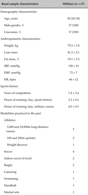
Table 1 - Basal study sample characteristics
Basal sample characteristics Military (n = 17)
Demographic characteristics
Age, years 20 (20-24)
Male gender, % 17 (100)
Caucasian, % 17 (100)
Anthropometric characteristics
Weight, kg 75.2 ± 7.8
Lean mass 41.3 ± 2.1
Fat mass, % 19.1 ± 3.3
SBP, mmHg 128 ± 10
DBP, mmHg 73 ± 7
HR, bpm 66 ± 12
Sports history
Years of competition 7.4 ± 3.4
Hours of training/day, sports history 2.3 ± 0.6
Hours of training/day, military course 4.0 ± 0.5
Modalities practiced in the past
Athletics
5,000 and 10,000m long-distance
runner 2
100 and 200m sprinter 2
Weight thrower 1
Soccer 4
Indoor soccer (Futsal) 2
Rugby 2
Canoeing 1
Swimming 1
Handball 1
Martial arts 1
SBP: systolic blood pressure; DBP: diastolic blood pressure; HR: heart rate.
Anthropometric evaluation
The anthropometric evaluation was performed under the nursing team coordination, and the military personnel was evaluated through a full body digital scale with impedance (HBF510W, OMRON®), which
allowed the assessment of body weight, percentage of fat mass (FM) and lean mass (LM) and height (using a tape measure). The systolic blood pressure (SBP) and diastolic blood pressure (DBP), as well as HR measurements were evaluated utilizing an arm blood pressure monitor (HEM 7113, OMRON®), according to the current recommendations.10
To predict the maximum HR, the indirect model was used, based on the equation Maximum HR = 220 – age.
The following variables were calculated: variation (Δ) weight, ΔLM, ΔFM; ΔSBP, ΔDBP, ΔHR through the
formula: [(final parameter - initial parameter)/initial parameter x 100].
Electrocardiographic evaluation
All 12-lead ECGs were performed by cardiopneumology technicians (electrocardiograph model 1200HR, NORAV®) and interpreted by two cardiologists according to the refined criteria,11 of which one of them was blinded to the study conditions.
Echocardiographic evaluation
All TTEs (Vivid 7, GE Healthcare®) were performed by a cardiologist and reviewed by an echocardiography specialist blinded to the study conditions. The e c h o c a r d i o g r a p h i c s t u d y w a s d e t a i l e d , a n d echocardiographic windows were obtained according to the current recommendations of the European Society of Cardiology.12,13 Data were digitally recorded for off-line analysis using the Echopac GE Healthcare software (Horton, Norway®). LV wall, interventricular septum (IVS) and LV posterior wall (LVPW) measurements, such as LV diastolic diameter (LVDD), were obtained at the parasternal long-axis window. The relative wall thickness (RWT) was calculated through the formula [(2 * LVPW)/LVDD].
The modified Simpson rule was used to determine the LV volumes and ejection fraction (LVEF) and left atrium (LA) volume. The results were indexed to the body surface area (BSA). LV mass was calculated using the Devereux’s formula.13
Pulsed Doppler was acquired using a four-chamber apical window. Tissue Doppler images of the mitral and tricuspid annuli were obtained, and the E and e’ waves were determined, as well as the S’ wave velocity, respectively.
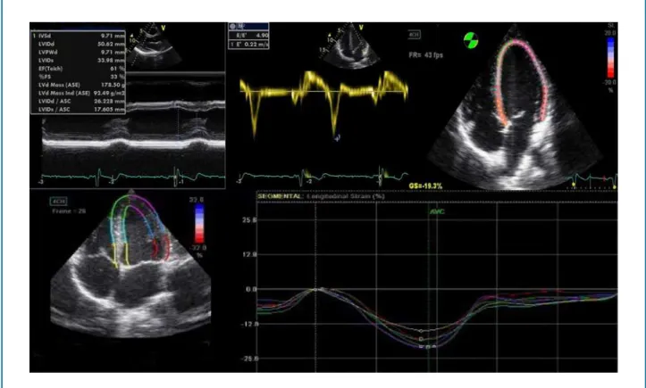
Figure 1 - Heart with left ventricular parietal thickness and dimension within limits considered to be normal, with supranormal values of diastolic function and normal global longitudinal strain.
longitudinal strain (GLS). The images were acquired in apical four-, two- and three-chamber windows, with electrocardiographic monitoring. In the off-line mode, with dedicated software (EchoPAC 9.0, GE Healthcare®, Horten, Norway), myocardial tracking and subsequent conversion to Lagrangian were performed using a semiautomatic endocardial edge detection program. This system allowed manual corrections to be performed to ensure the correct tracing between the cavity and the LV wall. Using the three apical windows of four, two and three chambers, the regional longitudinal strain mean was calculated, so that, subsequently, it was possible to calculate the GLS mean. The exam quality was considered good when up to two segments were excluded and excellent when all segments were analyzed.
The following variables were calculated: ΔIVS, ΔLV mass, ΔRWT, ΔLVDD, ΔLA volume, ΔLVEF, ΔS’, Delta of the Tricuspid Annular Plane Systolic Excursion (ΔTAPSE) and ΔGLS. The calculation of each of these variables
was performed by the formula [(final parameter – initial parameter)/initial parameter x 100].
Figure 1 depicts the TTE of a military after the special operations course.
Statistical analysis
The distribution of the continuous variables was verified by the Kolmogorov-Smirnov test. The homogeneity of the individual variables was evaluated by the Levene test. Variables with normal distribution were expressed as mean and standard deviation, and paired Student’s t-test with two-tailed analysis was used to compare groups. Variables with non-normal distribution were expressed as median and interquartile ranges, and the groups were compared using the paired Wilcoxon’s test. Categorical variables were shown as frequency and percentage, and chi-square test and Fisher’s test were used when appropriate.
Table 2 - Anthropometric data
Anthropometric data
Military (n = 17)
Initial Final p value
Weight, kg 75.2 ± 7.8 77.4 ± 6.6 < 0.01*
Lean mass, % 41.3 ± 2.1 44.4 ± 1.8 < 0.01*
Fat mass, % 19.1 ± 3.3 13.1 ± 3.5 < 0.01*
Systolic blood pressure, mmHg 128 ± 10 122 ± 7 < 0.01*
Diastolic blood pressure, mmHg 73 ± 7 66 ± 5 < 0.01*
Heart rate, bpm 62 (57-73) 60 (53-62) < 0.01+
*Paired Student's t-test; +Paired Wilcoxon test.
Results
Population description
The sample characteristics, anthropometric data, the sports history and the different modalities practiced are shown in Table 1. The sample consisted of young Caucasian males, with a median age of 20 (20-24) years and body mass index (BMI) of 25.3 ± 2.7 kg/m2.
Almost a fifth of the sample (20.6%) had at least one CVRF, with smoking being the most frequent (17.6%), followed by dyslipidemia (5.9%). None of the subjects included in the study had a family history of cardiovascular disease, hypertension or diabetes mellitus.
Anthropometric data variations
After the special operations course, the military had significant increase in body weight, with gain of LM and decrease in FM. There were also differences in relation to the blood pressure profile, with a decrease in mean SBP, DBP and HR (Table 2).
Electrocardiographic evaluation
All ECGs showed sinus rhythm and were considered normal or with only physiological alterations. The most prevalent physiological alteration was the early repolarization pattern (41.2%), followed by LV hypertrophy (29.4%), sinus bradycardia (17.6%) and incomplete right bundle branch block (17.6%).
Echocardiographic evaluation
The echocardiographic data are shown in Table 3. There was an increase, with statistical significance, in the
left cavity dimensions, both in the LV diastolic diameter and in the LA volume, in addition to an increase in LV mass and a decrease in RWT. In the functional variables, there was reduction in the LVEF at rest and increase in the S’ wave. Although the GLS did not show any significant differences, there was a tendency to decrease its absolute value.
Percentage differences between the beginning and end of the course
The differences in remodeling observed between the beginning and end of the special operations course in relation to anthropometric and echocardiographic data are shown in Figure 2. It was observed an increase in weight (3.1 ± 3.3%, p < 0.01) and LM (7.7 ± 4.1%, p < 0.01) and a decrease in FM (-30 ± 15.7%, p < 0.01). The pressure profile was altered with a decrease in SBP (-4.8 ± 3.0%, p < 0.01) and DBP (-8.6 ± 7.4%, p < 0.01). Among the echocardiographic findings, there was an increase in LV mass (10.2 ± 10.8%, p < 0.01) and LVDD (6.4 ± 14.3%, p < 0.01) and LA volume (10.6 ± 21.1%, p = 0.02), and a decrease in RWP (-7.0 ± 14.3%, p = 0.05). Functionally, there was a decrease in LVEF (-11.0 ± 12.8%, p < 0.01) and an increase in S’ [13.3 (6.3-19.4)%; p < 0.01].
Discussion
Table 3 - Variation of echocardiographic parameters
Parameters
Military (n = 17)
Initial Final p value
Interventricular septum, mm 9.7 ± 1.0 9.9 ± 1.0 0.39*
Posterior wall, mm 9.7 ± 0.9 9.6 ± 0.8 0.39*
Left ventricular mass, indexed, g/m2 93.1 ± 7.7 100.2 ± 11.4 < 0.01*
Relative wall thickness 0.40 ± 0.1 0.36 ± 0.1 0.05*
Left ventricular diastolic diameter, mm 49.7 ± 3.2 52.8 ± 3.4 < 0.01*
Left ventricular systolic diameter, mm 33.2 ± 3.3 35.1 ± 2.6 0.04*
Left atrial volume (mL/m2) 27.3 ± 4.5 28.2 ± 4.1 0.07*
Left ventricular ejection fraction (%) 60 ± 6 54 ± 6 < 0.01*
Lateral E’ (cm/s) 19 ± 3 19 ± 3 0.92*
E/E’ 5.3 ± 1.0 5.3 ± 0.9 0.61*
S’ (cm/sec) 15 (13-16) 17 (16-18) < 0.01+
Tricuspid annular plane systolic excursion, mm 25 ± 4 26 ± 5 0.34*
Left ventricular longitudinal strain (%) -21.3 ± 0.9 -20.5 ± 1.9 0.11*
*Paired Student's t-test; +Paired Wilcoxon test.
Figure 2 - Anthropometric and echocardiographic variables.
Δ weight Δ LM Δ SBP Δ DBP Δ LV Δ RWT
mass
Δ LVEF Δ LVDD ΔLA
volume
Military
Regarding vital signs, there was a decrease in mean SBP, DBP and HR at rest. This fact is due to the decrease in the activation of the sympathetic nervous system and the increase of the parasympathetic system at rest in the individuals submitted to the training.14,15 As for the anthropometric data, a mean increase in body weight was observed, with an increase in LM and marked reduction in FM. At the beginning of the training program, the military had FM values considered above the desirable for their age and gender,16 and these values decreased to levels considered appropriate after the military training program. The decrease in FM is related to the increase in intensity and/or duration of training,17 also demonstrated in this study.
The cardiac adaptations were compatible with eccentric cardiac remodeling. There was a significant increase in LV dimensions and mass, and tendency to increase the LA volume. It is known that cardiac remodeling resulting from physical exercise is related to the increase and harmonious hypertrophy of the cardiac cavities, also influencing the right cavities.17,18 During the special operations course, the percentage of military personnel with eccentric hypertrophy (RWT < 0.42 and LVMI > 115g/m2)19 practically doubled (from 5.9% to 11.8%). The verified changes result from the training intensity, frequency and method. Although the special operations training program consists of dynamic and static exercises, the dynamic training predominates. Frequency and intensity are other factors that contribute to high cardiac output, with corresponding volume overload and cardiac chamber dilatation.20
At the functional level, there was a decrease in LVEF, which agrees with what has been previously described.21-23 This occurs because athletes submitted to intense and prolonged exertion may show a slight decrease in resting LVEF. Several factors may contribute to this phenomenon, such as the athletes’ ability to significantly increase LVEF during exercise at the expense of increased LV diastolic dimensions, which, through the Frank-Starling mechanism, is associated with increased preload, more effective diastolic filling and lower LV systolic volume, allowing a considerable increase in ejection volume.22 Another possible explanation is that LVEF represents an estimate of LV function and not directly of myocardial contractility, which may underestimate its capacity when submitted to intense exertion. Also, the mathematical formula used to calculate LVEF (100 x ejection volume/ LVDD) in hearts with increased cavities underestimates LV performance.22 In this study, despite the verified
adaptations, one can say the latter were within ranges considered normal (none of the military had LVEF < 50% at the initial or final evaluations). However, after the military training program, 12 individuals (approximately 70% of the population) had LVEF at rest between 50 and 55%.
Myocardial GLS was studied using the speckle-tracking technique, and it was verified that the military had values within the ranges considered normal. There was a decrease in the absolute value of GLS, but it did not reach a statistically significant difference, probably due to the small sample size. This parameter may be important to differentiate left ventricular hypertrophy associated with exercise and disease, namely hypertrophic cardiomyopathy, since values of myocardial mechanics, within normal values, suggest physiological alterations.24-26
The literature on cardiac remodeling resulting from physical exercise in athletes is vast,27-30 but the existing bibliography in the military population is smaller. The authors did not find in the literature a study that addressed cardiovascular variations in competitive athletes when submitted to an intense military training program. Particularly in Portugal, in the Army special operations group, this is the first study that associated the military physical exercise training with the cardiovascular remodeling resulting from it.
Limitations
1. Henschen S. Skilanglauf und skiwettlauf: Eine Medizinische Sportstudie. Mitt Med Klin Upsala (Jena). 1899;2:15-8.
2. Maron BJ, Pelliccia A. The heart of trained athletes: cardiac remodeling and the risk of sports, including sudden death. Circulation. 2006;114(15):1633-44. doi: 10.1161/CIRCULATIONAHA.106.613562.
3. Morganroth J, Maron BJ, Henry WL, Epstein SE. Comparative left ventricular dimensions in trained athletes. Ann Intern Med. 1975;82(4):521-4. doi: 10.7326/0003-4819-82-4-521.
4. Dores H, Freitas A, Malhotra A, Mendes A, Sharma S. The hearts of competitive athletes: An up-to-date overview of exercise-induced cardiac adaptations. Rev Port Cardiol. 2015;34(1):51-64. doi: 10.1016/j. repc.2014.07.010.
5. Paterick TE, Gordon T, Spiegel D. Echocardiography: profiling of the athlete’s heart. J Am Soc Echocardiogr. 2014;27(9):940-8. doi: 10.1016/j. echo.2014.06.008.
6. Grazioli G, Sanz M, Montserrat S, Vidal B, Stiges M. Echocardiography in the evaluation of athletes. F1000Res. 2015 Jun 15;4:151. doi: 10.12688/ f1000research.6595.1.
7. Arbab-Zadeh A, Perhonen M, Howden E, Peshock RM, Zhang R, Adams-Huet B, et al. Cardiac remodeling in response to 1 year of intensive endurance training. Circulation. 2014;130(24):2152-61. doi: 10.1161/ CIRCULATIONAHA.114.010775.
8. Kemi OJ, Haram PM, Loennechen JP, Osnes JB, Skomedal T, Wisloff U, et al. Moderate vs. high exercise intensity: differential effects on aerobic fitness, cardiomyocyte contractility, and endothelial function. Cardiovasc Res. 2005;67(1):161-72. doi: https://doi.org/10.1016/j.cardiores.2005.03.010.
9. Garber CE, Blissmer B, Deschenes MR, Franklin BA, Lamonte MJ, Lee IM, et al; American College of Sports Medicine. American College of Sports Medicine position stand. Quantity and quality of exercise for developing and maintaining cardiorespiratory, musculoskeletal, and neuromotor fitness in apparently healthy adults: guidance for prescribing exercise. Med Sci Sports Exerc. 2011;43(7):1334-59. doi: 10.1249/ MSS.0b013e318213fefb.
10. Mancia G, Fagard R, Narkiewicz, Redón J, Zanchetti A, Bohm M, et al. 2013 ESH/ESC Guidelines for the management of arterial hypertension: the Task Force for the management of arterial hypertension of the European Society of Hypertension (ESH) and of the European Society of Cardiology. J Hypertens. 2013;31(7):1281-357. doi: 10.1097/01. hjh.0000431740.32696.cc.
11. Sheikh N, Papadakis M, Ghani S, Zaidi A, Gati S, Adami PE, et al. Comparison of electrocardiographic criteria for the detection of cardiac abnormalities in elite black and white athletes. Circulation. 2014;129(16):1637-49. doi: 10.1161/CIRCULATIONAHA.113.006179.
12. Evangelista A, Flachskampf F, Lancellotti P, Badano L, Aguilar R, Monaghan M, et al; European Association of Echocardiography. European Association of Echocardiography recommendations for standardization of performance, digital storage and reporting of echocardiographic studies. Eur J Echocardiogr. 2008;9(4):438-48. doi: 10.1093/ejechocard/jen174.
13. Lang RM, Badano LP, Mor-Avi V, Afilalo J, Armstrong A, Ernande L, et al. Recommendations for cardiac chamber quantification by echocardiography in adults: an update from the American Society of Echocardiography and the European Association of Cardiovascular Imaging. J Am Soc Echocardiogr. 2015;28(1):1-39.e14. doi: 10.1016/j.echo.2014.10.003.
References
in activities and evaluations in which the physical component is central, in addition to conventional or scheduled physical exercise practice. Fifthly, the study protocol did not include the hydration level assessment of the evaluated individuals to verify whether it was similar at both moments of the evaluation and, finally, although they denied taking any stimulants, this fact was not proven either.
Conclusion
Intense military physical training causes additional cardiac remodeling in athletes of competitive level, both structural and functional. The characteristics of the physical exercise are conditioning factors of cardiac remodeling, and should be considered during the evaluation of these individuals.
Author contributions
Conception and design of the research: Dinis P, Dores H, Teixeira R. Acquisition of data: Dinis P, Moreno L, Mónico J, Bergman M, Lekedal H. Analysis and interpretation of the data: Dinis P. Statistical analysis: Dinis P. Writing of the manuscript: Dinis P, Dores
H, Teixeira R. Critical revision of the manuscript for intellectual content: Dinis P, Dores H, Teixeira R, Cachulo MC, Cardoso J, Gonçalves L.
Potential Conflict of Interest
No potential conflict of interest relevant to this article was reported.
Sources of Funding
There were no external funding sources for this study.
Study Association
This article is part of the thesis of master submitted by Paulo Dinis, from Universidade de Coimbra.
Ethics approval and consent to participate
14. Aubert A, Seps B, Beckers F. Heart rate variability in athletes. Sports Med. 2003;33(12):889-919. PMID: 12974657.
15. Fernandes T, Oliveira M. Eccentric and concentric cardiac hypertrophy induced by exercise training: microRNAs and molecular determinants. Braz J Med Biol Res. 2011;44(9):836-47. doi: http://dx.doi.org/10.1590/ S0100-879X2011007500112.
16. American College of Sports Medicine. (editor). ACSM’s health-related physical fitness assessment manual. 4th ed. Baltimore: Lippincott Williams & Wilkins; 2013. ISBN: 978-1-4511-568-0.
17. Zaidi A, Ghani S, Sharma R, Oxborough D, Panoulas VF, Sheikh N, et al. physiological right ventricular adaptation in elite athletes of African and Afro-Caribbean origin. Circulation. 2013;127(17):1783-92. doi: 10.1161/ CIRCULATIONAHA.112.000270.
18. Ujka K, Bruno RM, Catuzzo B, Bastiani L, Tonacci A, D’angelo G, et al. Right cardiac chambers remodeling in marathon and ultra-trail athletes detected by speckle tracking echocardiography. Eur Heart J Cardiovasc Imaging. 2016;17(suppl_2):ii45-ii48. doi: 10.1093/ehjci/jew236.003.
19. Lang RM, Bierig M, Devereux RB, Flachskampf FA, Foster E, Pellikka PA, et al; American Society of Echocardiography's Nomenclature and Standards Committee; Task Force on Chamber Quantification; American College of Cardiology Echocardiography Committee; American Heart Association; European Association of Echocardiography, European Society of Cardiology. Recommendations for cardiac chamber quantification. Eur J Echocardiogr. 2006;7(2):79-108. doi: 10.1016/j.euje.2005.12.014.
20. Pluim BM, Zwinderman AH, van der Laarse A, van der Wall EE. The athlete’s heart. A meta-analysis of cardiac structure and function. Circulation. 2000;101(3):336-44. doi: https://doi.org/10.1161/01.CIR.101.3.336.
21. Abernethy WB, Choo JK, Hutter AM Jr. Echocardiographic characteristics of professional football players. J Am Coll Cardiol. 2003;41(2):280-4. doi: 10.1016/S0735-1097(02)02633-5.
22. Galderisi M, Cardim N, D’Andrea A, Bruder O, Cosyns B, Davin L, et al. The multi-modality cardiac imaging approach to the athlete’s heart: an expert consensus of the European Association of Cardiovascular Imaging. Eur Heart J Cardiovasc Imaging. 2015;16(4): 353. doi: 10.1093/ehjci/jeu323.
23. Gilbert CA, Nutter DO, Felner JM, Perkins JV, Heymsfield SB, Schlant RC. Echocardiographic study of cardiac dimensions and function in the endurance-trained athlete. Am J Cardiol. 1977;40(4):528-33. doi: https:// doi.org/10.1016/0002-9149(77)90067-4.
24. Beaumont A, Grace F, Richards J, Hough J, Oxborough D, Sculthorpe N. Left ventricular speckle tracking-derived cardiac strain and cardiac twist mechanics in athletes: a systematic review and meta-analysis of controlled studies. Sports Med. 2017;47(6):1145-70. doi: 10.1007/s40279-016-0644-4.
25. Galanti G, Stefani L, Mascherini G, Di Tante V, Tonncelli L. Left ventricular remodeling and the athlete’s heart, irrespective of quality load training. Cardiovasc Ultrasound. 2016;14(1):46. doi: 10.1186/s12947-016-0088-x.
26. Caselli S, Montesanti D, Autore C, Di Paolo FM, Pisicchio C, Squeo MR, et al. Patterns of left ventricular longitudinal strain and strain rate in Olympic athletes. J Am Soc Echocardiogr. 2015;28(2):245-53. doi: 10.1016/j.echo.2014.10.010.
27. Sharma S, Maron BJ, Whyte G, Firoozi S, Elliott PM, Mckenna WJ. Physiologic limits of left ventricular hypertrophy in elite junior athletes: relevance to differential diagnosis of athlete’s heart and hypertrophic cardiomyopathy. J Am Coll Cardiol. 2002;40(8):1431-6. doi: https://doi. org/10.1016/S0735-1097(02)02270-2.
28. Basavarajaiah S, Boraita A, Whyte G, Wilson M, Carby L, Shah A, et al. Ethnic differences in left ventricular remodeling in highly-trained athletes: relevance to differentiating physiologic left ventricular hypertrophy from hypertrophic cardiomyopathy. J Am Coll Cardiol. 2008;51(23):2256-62. doi: 10.1016/j.jacc.2007.12.061.
29. Pelliccia A, Culasso F, Di Paolo F, Maron BJ. Physiologic left ventricular cavity dilatation in elite athletes. Ann Intern Med. 1999;130(1):23-31. doi: 10.7326/0003-4819-130-1-199901050-00005.
30. Abergel E, Chatellier G, Hagege AA, Oblak A, Linhart A, Ducardonnet A, et al. Serial left ventricular adaptations in world-class professional cyclists: implications for disease screening and follow-up. J Am Coll Cardiol. 2004;44(1):144-9. doi: 10.1016/j.jacc.2004.02.057.