Sulfate Proteoglycan Sulfation Regulates WNT and BMP
Trans-Synaptic Signaling
Neil Dani1, Minyeop Nahm2, Seungbok Lee2, Kendal Broadie1*
1Department of Biological Sciences and Department of Cell and Developmental Biology, Kennedy Center for Research on Human Development, Vanderbilt University, Nashville, Tennessee, United States of America,2Department of Cell and Developmental Biology, Seoul National University, Seoul, Republic of Korea
Abstract
ADrosophilatransgenic RNAi screen targeting the glycan genome, including all N/O/GAG-glycan biosynthesis/modification enzymes and glycan-binding lectins, was conducted to discover novel glycan functions in synaptogenesis. As proof-of-product, we characterized functionally paired heparan sulfate (HS) 6-O-sulfotransferase (hs6st) and sulfatase (sulf1), which bidirectionally control HS proteoglycan (HSPG) sulfation. RNAi knockdown ofhs6standsulf1 causes opposite effects on functional synapse development, with decreased (hs6st) and increased (sulf1) neurotransmission strength confirmed in null mutants. HSPG co-receptors for WNT and BMP intercellular signaling, Dally-like Protein and Syndecan, are differentially misregulated in the synaptomatrix of these mutants. Consistently, hs6stand sulf1 nulls differentially elevate both WNT (Wingless; Wg) and BMP (Glass Bottom Boat; Gbb) ligand abundance in the synaptomatrix. Anterograde Wg signaling via Wg receptor dFrizzled2 C-terminus nuclear import and retrograde Gbb signaling via synaptic MAD phosphorylation and nuclear import are differentially activated inhs6standsulf1mutants. Consequently, transcriptional control of presynaptic glutamate release machinery and postsynaptic glutamate receptors is bidirectionally altered inhs6st andsulf1 mutants, explaining the bidirectional change in synaptic functional strength. Genetic correction of the altered WNT/BMP signaling restores normal synaptic development in both mutant conditions, proving that alteredtrans-synaptic signaling causes functional differentiation defects.
Citation:Dani N, Nahm M, Lee S, Broadie K (2012) A Targeted Glycan-Related Gene Screen Reveals Heparan Sulfate Proteoglycan Sulfation Regulates WNT and BMP Trans-Synaptic Signaling. PLoS Genet 8(11): e1003031. doi:10.1371/journal.pgen.1003031
Editor:Norbert Perrimon, Harvard Medical School, Howard Hughes Medical Institute, United States of America
ReceivedMay 10, 2012;AcceptedAugust 26, 2012;PublishedNovember 8, 2012
Copyright:ß2012 Dani et al. This is an open-access article distributed under the terms of the Creative Commons Attribution License, which permits unrestricted use, distribution, and reproduction in any medium, provided the original author and source are credited.
Funding:These studies were funded by NIH RO1 grants GM54544, MH084989, and MH096832 to KB (www.nih.gov). The funders had no role in study design, data collection and analysis, decision to publish, or preparation of the manuscript.
Competing Interests:The authors have declared that no competing interests exist. * E-mail: [email protected]
Introduction
Glycans coat cell surfaces, and glycosylation decorates secreted molecules of the pericellular space and extracellular matrix (ECM) [1,2]. It is well known that glycan modifications mediate critical functions of intercellular signaling and regulate interactions of numerous growth factors with the ECM [3,4]. The synthesis, modification and degradation of glycoconjugates, including O/N-linked glycoproteins, glycosaminoglycan (GAG) proteoglycans and glycan-binding lectins, is controlled by a dedicated cadre of genes [5,6]. In the nervous system, these glycan-related genes play key roles in development, including neuron fate specification, migra-tion, formation of axon tracts and synapse maturation [7]. At synapses, glycosylated ECM molecules, membrane receptors and outer-leaflet glycolipids together form the highly specialized synaptomatrix interface [4,8], which interacts with trans-synaptic signals to modulate synaptogenesis [9].
A prime example is the classic Agrin proteoglycan, which bears heparan sulfate (HS) chains, O/N-linked glycans and also a glycan-binding lectin domain that binds other glycoconjugates [10,11,12]. Reduction of GAG sulfation perturbs the Agrin signaling that drives postsynaptic acetylcholine receptor (AChR)
cluster maintenance at the neuromuscular synapse [13]. Likewise, Galbeta1,4GlcNAc and Galbeta1,3GalNAc glycans inhibit Agrin signaling by suppressing muscle specific kinase (MuSK) autophos-phorylation, a key step during synaptogenesis [14]. Analogous glycan-dependent mechanisms at the Drosophila neuromuscular synapse involve the secreted Mind-the-Gap (Mtg) lectin, which assembles the glycosylated synaptomatrix between presynaptic active zone and postsynaptic glutamate receptor (GluR) domains [15]. This glycan mechanism induces GluR clustering, synaptic localization of integrin ECM receptors, and shapestrans-synaptic signaling by controlling ligand/receptor abundance [16,17,18]. Thus, many long-term studies in vertebrate and invertebrate genetic models suggest that glycan mechanisms are a core foundation of synapse development.
In the current study, we conducted a broad transgenic RNA interference (RNAi) screen of synaptic glycan function, assaying requirements in both structural and functional development of the
RNAi-knockdown of genes in all eight categories affects synaptic morphological development, with gene-specific effects on branch-ing, bouton differentiation and synapse area. Likewise, all eight categories regulate synaptic functional development, with gene-specific effects both weakening and strengthening neurotransmis-sion. Interestingly, only a few genes affect both structure and function, suggesting separable roles for glycans in regulating these synaptogenic pathways. The results of this genomic transgenic screen are presented as a platform from which to pursue systematic investigation of glycan mechanisms in synaptic development.
Two genes were selected for screen validation and mechanistic characterization; functionally-paired HS 6-O-endosulfatase (sulf1) and HS 6-O-sulfotransferase (hs6st). RNAi knockdown and null mutants identically alter synaptic functional development in a bidirectional manner; loss of sulf1 elevates neurotransmission strength, whereas loss of hs6st weakens it. Heparan sulfate proteoglycan (HSPG) targets Dally-like Protein (Dlp) and Syndecan (Sdc) [19,20] are mislocalized in sulf1 and hs6st null synapses. In other developmental contexts, the sulfation state of these HSPG co-receptors strongly regulates WNT and BMP intercellular signaling [20,21,22]. At Drosophila synapses, WNT (Wg) is a key anterograde [23,24] and BMP (Gbb) a key retrograde [25,26] trans-synaptic signal. Consistently, loss of sulf1 and hs6st
differentially changes synaptomatrix levels of Wg and Gbb, and downstream signaling into muscle and motor neuron nuclei, respectively. Glutamate release and receptor machinery is thereby bidirectionally altered in the two nulls. Genetic restoration of Wg/ Gbb signaling to control levels restores the bidirectional changes in synaptic functional strength and pre-/post- synaptic differentiation in bothsulf1andhs6stnulls. We conclude that extracellular HSPG sulfation state in the synaptomatrix is a point of intersection between WNT/BMPtrans-synaptic signaling pathways that drive functional development of the neuromuscular synapse.
Results
RNAi screen of glycan-related genes identifies multiple synaptogenesis defects
Synaptic glycans play important roles as ligands, modulators and co-receptors regulating cell-matrix and intercellular commu-nication [3,27,28]. Differential glycan distribution on pre- and postsynaptic surfaces, and in the cleft, of numerous protein classes, strongly suggests that glycan mechanisms mediate synaptic structural and functional development [29,30,31]. To test the genomic scope of this requirement, we used confocal imaging and electrophysiological recording at the well-characterizedDrosophila
glutamatergic neuromuscular junction (NMJ) [32,33,34] to screen the Vienna Drosophila RNAi Center (VDRC) library of glycan-related genes [35]. We induced UAS-RNAi knockdown using the ubiquitous UH1-GAL4 driver [15,36]. We assayed morphological defects by co-labeling for pre- and postsynaptic markers, and assayed functional defects with two-electrode voltage clamp (TEVC) recording of neurotransmission strength. A summary of the screen results is shown in Figure 1. Full numerical results of the screen are shown in Table S1.
Candidate glycan-related genes were identified and classified into eight functional categories using the Kyoto Encyclopedia of Genes and Genomes (KEGG) database [37] (Figure 1). Additional genes were added to the screen based on ortholog identification using the Information Hyperlinked over Proteins (iHOP) database [38]. The candidate gene list was expanded and verified using Flybase [39]. From this list, genes were cross-referenced with available VDRC UAS-RNAi transgenic lines to generate a final candidate list containing 130 genes within eight functionally-defined categories (Figure 1): N-glycan, O-glycan and glycosami-noglycan (GAG) biosynthesis; glycan core proteins (HSPG core proteins/glycoproteins); sugar transporters; glycosyltranferases; glycan modification genes (modification and degradation of glycans); and glycan-binding lectins. On genetic knockdown, 103 lines were viable until the wandering 3rdinstar, whereas 27 lines showed developmental lethality at embryonic and early larval stages of development. From the 103 genetic lines characterized by confocal microscopy and TEVC electrophysiology in the 3rdinstar (Figure 1), 21 exhibited pupal stage developmental lethality. Interestingly, .50% of pupal lethal lines displayed statistically significant defects in NMJ synaptic morphology and function.
For all 103 larval-viable lines, synapse morphology and function was quantified at the wandering 3rdinstar NMJ (Figure 1; Table S1). Each UAS-RNAi line driven by UH1-GAL4 in the w1118
background was compared to the genetic control ofw1118crossed to UH1-GAL4 (UH1-GAL46w1118) [35]. All morphological and functional assays were done blind to genotype, with values reported as fold-change compared to genetic control, as well as statistical significance calculated using one-way ANOVA analyses (see color scheme; P,0.05 (*), P,0.01 (**); Figure 1). The data represents $6 NMJs from $3 animals from every genotype. Synapse morphology was imaged by co-labeling with presynaptic marker anti-horse radish peroxidase (HRP) and postsynaptic marker anti-Discs Large (DLG). A synaptic bouton was defined as a varicosity of$2mm in minimum diameter labeled by both HRP and DLG, and a synaptic branch was defined as a process containing at least two boutons [40]. NMJ branch number was the least affected morphological parameter, with only 2 of 103 genes showing a statistically significant change (Figure 1). Many more genes were involved in bouton development. All 27 genes showing a statistically significant change compared to genetic control exhibited elevated bouton numbers (Figure 1), suggesting that glycan mechanisms primarily limit morphological growth. Synapse Author Summary
area was determined by outlining the terminal area labeled by DLG using the thresholding function in ImageJ. The majority of gene knockdown conditions showed a decrease in NMJ area compared to control (Figure 1). 7 RNAi lines exhibited a statistically significant decrease in area, whereas only 2 lines exhibited a statistically significant increase in synaptic area. All raw values of measured morphological parameters are included in Table S1.
To assay functional differentiation, the motor nerve was stimulated with a suction electrode while the evoked excitatory junctional current (EJC) was recorded in the muscle (Figure 1) [41]. Nerve stimulation was applied at 4 V for 0.5 ms at a frequency of 0.2 Hz, with the muscle clamped at260 mV. EJC amplitudes were calculated from recorded traces in the ubiqui-tously-driven RNAi lines (w1118 background) compared to the
w1118; UH1-GAL4/+control. Recordings were obtained from$3 independent trials for each RNAi knockdown condition. All electrophysiological screening was done blind to genotype, with values reported as fold-change and statistical significance calcu-lated by one-way ANOVA analyses (see color scheme; P,0.05 (*), P,0.01 (**); Figure 1). Genes from all eight glycan classes were identified to produce changes in neurotransmission strength upon genetic knockdown. For the 103 larval-viable lines tested, 26 lines showed a trend towards increased transmission strength, and 12 were statistically elevated compared to genetic control (Figure 1). 4 gene knockdowns showed a trend towards decreased transmission strength, of which only 1 line reached statistical significance. 73 of the 103 lines tested showed no change in functional strength (Figure 1). Interestingly, only 6 RNAi lines showed statistically significant effects on both NMJ morphology parameters and EJC amplitude: CG1597, CG6657, CG7480, CG4451, CG6725 and CG11874 (Figure 1). This suggests that glycan effects on synapse morphological and functional development are largely separable. All raw values of EJC measurements are included in Table S1.
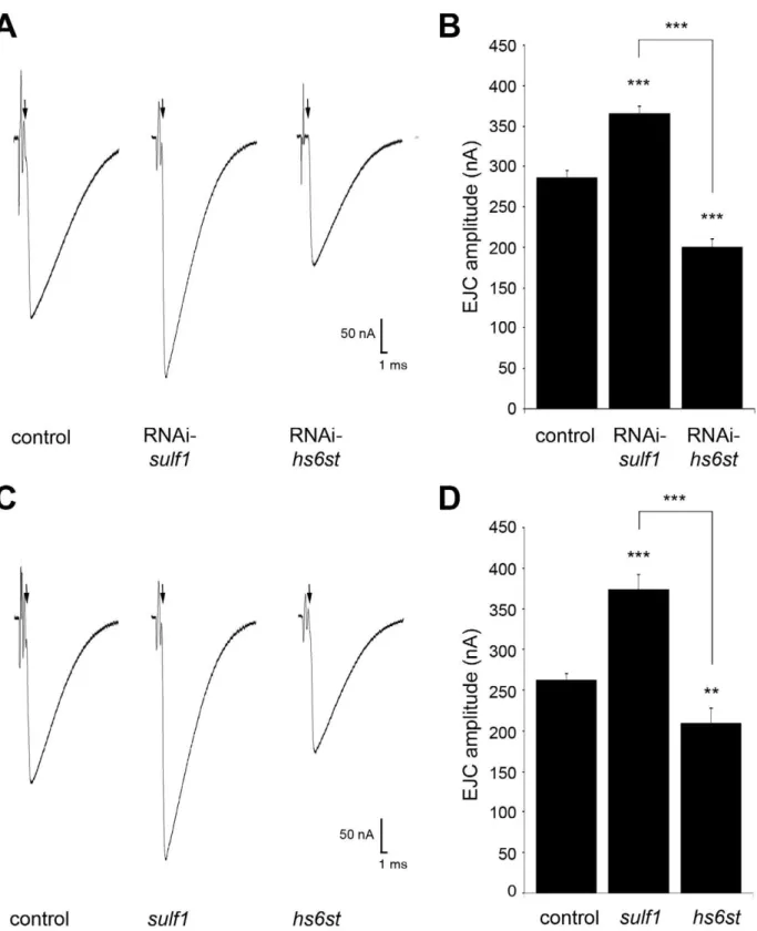
To validate results, a secondary screen was conducted using independent RNAi lines obtained from the VDRC and Harvard TRiP collections (Table S2). Of the 44 genes that showed morphological and functional defects in the primary screen, 33 were retested using independent RNAi lines, with the others lacking available secondary lines from any source. Using the same screen of morphological and functional characterization, we determined that ,80% of retested secondary lines showed the reported structural (bouton number) and functional (EJC) pheno-types consistent with primary screen (Table S2). These primary and secondary RNAi screen results now represent a resource for the systematic characterization of glycan mechanisms underlying synaptic structural and functional development. Screen results were further studied by comparing synaptogenesis phenotypes of RNAi knockdown with defined genetic nulls for two genes, CG6725 and CG4451, from the glycosaminoglycan biosynthesis class (Figure 1). The RNAi screen of functional strength as measured by EJC amplitudes indicated opposite effects for these two lines, with CG6725 (RNAi-sulf1) knockdown exhibiting an increase in transmission strength and CG4451 (RNAi-hs6st) knockdown producing a decrease (Figure 1). Along with our goal to identify interesting glycan-related genes involved in synapse development, we show here characterization of null alleles of two genes obtained from screen results and define the associated
mechanisms driving the bidirectional regulation of synaptic functional development.
Synaptogenesis is bidirectionally regulated by paired sulf1andhs6stgenes
The RNAi screen identified two functionally-paired genes,sulf1
(CG6725) andhs6st(CG4451), with similar effects on morpholog-ical development but opposite effects on synaptic functional differentiation (Figure 1). Our goal was to use these genes as a test case from the completed glycan screen, by assaying phenotypes in recently characterized null mutants of both genes [42,43]. The gene products Sulfated (Sulf1), an HS 6-endosulfatase, and Hs6st, an HS 6-O-sulfotransferase, drive opposing changes in sulfation state of the same C6carbon of the repeated glucosamine unit in
GAG modified heparan sulfate proteoglycans [43,44]. Viable null mutants are available for both genes, e.g.sulf1 (sulf1D1) andhs6st
(hs6std770) [42,43], but requirements have never been assayed in the nervous system or neuromusculature. We therefore first compared phenotypes of RNAi knockdown and null alleles at the NMJ synapse by confocal imaging of synaptic morphogenesis and TEVC recording of synaptic functional neurotransmission.
Using double-labeling for HRP (presynaptic) and DLG (post-synaptic), NMJ structural parameters including bouton number, branch number and synaptic area were quantified in sulf1 and
hs6st null alleles. The mutant results closely recapitulated the RNAi knockdown findings from the screen (Table S1). To consistently compare RNAi and null mutant conditions, both animal groups were simultaneously reared and processed to visualize the NMJ (Figure S1). Structural quantification showed an increased bouton number with RNAi-mediatedsulf1knockdown (sulf1-RNAi6UH1-GAL4; 36.461.6, n = 10) andhs6stknockdown (hs6st-RNAi6UH1-GAL4; 35.161.96, n = 10) compared to the transgenic control (w11186UH1-GAL4; 21.961.84, p,0.001, n = 10; Figure S1A, S1B). Consistently, increased bouton number was observed in both sulf1 (31.961.37, n = 10) and hs6st
(36.2562.58, n = 8) null mutants compared to genetic control (w1118, 19.361.69, p,0.001, n = 10; Figure S1C, S1D). In contrast, no significant change in branch number was exhibited with sulf1 knockdown (3.2260.28, p.0.05, n = 9) or hs6st
knockdown (3.2260.22, p.0.05, n = 9) compared to control (w11186UH1-GAL4; 2.6460.06, n = 11). Similarly, no significant change was observed in the synaptic branch number in sulf1
(2.860.33, p = 0.27, n = 10,) and hs6st (3.6360.38, p = 0.115, n = 10) nulls compared to control (w1118; 3.460.46, n = 8). Further, there was no significant difference in synaptic area in
sulf1 (138.1665.82, p.0.05, n = 10,) and hs6st (138.48613.38, p.0.05, n = 8,) mutants compared to the control (w1118; 118.0468,38, n = 10), however a slight increase in synaptic area was observed insulf1knockdown (178.68610.64, p,0.05, n = 9), while no change was observed forhs6st knockdown (16468.47, p.0.05, n = 10) as compared to control (w11186UH1-GAL4; 134.57611.95, n = 10). Based on these imaging studies, we conclude morphological differences in synaptic architecture observed in bothsulf1andhs6stnull allele conditions are consistent with both RNAi knockdown conditions.
Functional development was next tested with electrophysiolog-ical recording to compare RNAi and null mutant phenotypes
crossed to the UH1-GAL4 driver line. Target genes are indicated byDrosophilagenome CG annotation number and categorized by function. Confocal imaging of co-labeled pre- and postsynaptic markers was used to quantify NMJ architecture, including branch number, bouton number and synaptic area. TEVC electrophysiology was used to quantify evoked excitatory junctional current (EJC) amplitudes. The magnitude of fold changes compared to control (w1118
6UH1-GAL4) is shown on a color scale (see legend below the two columns). Statistical significance was calculated using one-way
(Figure 2). Representative TEVC records are shown as an average of 10 consecutive nerve stimulus responses in 1.0 mM extracellular Ca2+
for each transgenic genotype in Figure 2A;sulf1knockdown (UH1-GAL46sulf1-RNAi), hs6st knockdown (UH1-GAL46 hs6st-RNAi) and genetic control (UH1-GAL46w1118). There was a striking,80% difference in EJC amplitude betweensulf1andhs6st knockdown conditions, with sulf1 elevated by ,30% and hs6st reduced by ,30% compared to control. Quantification of EJC amplitudes showed both knockdown conditions to be highly significantly different from control and each other (control, 286.2268.56 nA; sulf1-RNAi, 365.0169.502 nA, p,0.001;
hs6st-RNAi, 199.19611.84 nA, p,0.001; sulf1-RNAi vs.
hs6st-RNAi, p,0.001; Figure 2B). These opposite effects on neuro-transmission strength were confirmed in characterized null alleles for both genes [42,43]. Representative traces from sulf1D1 and
hs6std770
null mutants compared to w1118
control are shown in Figure 2C. Quantification of EJC amplitudes showed null mutants to be highly significantly different from control and each other (w1118, 256.1467.38 nA; sulf1D1, 372.86618.49 nA, n = 11, p,0.001; hs6st, 209.66613.44 nA, n = 14, p,0.01; sulf1D1 vs.
hs6st, p,0.001; Figure 2D). These results were confirmed in an independent sulf1 null allele (sulf1DP1), which shows comparable elevation compared to control (w1118
, 244.9169.04 nA;sulf1DP1, 282.28613.59, p,0.05, n = 22), as well as thehs6stnull (hs6std770) over deficiency (Df(3R)ED6027), which shows comparable de-pression compared to control (w1118, 256.1467.38 nA; hs6st/ Df(3R)ED6027, 224.0667.65 nA, p,0.05, n = 18). These results reveal a critical role forsulf1andhs6stgenes in synaptic functional development.
Given the functionally-paired nature ofsulf1andhs6stactivities on 6-O-S modification, and the epistatic function ofhs6sttosulf1, we predicted that knocking both genes down would produce a phenotype similar to knockdown ofhs6stalone. Consistently,hs6st
andsulf1double knockdown produced EJC amplitudes significantly lower than control (w1118
6hs6st-RNAi; sulf1-RNAi (control), 225.1766.28 nA, n = 12; hs6st-RNAi, sulf1-RNAi6UH1-GAL4, 198.2269.77 nA, n = 15, p,0.05; Figure S2). Cell-specific knock-down in neural (elav-GAL4), muscle (24B-GAL4) and glia (repo -GAL4) also support the observed opposite effects in neurotransmis-sion strength. Withsulf1knockdown in muscle, EJC amplitude was significantly elevated compared to control (w11186sulf1-RNAi (control), 199.97621.86 nA; 24B-GAL46sulf1-RNAi (knockdown), 222.88625.78 nA, p,0.01, n = 10), but no change occurred with neural knockdown (elav-GAL46sulf1-RNAi, 196.09625.08 nA, p = 0.72, n = 10) or glial knockdown (repo-GAL46sulf1-RNAi, 208.40632.45 nA, p = 0.53, n = 7). Moreover, only neural knock-down ofhs6st caused a decrease in EJC amplitude (w11186hs6st -RNAi (control), 211.496622.142 nA, elav-GAL46hs6st-RNAi (knockdown), 184.68628.97 nA, p,0.05, n = 16), while no change occurred with muscle knockdown (24B-GAL46hs6st-RNAi, 209.92624.74 nA, p = 0.88, n = 9) or glial knockdown (repo -GAL46hs6st-RNAi, 216.38637.80 nA, p = 0.32, n = 7). We con-clude that HSPG sulfation state strongly modulates NMJ functional development, with contributions from both motor neuron and muscle, but not glia. The clear next step was to test for differences in the localization and abundance of synaptic HSPG targets known to regulate NMJ synaptogenesis.
HSPG abundance at the synaptic interface is dependent on sulf1andhs6st
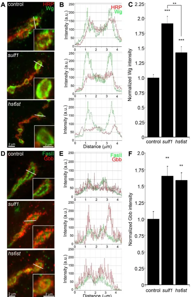
Both GPI-anchored HSPG glypican Dally-like (Dlp) and transmembrane HSPG Syndecan (Sdc) are clearly expressed at theDrosophilaNMJ (Figure S3), where they are known to regulate synaptogenesis [45]. We detect no enrichment of the secreted
HSPG perelcan (Trol) at the NMJ, although it is abundantly expressed in the motor nerve leading up to the synaptic terminal and present in lower levels throughout the muscle (Figure S4). We therefore hypothesized that membrane-associated Dlp and Sdc HSPGs are targeted by sulf1 and hs6st activity to regulate their synaptic distribution and/or function. To test this hypothesis, we assayed both Dlp and Sdc under non-permeabilized, detergent-free conditions to examine their cell surface expression at the NMJ synaptic interface of sulf1 and hs6st null mutants compared to control. These data are summarized in Figure 3.
In the genetic background control (w1118
), Dlp shows a punctate expression pattern strongly concentrated in a halo-like array around the anti-HRP labeled presynaptic membrane (Figure 3A, top; Figure S3). Insulf1mutants there was a clear and consistent increase in Dlp abundance, with more numerous and intense punctae at the synaptic interface surrounding NMJ boutons, while aths6stmutant synapses there was an opposing decrease in Dlp abundance (Figure 3A). This bidirectional and differential effect on Dlp abundance was quantified as fluorescence intensity normalized to the internal HRP labeling control. There was a significant Dlp increase in sulf1 compared to control (,40% elevated over control; p,0.05; n = 11), and a significant Dlp decrease in thehs6st null synapse (,15% reduced compared to control; p,0.05; n = 11; Figure 3B). Importantly, the difference betweensulf1andhs6stnulls was very highly significant (p,0.001). In comparison, cell surface Sdc labeling also showed a dense halo-like localization around NMJ synaptic boutons labeled with cell adhesion marker Fasciclin II (FasII; Figure 3C; Figure S3). Synaptic Sdc labeling intensity was consistently greater in both
sulf1 and hs6stnulls compared to control (Figure 3C). Quantifi-cation of fluorescence intensity normalized to HRP revealed that Sdc abundance was greatly increased in sulf1 null synapses compared to control (,35% elevated over control; p,0.01; n = 17) and, to a greater degree, also inhs6stnulls (,50% elevated over control; p,0.001; n = 12; Figure 3D). Thus, both Dlp and Sdc HSPGs are strongly altered in sulf1 and hs6st null NMJ synapses, with Dlp bidirectionally misregulated and Sdc differen-tially elevated in the two mutant conditions.
HSPGs act as co-receptors for WNT and BMP intercellular signaling ligands in many developmental contexts, acting to modulate extracellular ligand abundance and downstream signal-ing [46,47]. Drosophila WNT Wingless (Wg) distribution and signaling is known to be modulated by Dlp, which retains Wg at the cell surface in a mechanism that is enhanced by HS GAG chains [48]. Specifically, Wg ligand abundance and signaling activity along the dorso-ventral axis of the developingDrosophila
wing disc is elevated insulf1mutants [22]. Likewise, BMP ligands in other cellular contexts are closely regulated by HSPG co-receptors [20]. Specifically, Dlp has been suggested to similarly regulate Drosophila BMP Glass Bottom Boat (Gbb) [20]. We therefore hypothesized that altered HSPG co-receptors Dlp and/ or Sdc in sulf1 and hs6st null synapses regulate Wg and Gbb abundance to drive differentially altered trans-synaptic signaling across the synaptic cleft.
HSPG sulfation regulates abundance of WNT/BMPtrans -synaptic ligands
Figure 2. Loss ofsulf1/hs6stcauses opposite effects on transmission strength.(A) Representative excitatory junctional current (EJC) traces from control (w1118
6UH1-GAL4),sulf1RNAi (UH1-GAL46UAS-CG6725) andhs6stRNAi (UH1-GAL46UAS-CG4451). The nerve was stimulated (arrows)
in 1.0 mM external Ca2+, with TEVC records (
260 mV holding potential) from muscle 6 in segment A3. Each trace averaged from 10 consecutive recordings. (B) Quantified mean EJC amplitudes (nA) for the three genotypes shown in panel A. (C) Representative traces from control (w1118
),sulf1D1 andhs6std770null alleles under the same conditions described in panel A. (D) Quantified mean EJC amplitudes (nA) for the three genotypes shown in panel C. Sample sizes are at least 11 animals per indicated genotype. Statistically significant differences calculated using student’s t-test, ** p,0.01, *** p,0.001. Error bars indicate S.E.M.
receptor [17], has no known interaction with HSPGs and therefore would not be expected to be affected insulf1and hs6st
nulls, providing a comparison for specificity. To test the hypothesis that the observed alterations of HSPG co-receptor abundance will drive specific changes in WNT and BMP intercellular pathways, we labeled NMJ synapses with antibodies under non-permeablized conditions to reveal extracellular trans-synaptic signaling ligands (Figure S5), and compared protein abundance and distribution in controls,sulf1andhs6stnull mutants. The data are summarized in Figure 4.
NMJ synapses were first labeled with Wg antibody (green) together with anti-HRP (red) to label the presynaptic membrane (Figure 4A). In control animals (w1118), external Wg localized at large type Ib synaptic boutons in a dynamic pattern of punctuate distribution at the synaptic interface between motor neuron and muscle (Figure 4A, top; Figure S5). Insulf1andhs6stmutants, Wg was consistently elevated and concentrated uniformly in the extracellular domain adjacent to, and overlapping with, the anti-HRP-labeled presynaptic membrane (Figure 4A, middle and bottom). The elevated Wg levels in mutants were clearly observed at the level of individual synaptic boutons, as shown in the magnified insets in Figure 4A. To examine changes in Wg spatial distribution, cross-sectional planes were examined in single confocal line scans through the diameter of individual synaptic boutons (Figure 4A, white lines). Representative distribution plots for membrane-marker HRP (red) and external Wg (green) are shown in Figure 4B. In all genotypes, extracellular Wg was closely associated with the HRP-labeled presynaptic membrane, but both
sulf1 and hs6st nulls displayed a consistent increase in Wg label intensity and broadening of the spatial domain occupied by the secreted Wg ligand (Figure 4B, middle and bottom). To quantify changes in extracellular Wg abundance, the mean fluorescent signal intensity was normalized to the internal HRP co-label, and then normalized to analogous control intensity ratios. In sulf1D1
nulls, there was very highly significant elevation of Wg compared to control (,90% increased; p,0.001; n = 16; Figure 4C). A similar increase was observed in the independent sulf1DP1 null (p,0.001; n = 11). Thehs6st null displayed a smaller significant increase in Wg abundance (,40% increased; p,0.001; n = 15; Figure 4C), which was again recapitulated in hs6st null over deficiency (Df(3R)ED6027) condition. Importantly, Wg abun-dance is differentially elevated insulf1 vs.hs6stmutants (p,0.01, Figure 4C).
To test whether thesulf1/hs6stmechanism might coordinately regulate multipletrans-synaptic signals, we next assayed the BMP Gbb, a muscle-derived retrograde signal [25]. A barrier to previous Gbb analyses has been the absence of an anti-Gbb antibody. We therefore generated a specific anti-Gbb antibody for this study (see Methods). As above, labeling was done under non-permeabilized conditions to reveal only the extracellular Gbb, together with labeling for HRP or the cell adhesion molecule marker FasII to reveal the presynaptic membrane (Figure S5). In the control (w1118), extracellular Gbb concentrated in a ring of punctate domains around boutons (Figure 4D, top). Gbb was similarly punctate insulf1 and hs6st nulls, but consistently more extensive and denser (Figure 4D, middle and bottom; see
magnified insets). To examine Gbb spatial distribution, cross-sectional planes of confocal line scans were made through individual synaptic boutons (Figure 4D, white lines). Representa-tive plots for FasII (green) and Gbb (red) show extracellular Gbb closely associated with the FasII-labeled presynaptic membrane in all genotypes (Figure 4E). However, sulf1 and hs6st nulls consistently displayed increased Gbb intensity and broadened expression compared to the control. Upon quantifying signal intensity of Gbb normalized to HRP co-label,sulf1D1exhibited a significantly higher Gbb abundance than control (65% increased; p,0.01; n = 12; Figure 4F). The independentsulf1DP1null allele showed a similar increase (p,0.001; n = 12). The hs6st null also showed Gbb elevation compared to control (59% increased; p,0.01; n = 11; Figure 4E), which was confirmed inhs6stnull over deficiency (Df(3R)ED6027; p,0.05; n = 23).
To test further whether extracellular Wg and Gbb abundance was sensitive to the sulfation state of GAGs, a biochemical approach was next used to determine effects on Wg and Gbb
trans-synaptic signals (Figure S6). Specifically, NMJs were acutely exposed to heparin, the most sulfated form of GAG [53], and then synaptic Wg and Gbb abundance was measured by immunolabel-ing as above. We found that both trans-synaptic signals were rapidly altered by heparin incubation in a dose-dependent manner. Specifically, incubation with increasing concentrations of heparin caused a reciprocal decrease in Wg labeling intensity in the NMJ synaptic domain (Figure S6A, S6C), with a significant decrease first detected with 0.315 mg/ml heparin incubation (,50% less than control, p,0.01, n = 4). Interestingly, increasing heparin concentrations caused a parallel increase in Gbb abundance in the NMJ synaptic domain (Figure S6B, S6C) in a dose-dependent manner, with significant increases again first detected at 0.315 mg/ml heparin (,25% greater than control, p,0.05) and rising further at 0.625 mg/ml heparin (,40% greater than control, p,0.001). These results indicate that HSPG sulfation state does indeed affecttrans-synaptic signal abundance, supporting the observed alterations in Wg and Gbb abundance in mutants of heparan sulfate modifying genes,sulf1andhs6st.
To examine effects on othertrans-synaptic signaling pathways in thesulf1andhs6stmutant synapses, we also assayed for changes in Jeb [17] and FGF [17] signaling. In both control and mutants, extracellular Jeb labeling was tightly associated with NMJ type Ib boutons and, like other trans-synaptic ligands, occupied an extracellular domain closely associated with the presynaptic membrane (Figure S7A). However, in stark contrast to Wg and Gbb ligands in the same extracellular synaptomatrix domain, no change was observed in Jeb abundance or spatial distribution in
sulf1null (p = 0.99, n = 10) orhs6stnull (p = 0.36, n = 8) compared to control (w1118) NMJ synapses (Figure S7B). FGF signaling is also well established to be affected by HSPGs [54], and one pioneering study has investigated roles for FGF signaling at the Drosophila
NMJ [55]. The probe used in the previous study was an antibody against the FGF receptor Heartless (Htl) [56]. Using this antibody, we confirmed that the Htl receptor beautifully localizes to NMJ boutons to mediate FGF signaling (Figure S8A). However, Htl receptor synaptic abundance and distribution was very similar for the sulf1 (p = 0.89, n = 9) and hs6st (p = 0.69, n = 7) mutants
Figure 3. Synaptic HSPG co-receptor abundance is modified by 6-O-S sulfation.(A) Representative NMJ synaptic boutons imaged from control (w1118
),sulf1andhs6stnulls, probed with presynaptic neural marker anti-HRP (green) and Dally-like (Dlp; red). Right: Dlp distribution without the HRP signal is shown for clarity. (B) Quantification of mean fluorescent intensity levels of anti-Dlp labeling normalized to the HRP co-label at the muscle 6 NMJ, normalized to genetic control. (C) Boutons labeled with neural marker anti-Fasciclin II (FasII, green) and anti-Syndecan (Sdc, red). Right: Sdc distribution is shown alone for clarity. (D) Quantification of the mean fluorescent intensity levels of anti-Sdc labeling at the muscle 6 NMJ, normalized to genetic control. Sample sizes are at least 12 independent NMJs of at least 7 animals per indicated genotypes. Statistically significant differences calculated using student’s t-test, * p,0.01, ** p,0.01, ***p,0.001. Error bars indicate S.E.M.
compared to control (w1118) (Figure S8B). Unfortunately, no antibody probes are available forDrosophilaFGF ligands, so these signals have not yet been queried. Together, these results show that both WNT (Wg) and BMP (Gbb) ligand abundance is coordinately upregulated by thesulf1andhs6stmechanism at the NMJ synapse, but that a spatially overlapping signaling ligand (Jeb) and at least FGF receptor expression are unaffected. These results strongly predict that Wg and Gbbtrans-synaptic signaling controlled bysulf1and hs6stactivity regulates synaptic functional development.
Trans-synaptic WNT/BMP signaling is regulated by HSPG sulfation
Wg and Gbb serve as anterograde and retrogradetrans-synaptic signals, respectively, activating cognate receptors to initiate downstream signaling cascades and nuclear import pathways in muscles and motor neurons, respectively [24,26,50,51]. The anterograde Wg signal drives dFrizzled-2 (dFz2) receptor inter-nalization in the postsynaptic domain followed by cleavage of the receptor C-terminus, which then enters the muscle nuclei [57]. The muscle-derived retrograde Gbb signal activates presynaptic receptors to drive phosphorylation of the Mothers Against Decapentaplegic (Mad) transcription factor, and then P-Mad enters the motor neuron nuclei to regulate transcription [25,26,58]. Given the differential change in both HSPG co-receptor and Wg/Gbb ligand abundance insulf1vs.hs6stmutants, we hypothesized that these signaling pathways would be differen-tially affected during synaptogenesis. We therefore quantitatively assayed the paired muscle and motor neuron nuclear import pathways to determine whether and howtrans-synaptic signaling may be modulated bysulf1andhs6stat the NMJ synapse.
Characterized antibodies specifically recognizing the N- and C-termini of the Wg dFz2 receptor allow measurements of the receptor at the NMJ synapse (dFz2N; Figure S9) and the cleaved fragment (dFz2C; Figure 5) imported into muscle nuclei [57,59]. We first assayed dFz2 receptor abundance at the NMJ with the N-terminal specific antibody. The dFz2 receptor is closely associated with the synaptic cell membrane marker FasII and occupies a domain that envelopes all type Ib boutons (Figure S9A). Inhs6st
nulls, the dFz2 receptor domain was spatially extended as compared to controls, howeversulf1alleles showed no detectable change in the receptor. Likewise, fluorescence intensity measure-ments showed no significant difference between control andsulf1
nulls, buths6stnull synapses displayed a,25% increase in dFz2 receptor abundance, a very significant elevation (p,0.01, n = 12; Figure S9B) in synaptic dFz2 abundance. Thus, importantly (see Discussion), significantly more dFz2 receptors occur in the hs6st
null compared tosulf1null synapse.
To assay downstream signal transduction, the cleaved Fz2C fragment imported into muscle nuclei was quantified using the established method of counting dFz2C-positive punctae in nuclei proximal to the NMJ (Figure 5) [59]. In genetic control (w1118), most muscle nuclei contained a small number (1–3) of detectable dFz2C punctae, but some nuclei contained more and others were
devoid of detectable dFz2C (Figure 5A, top). More than 100 muscle nuclei were quantified in .7 different animals to determine the control level of dFz2C nuclear import. In sulf1
andhs6stmutants, there was a clear and consistent bidirectional difference in the number and size of dFz2C punctae in muscle nuclei (Figure 5A, middle and bottom). Nullsulf1nuclei showed a highly significant decrease in number of dFz2C punctae per nuclei (.50% decreased; p,0.01; n = 163; Figure 5B). In contrast,hs6st
nulls had an opposing highly significant increase in dFz2C punctae per nuclei (.60% increased; p,0.01; n = 163; Figure 5B). The difference betweensulf1 and hs6stnull mutants was very highly significant (p,0.001), with a differential change in signaling paralleling the bidirectional change in synaptic functional differ-entiation (Figure 2).
A characterized antibody specifically recognizing phosphorylated Mad (P-Mad) allowed independent measurements of Gbb signaling in the presynaptic terminal and P-Mad import into the motor neuron nuclei as a transcriptional regulator (Figure 6) [25,60]. To assay this transduction pathway, P-Mad fluorescent intensity normalized to FasII was first assayed in presynaptic boutons [61,62]. In the genetic control (w1118
), P-Mad labeling was bounded by the synaptic cell adhesion molecule marker FasII, with P-Mad localized in numerous punctate domains (Figure 6A, arrows). In
sulf1andhs6stnulls, both the intensity and size of P-Mad positive punctae were obviously and consistently greater than in controls (Figure 6A, middle and bottom). In fluorescence intensity quanti-fication, sulf1 null synapses displayed a significant increase in synaptic P-Mad (45% increased; p,0.05; n = 10; Figure 6C). An increase in P-Mad was also observed in thehs6stnull boutons (42% greater than control; p,0.01; n = 15; Figure 6C). The motor neuron nuclei at the ventral nerve cord (VNC) midline accumulate P-Mad transcription factor downstream of Gbb signaling at the NMJ [25,61,62]. In genetic control (w1118), P-Mad nuclear labeling was consistently detected in these motor neuron nuclei (Figure 6B, arrows). A similar P-Mad distribution was observed in motor neuron nuclei ofsulf1and hs6stnulls, but the intensity of P-mad expression was clearly and consistently elevated in both mutants compared to control (Figure 6B, middle and bottom). In fluores-cence intensity quantification,sulf1null neuronal nuclei displayed a very significant increase in P-Mad accumulation (15% increased; p,0.01; n = 14; Figure 6D), paralleling increased P-Mad signaling at the NMJ (Figure 6C). Likewise,hs6st null motoneuron nuclei exhibited a smaller but still significant elevation in P-Mad accumulation (9% elevated over control; p,0.05; n = 21; Figure 6D), again paralleling the observed P-Mad signaling change at the NMJ (Figure 6C). We conclude that both anterograde WNT (Wg) and retrograde BMP (Gbb)trans-synaptic signaling in muscle and motor neuron nuclei, respectively, is differentially regulated by thesulf1andhs6stHSPG sulfation mechanism.
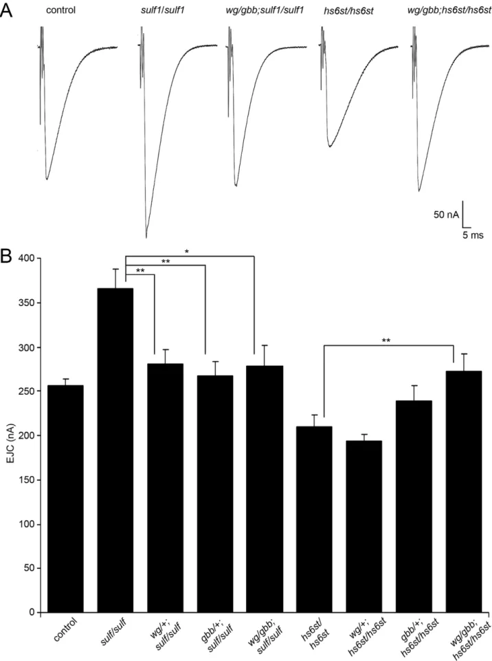
Trans-synaptic WNT/BMP signals genetically interact with sulf1andhs6stnulls
In thesulf1andhs6stnulls we identified a bi-directional change in synaptic functional differentiation, measured as evoked junction
measurements. Insets indicate single synaptic boutons at higher magnification. (A) Representative NMJ boutons from control (w1118
),sulf1andhs6st
current amplitudes increased in sulf1and decreased inhs6stnull synapses (Figure 2). We therefore hypothesized that these functional changes are driven by the differential Wg and Gbb
trans-synaptic signaling defects characterized above in sulf1 and
hs6stmutants (Figure 3, Figure 4, Figure 5, Figure 6). We reasoned that correcting Wg and Gbb levels insulf1andhs6stnulls should restore neurotransmission to control levels. To test this hypothesis, we crossed heterozygouswg/+andgbb/+mutants into bothsulf1
and hs6st homozygous null backgrounds, both singly and in combination, and compared them to both positive and negative controls. The resulting 9 genotypes were all assayed with TEVC electrophysiology to compare EJC transmission strength. A summary of these data is given in Figure 7.
Representative transmission records are shown as an average of 10 consecutive EJC responses (1.0 mM extracellular Ca2+
) for the genotypes in Figure 7A, with quantification of mean peak amplitudes in all genotypes shown in Figure 7B. First testingsulf1
nulls, we examined the consequences of heterozygous genetic
reduction of Wg and Gbb, alone and in combination. Compared to the elevated EJC amplitude of the sulf1 null condition (381.2861 62.24 nA, p,0.01, n = 9; Figure 7B), genetic reduction of Wg (wg/+; sulf1/sulf1) caused very significantly reduced transmission, similar to genetic reduction of Gbb (gbb/+; sulf1/ sulf1) with a comparable effect, restoring EJC amplitude to control levels (267.16616.33, p,0.01, n = 9; Figure 7B). Combinatorial genetic reduction of both Wg and Gbb in the sulf1 null (wg/ gbb;sulf1/sulf1) similarly returned EJC amplitudes to control levels (278.78623.17, n = 7; Figure 7B). Secondly testing hs6st nulls, genetic reduction of either Wg or Gbb alone was not sufficient to significantly change the depressed synaptic function (Figure 7B). In this case, combinatorial genetic reduction of both Wg and Gbb in the hs6st null (wg/gbb;hs6st/hs6st) was required to raise the depressed EJC amplitude, a very significant increase back to control levels (272.98618.58, p,0.01, n = 8; Figure 7B). There-fore, we conclude that combinatorial Wg and Gbbtrans-synaptic signaling defects are causative for the observed bi-directional
Figure 5. Loss ofsulf1andhs6stcauses opposite effects on WNT signaling.(A) Representative images of muscle nuclei from control (w1118),
sulf1andhs6stnulls, labeled with nuclear marker propidium iodide (PI, red) and for the C-terminus of the Wingless receptor Frizzled 2 (dFz2-C, green). Arrows indicate punctate dFz2-C nuclear labeling. Nuclei shown from muscle 6 in segment A3. (B) Quantification of the number of dFz2-C punctae per nuclei, normalized to genetic control. The total number of nuclei analyzed is indicated in each column; 119 for control (w1118
) and 163 nuclei each forsulf1andhs6stnull mutants. Sample sizes are$9 animals per indicated genotypes. Statistically significant differences calculated using student’s t-test; ** p,0.01 *** p,0.001. Error bars indicate S.E.M.
effects on synaptic functional differentiation in thesulf1andhs6st
null mutant conditions.
Thesulf1 andhs6stmechanism regulates pre- and postsynaptic differentiation
The consequence of WNT (Wg) and BMP (Gbb)trans-synaptic signaling is nuclear import and transcriptional regulation in both synaptic partner cells [49,51]. We therefore hypothesized thatsulf1
and hs6stnull mutants would show bidirectional changes in pre-and postsynaptic molecular components that would explain the bidirectional change in synaptic functional differentiation (Figure 2 and Figure 7). To test this hypothesis, we examined a key component of the presynaptic active zone (Bruchpilot; Brp) [63],
and an essential subunit of the postsynaptic glutamate receptor (Bad Reception (Brec); GluRIID) [64]. In parallel, we also performed a miniature EJC (mEJC) analysis to compare functional presynaptic vesicle release probability and postsynaptic response amplitude. A summary of these data is shown in Figure 8.
First, NMJ synapses were double-labeled for GluRIID recog-nized with anti-Brec (green) and Brp recogrecog-nized with anti-nc82 (red) to compare genetic control (w1118) withsulf1andhs6stnulls (Figure 8A). We found that GluRIID was very significantly elevated atsulf1synapses compared to control (,30% increased; p,0.01, n = 20; Figure 8B). In the opposing direction,hs6stnull synapses showed a significant decrease in GluRIID abundance (,15% reduced; p,0.05, n = 21; Figure 8B). The GluRIID field Figure 6. Loss ofsulf1andhs6stcauses differential effects on BMP signaling.(A) Representative NMJ synaptic boutons on muscle 6 in segment A3 from control (w1118
),sulf1andhs6stnulls, labeled with neural marker anti-Fasciclin II (FasII, red) and for phosphorylated Mothers against decapentaplegic (P-Mad; green) activated downstream of Gbb signaling. Arrows indicate representative P-Mad punctae in the indicated genotypes. (B) Representative ventral nerve cord (VNC) midlines from the same 3 genotypes, labeled with anti-FasII (red) and P-Mad (green). Labeled motor neuron nuclei are indicated by arrows. Quantification of the mean fluorescent intensity level of P-Mad labeling normalized to FasII co-label at the NMJ synapse (C) and in motor neuron nuclei (D), normalized to genetic control. Sample sizes are$14 animals per indicated genotypes. Statistically significant differences calculated using the Mann-Whitney test for non-parametric data, * p,0.05, ** p,0.01. Error bars indicate S.E.M.
Figure 7. WNT and BMP signals genetically interact withsulf1andhs6stnulls.Genetic reduction of Wg and Gbb levels insulf1andhs6st
area per bouton and number of GluRIID punctae normalized to field area per synaptic bouton were also bidirectionally altered in thesulf1and hs6stnulls (Figure 8C, 8D). GluRIID receptor field area was increased in sulf1(,30% greater; p,0.01, n = 47) but decreased inhs6st(,25% reduced; p,0.01, n = 51). Conversely, measurements of GluRIID puncta normalized to field area per synaptic bouton were decreased in sulf1(,15% lower; p,0.05, n = 47), but increased in hs6st nulls (,40% greater; p,0.01, n = 51, Figure 8D). The bi-directional differences between sulf1
andhs6stwere very highly significant (p,0.001). The active zone protein Brp also showed opposite effects (Figure 8A). Although the difference betweensulf1null and control was not quite significant (p.0.05, n = 20), hs6st null synapses showed a very significant decrease in Brp compared to control (,20% reduced; p,0.01, n = 21; Figure 8A).
Based on these results, we next tested pre- (Brp) and postsynaptic (Brec/GluRIID) changes in sulf1 and hs6stmutants with genetic reduction of Wg and Gbb (wg/gbb;sulf1/sulf1andwg/ gbb;hs6st/hs6st), as in Figure 7. Distribution changes of both pre-and postsynaptic components were assayed as measurements of glutamate receptor field and active zone areas (Figure S10A). To measure glutamate receptor distribution comparingwg/gbb;sulf1/ sulf1 to matched control, we counted the number of GluRIID punctae per bouton (p = 0.73, n = 48; Figure S10B) and GluRIID area (p = 0.92, n = 48; Figure S10C), and found both corrected back to control levels. Likewise, forwg/gbb;hs6st/hs6stcompared to control, GluRIID puncta number (p = 0.88, n = 48) and area (p = 0.41, n = 58) were both corrected to control levels. To measure Brp-positive presynaptic active zones comparing wg/ gbb;sulf1/sulf1to matched control, we counted the number of Brp punctae per bouton (p = 0.43, n = 48; Figure S10D) and Brp area (p = 0.39, n = 48; Figure S10D), and found both corrected back to control levels. Likewise, forwg/gbb;hs6st/hs6stcompared to control, Brp number (p = 0.54, n = 58) and area (p = 0.19, n = 58) were also corrected back to control levels. These results provide strong genetic evidence that Wg and Gbbtrans-synaptic signaling changes are causative for the pre- and postsynaptic molecular differenti-ation defects in thesulf1andhs6stnull mutants.
These bidirectional pre- and postsynaptic molecular changes parallel functional transmission changes insulf1andhs6stmutants (Figure 2). To assay function at the single synapse level, we finally assayed spontaneous synaptic vesicle fusion events. Representative mEJC traces for control compared to sulf1 and hs6st nulls are shown in Figure 8E. Consistent with observed bidirectional changes in evoked transmission, mEJC amplitudes inhs6st were ,25% lower than in sulf1 nulls (hs6st, 0.6060.02 nA vs. sulf1, 0.7660.05 nA; p,0.5, n = 34; Figure 8F). Moreover, hs6st nulls had a,100% elevated mEJC frequency compared tosulf1nulls (hs6st, 2.5660.27 vs. sulf1, 1.3060.09; p,0.001, n = 34; Figure 8G). Based on these mEJC measurements, there was a highly significant bidirectional change in quantal content between the two mutant conditions, with sulf1 quantal content ,50% greater than hs6st (sulf1, 539.98622.02 vs. hs6st, 350.6968.92; p,0.001, n = 34; Figure 8H). Taken together, these results show a bi-directional change in presynaptic glutamate release machinery and vesicle fusion probability, as well as postsynaptic glutamate receptor levels and functional responsiveness. We conclude that
these changes underlie the bi-directional switch in neurotransmis-sion strength characterizingsulf1andhs6stmutants.
Discussion
It is well known that synaptic interfaces harbor heavily-glycosylated membrane proteins, glycolipids and ECM molecules, but understanding of glycan-mediated mechanisms within this synaptomatrix is limited [9]. Our genomic screen aimed to systematically interrogate glycan roles in both structural and functional development in the genetically-tractableDrosophilaNMJ synapse. 130 candidate genes were screened, classified into 8 functional families: N-glycan biosynthesis, O-glycan biosynthesis, GAG biosynthesis, glycoprotein/proteoglycan core proteins, glycan modifying/degrading enzymes, glycosyltransferases, sugar transporters and glycan-binding lectins. From this screen, 103 RNAi knockdown conditions were larval viable, whereas 27 others produced early developmental lethality. 35 genes had statistically significant effects on different measures of morphological devel-opment: 27 RNAi-mediated knockdowns increased synaptic bouton number, 9 affected synapse area (2 increased, 7 decreased) and 2 genes increased synaptic branch number. These data suggest that overall glycan mechanisms predominantly serve to limit synaptic morphogenesis. 13 genes had significant effects on the functional differentiation of the synapse, with 12 increasing transmission strength and only 1 decreasing function upon RNAi knockdown. Thus, glycan-mediated mechanisms also predomi-nantly limit synaptic functional development. A very small fraction of tested genes (CG1597;pgant35A, CG7480;veg, CG6657;hs6st, CG4451; sulf1, CG6725 and CG11874) had effects on both morphology and function. A large percentage of genes (,30%) showed morphological defects with no corresponding effect on function, while only 7% of genes showed functional alterations without morphological defects, and,5% of all genes affect both. These results suggest that glycans have clearly separable roles in modulating morphological and functional development of the NMJ synapse.
A growing list of neurological disorders linked to the synapse are attributed to dysfunctional glycan mechanisms, including muscular dystrophies, cognitive impairment and autism spectrum disorders [65,66,67].Drosophila homologs of glycosylation genes implicated in neural disease states includeALG3(CG4084),ALG6(CG5091),
DPM1(CG10166), FUCT1 (CG9620),GCS1(CG1597), MGAT2
(CG7921), MPDU1 (CG3792), PMI (CG33718) and PPM2
(CG12151) [65]. Two of these genes, Gfr (CG9620) and CG1597, showed synaptic morphology phenotypes in our RNAi screen. Given that connectivity defects are clearly implicated in cognitive impairment and autism spectrum disorders [68,69], it would be of interest to explore the glycan mechanism affecting synapse morphology inDrosophila models of these disease states. Glycans are well known to modulate extracellular signaling, including ligands of integrin receptors, to regulate intercellular communication [70,71]. In our genetic screen, several O-glycosyltransferases mediating this mechanism were identified to show morphological (GalNAc-T2, CG6394;pgant35A,CG7480, O-fut2,CG14789;rumi,CG31152) and functional (pgant5,CG31651;
pgant35A, CG7480) synaptic defects upon RNAi knockdown. These findings suggest that known integrin-mediated signaling
homozygous sulf1D1
null, heterozygous wg/+and gbb/+ insulf1 null background (wgI-12/gbb2 ; sulf1D1
/sulf1D1
), homozygous hs6std770 null and heterozygouswg/+andgbb/+inhs6stnull background (wgI-12/gbb2;hs6std770/hs6std770). The nerve was stimulated (arrows) in 1.0 mM external Ca2+,
and TEVC records (260 mV holding potential) made from muscle 6 in segment A3. Each trace was averaged from 10 consecutive evoked EJC recordings. (B) Quantified mean EJC amplitudes (nA) for the nine genotypes shown. Sample sizes are$7 animals per indicated genotype. Statistically significant differences calculated using student’s t-test, * p,0.05, ** p,0.01. Error bars indicate S.E.M.
pathways controlling NMJ synaptic structural and functional development [16,41,72,73] are modulated by glycan mechanisms. Our screen showed CG6657 RNAi knockdown affects functional differentiation, consistent with reports that this gene regulates peripheral nervous system development [74]. The corroboration of our screen results with published reports underscores the utility of RNAi-mediated screening to identify glycan mechanisms, and supports use of our screen results for bioinformatic/meta-analysis to link observed phenotypes to neurophysiological/pathological disease states and to direct future glycan mechanism studies at the synapse.
From our screen, the two functionally-paired genessulf1 and
hs6st were selected for further characterization. As in the RNAi screen, null alleles of these two genes had opposite effects on synaptic functional differentiation but similar effects on synapse morphogenesis, validating the corresponding screen results. The two gene products have functionally-paired roles; Hs6st is a heparan sulfate (HS) 6-O-sulfotransferase [43], and Sulf1 is a HS 6-O-endosulfatase [75]. These activities control sulfation of the same C6on the repeated glucosamine moiety in HS GAG chains
found on heparan sulfate proteoglycans (HSPGs). At theDrosophila
NMJ, two HSPGs are known to regulate synapse assembly; the GPI-anchored glypican Dally-like protein (Dlp), and the trans-membrane Syndecan (Sdc) [45]. In contrast, the secreted HSPG Perlecan (Trol) is not detectably enriched at the NMJ [76], and indeed appears to be selectively excluded from the perisynaptic domain. In other developmental contexts, the membrane HSPGs Dlp and Sdc are known to act as co-receptors for WNT and BMP ligands, regulating ligand abundance, presentation to cognate receptors and therefore signaling [20,48]. Importantly, the regulation of HSPG co-receptor abundance has been shown to be dependent on sulfation state mediated by extracellular sulfatases [77]. Consistently, we observed upregulation of Dlp and Sdc insulf1null synapses, whereas Dlp was reduced inhs6st
null synapses. In the developingDrosophila wing disc, HSPG co-receptors increase levels of the Wg ligand due to extracellular stabilization [78], and the primary function of Dlp in this developmental context is to retain Wg at the cell surface [21]. Likewise, in developingDrosophilaembryos, a significant fraction of Wg ligand is retained on the cell surfaces in a HSPG-dependent manner [79], with the HSPG acting as an extracellular co-receptor. Syndecan also modulates ligand-dependent activation of cell-surface receptors by acting as a co-receptor [19,20]. At the NMJ, regulation of both these HSPG co-receptors occurs in the closely juxtaposed region between presynaptic bouton and muscle subsynaptic reticulum, in the exact same extracellular space traversed by the secretedtrans-synaptic Wg and Gbb signals [45]. We therefore proposed that altered Dlp and Sdc HSPG co-receptors insulf1andhs6stmutants differentially trap/stabilize Wg and Gbb trans-synaptic signals at the interface between motor neuron and muscle, to modulate the extent and efficacy of intercellular signaling driving synaptic development.
HS sulfation modification is linked to modulating the intercel-lular signaling driving neuronal differentiation [80]. In particular, WNT and BMP ligands are both regulated via HS sulfation of
their extracellular co-receptors, and both signals have multiple functions directing neuronal differentiation, including synaptogen-esis [49,50,51]. In the Drosophila wing disc, extracellular WNT (Wg) ligand abundance and distribution was recently shown to be strongly elevated insulf1 null mutants [22]. Moreover,sulf1has also recently been shown to modulate BMP signaling in other cellular contexts [81]. Consistently, we have shown here increased WNT Wg and the BMP Gbb abundance and distribution insulf1
null NMJ synapses. Thehs6stnull also exhibits elevated Wg and Gbb at the synaptic interface, albeit the increase is lower and results in differential signaling consequences. In support of this contrasting effect, extracellular signaling ligands are known to bind HSPG HS chains differentially dependent on specific sulfation patterns [82,83,84]. It is important to note that thesulf1andhs6st
modulation oftrans-synaptic signals is not universal, as Jelly Belly (Jeb) ligand abundance and distribution was not altered in thesulf1
andhs6stnull conditions [17]. This indicates that discrete classes of secretedtrans-synaptic molecules are modulated by distinct glycan mechanisms to control NMJ structure and function.
At the Drosophila NMJ, Wg is very well characterized as an anterogradetrans-synaptic signal [23,24,85] and Gbb is very well characterized as a retrograde trans-synaptic signal [25,26,50,86]. In Wg signaling, the dFz2 receptor is internalized upon Wg binding and then cleaved so that the dFz2-C fragment is imported into muscle nuclei [57,59,85]. Inhs6stnulls, increased Wg ligand abundance at the synaptic terminal corresponds to an increase in dFz2C punctae in muscle nuclei as expected. In contrast, the increase in Wg at thesulf1null synapse did not correspond to an increase in the dFz2C-terminus nuclear internalization, but rather a significant decrease. One explanation for this apparent discrepancy is the ‘exchange factor’ model based on the biphasic ability of the HSPG co-receptor Dlp to modulate Wg signaling [48]. In the Drosophila wing disc, this model suggests that the transition of Dlp co-receptor from an activator to repressor of signaling depends on Wg cognate receptor dFz2 levels, such that a low ratio of Dlp:dFz2 potentiates Wg-dFz2 interaction, whereas a high ratio of Dlp:dFz2 prevents dFz2 from capturing Wg [48]. In
sulf1 null synapses, we observe a very great increase in Dlp abundance (,40% elevated) with no significant change in the dFz2 receptor. In contrast, aths6stnull synapses there is a decrease in Dlp abundance (15% decreased) together with a significant increase in dFz2 receptor abundance (,25% elevated). Thus, the higher Dlp:dFz2 ratio insulf1nulls could explain the decrease in Wg signal activation, evidenced by decreased dFz2-C terminus import into the muscle nucleus. In contrast, the Dlp:Fz2 ratio in
hs6stis much lower, supporting activation of the dFz2-C terminus nuclear internalization pathway. This previously proposed com-petitive binding mechanism dependent on Dlp co-receptor and dFz2 receptor ratios predicts the observed synaptic Wg signaling pathway modulation insulf1andhs6stdependent manner [48].
At the Drosophila NMJ, Gbb is very well characterized as a retrogradetrans-synaptic signal, with muscle-derived Gbb causing the receptor complex Wishful thinking (Wit), Thickveins (Tkv) and Saxaphone (Sax) to induce phosphorylation of the transcription factor mothers against Mothers against decapentaplegic (P-Mad)
Figure 8. Bi-directional effects ofsulf1andhs6stnulls on synaptic assembly.(A) Representative NMJ boutons from control (w1118
),sulf1and
hs6stnull genotypes, labeled for postsynaptic Bad Reception (Brec) glutamate receptor IID subunit (GluRIID, green) and presynaptic active zone Bruchpilot (anti-nc82, red). Quantification of GluRIID mean fluorescent intensity ($18 animals per indicated genotype) (B), GluRIID field area ($40 boutons from$9 animals per indicated genotype) (C), and GluRIID punctae number per synaptic bouton ($40 boutons from$9 animals per indicated genotype) (D), all normalized to genetic control. (E) Representative mEJC traces from control (w1118),sulf1D1
andhs6std770null alleles. Quantified mean mEJC amplitude (nA) (F), mean mEJC frequency (Hz) (G) and mean quantal content (H), with genetic control levels indicated as a dotted red line in each case. Sample sizes$15 recordings per indicated genotype. Statistically significant differences calculated using student’s t-test or Mann-Whitney test for non-parametric data and indicated as, * p,0.05, ** p,0.01, *** p,0.001. Error bars indicate S.E.M.
[25,26,87]. Mutation of Gbb ligand, receptors or regulators of this pathway have shown that Gbb-mediated retrograde signaling is required for proper synaptic differentiation and functional development [25,52,61,86,88]. Further, loss of Gbb signaling results in significantly decreased levels of P-Mad in the motor neurons [25]. We show here that accumulation of Gbb insulf1and
hs6stnull synapses causes elevated P-Mad signaling at the synapse and P-Mad accumulation in motor neuron nuclei. Importantly,
sulf1 null synapses show a significantly higher level of P-Mad signaling compared tohs6stnull synapses, and this same change is proportionally found in P-Mad accumulation within the motor neuron nuclei. These findings indicate differential activation of Gbbtrans-synaptic signaling dependent on the HS sulfation state is controlled by the sulf1 and hs6st mechanism, similar to the differential effect observed on Wg trans-synaptic signaling. Our genetic interaction studies show that these differential effects on
trans-synaptic signaling have functional consequences, and exert a causative action on the observed bi-directional functional differ-entiation phenotypes insulf1andhs6stnulls. Genetic correction of Wg and Gbb defects in thesulf1null background restores elevated transmission back to control levels. Similarly, genetic correction of Wg and Gbb in hs6st nulls restores the decreased transmission strength back to control levels. These results demonstrate that the Wg and Gbbtrans-synaptic signaling pathways are differentially regulated and, in combination, induce opposite effects on synaptic differentiation.
Both wg and gbb pathway mutants display disorganized and mislocalized presynaptic components at the active zone (e.g. Bruchpilot; Brp) and postsynaptic components including gluta-mate receptors (e.g. Bad reception; Brec/GluRIID) [23,86,89]. Consistently, the bi-directional effects on neurotransmission strength insulf1andhs6stmutants are paralleled by dysregulation of these same synaptic components. Changes in presynaptic Brp and postsynaptic GluR abundance/distribution causally explain the bi-directional effects on synaptic functional strength between
sulf1 andhs6st null mutant states. Alterations in active zone Brp and postsynaptic GluRs also agree with assessment of spontaneous synaptic activity. Null sulf1 and hs6st synapses showed opposite effects on miniature evoked junctional current (mEJC) frequency (presynaptic component) and amplitude (postsynaptic component). Further, quantal content measurements also support the observa-tion of bidirecobserva-tional synaptic funcobserva-tion in the two funcobserva-tionally paired nulls. Genetic correction of Wg and Gbb defects in both
sulf1andhs6stnulls restores the molecular composition of the pre-and postsynaptic compartments back to wildtype levels. When both trans-synaptic signaling pathways are considered together, these data suggest that HSPG sulfate modification under the control of functionally-pairedsulf1andhs6stjointly regulates both WNT and BMPtrans-synaptic signaling pathways in a differential manner to modulate synaptic functional development on both sides of the cleft.
We present here the first systematic investigation of glycan roles in the modulation of synaptic structural and functional develop-ment. We have identified a host of glycan-related genes that are important for modulating neuromuscular synaptogenesis, and these genes are now available for future investigations, to determine mechanistic requirements at the synapse, and to explore links to neurological disorders. As proof for the utilization of these screen results, this study has identified extracellular heparan sulfate modification as a critical platform of the intersection for two secretedtrans-synaptic signals, and differential control of their downstream signaling pathways that drive synaptic development. Other trans-synaptic signaling pathways are inde-pendent and unaffected by this mechanism, although it is of course
possible that a larger assortment of signals could be modulated by this or similar mechanisms. This study supports the core hypothesis that the extracellular space of the synaptic interface, the heavily-glycosylated synaptomatrix, forms a domain where glycans coordinately mediate regulation oftrans-synaptic pathways to modulate synaptogenesis and subsequent functional maturation.
Materials and Methods
Drosophilastocks and genetics
The glycan-related gene collection was generated using the KEGG glycan databases and Flybase annotation. The 163 UAS-RNAi lines tested were obtained from the ViennaDrosophilaRNAi Center (VDRC) and Harvard TriP collection. Transgenic UAS-RNAi males were crossed to GAL4 driver females, with progeny raised at 25uC on standard food, controlling for density (3 R
crossed to 2=). The UH1-GAL4 driver was used for ubiquitous knockdown of target gene expression [15]. Neural specificelav -GAL4 [90], muscle specific 24B--GAL4 [91] and glia specificrepo -GAL4 lines [92] from Bloomington stock center were used to assay cell-targeted knockdown. The two sulf1 null alleles used were
sulf1D1[42] andsulf1DP1[43]. The twohs6stnull alleles used were
hs6std770 and the deficiency Df(3R)ED6027 [93]. The wg allele
wgI-12[94] andgbballelesgbb1andgbb2were used [25,87]. Multiply mutant animals were made using standard genetic crosses. The trol-GFP line was obtained from Flytrap [76].
Antibody production
We generated a rabbit polyclonal anti-Gbb antibody using a 1:1 combination of two Gbb-specific peptides (SHHRSKRSASHP, NDENVNLKKYRNMIVKSC) corresponding to amino acids 319–330 and 435–452 of Gbb (Young-In Frontier, Seoul, Korea). The antibody was purified by Protein A affinity chromatography, and antibody specificity demonstrated by examining immunore-activity in the wandering third instar neuromusculature withgbb
mutants and by expressing UAS-gbb9.1
under the control of the muscle driver BG57-GAL4 (Figure S11). Immunoreactivity in the wandering third instar neuromusculature was severely reduced in a strong hypomorphicgbballele (gbb1/gbb2,UAS-gbb9.9), which has leaky expression of UAS-gbb9.9 in a null allelic combination [25,87,95]. In sharp contrast, the anti-Gbb signal was strongly elevated inBG57-GAL4/UAS-gbb9.1relative to wildtype larvae.
Immunocytochemistry
Wandering third instars were dissected in Ca2+-free saline and
done at 1:100 dilution of 1 mg/ml propidium iodide incubated for 30 minutes at room temperature.
Imaging quantification
Images were taken with on an upright Zeiss LSM 510 META laser-scanning confocal using a Plan Apo 636oil objective. For structural quantification, including NMJ synapse branch number, bouton number and area, preparations were double-labeled with anti-HRP and anti-DLG, with counts made at muscle 4 in segment A3. For nuclear import studies, nuclei were identified by propidium iodide staining with fluorescent punctae counted and intensity quantified [59]. For synaptic functional protein quanti-tation, glutamate receptor and Brp punctae were quantified for muscle 4, segment 3. Glutamate receptor number and field area was quantified in consecutive boutons of .3mm diameter. All preparations were fixed, stained and processed simultaneously to allow for intensity comparisons. All analyses were done with ImageJ software (National Institutes of Health) using the threshold function to outline areas and Z-stacks made using the maximum projection function. Statistics were done either with one-way ANOVA analysis followed by Dunnett’s post-test, student’s t-test or Mann-Whitney test for non-parametric data. All analyses were done blind to genotypes during all stages of experimentation and analysis. All figure images were projected in LSM Image Examiner (Zeiss) and exported to Adobe photoshop.
Heparin treatment
Stock solution of heparin (Sigma, H3393) in 16PBS was prepared and serially diluted to obtain concentrations (e.g. 0.625, 0.315 and 0.156 mg/ml). Dissected wandering third instar larvae were incubated with these heparin concentrations for 5 minutes at RT, followed by a 1 minute wash with 16PBS and then 10 minute fix with 4% paraformaldehyde in 16PBS. After fixation, anti-Wg or anti-Gbb antibodies were used as above with appropriate secondary antibodies. Processed animals were ana-lyzed for changes in intensity measurements as above in the image quantification section. All fluorescence intensity measurements were compared to preparations treated identically with only 16PBS and no heparin, and the processed simultaneously for immunolabeling, microscopy and quantification.
Electrophysiology
Two-electrode voltage-clamp (TEVC) records were made from the wandering third instar NMJ as previously described [41]. In brief, staged control, mutant and transgenic RNAi animals were secured on sylgard-coated coverslips with surgical glue (liquid suture), dissected longitudinally along the dorsal midline, and glued flat. The segmental nerves were cut near the base of the ventral nerve cord. Recording was performed in 128 mM NaCl, 2 mM KCl, 4 mM MgCl2, 1.0 mM CaCl2, 70 mM sucrose, and
5 mM Hepes. Recording electrodes (1-mm outer diameter capillaries; World Precision Instruments) were filled with 3 M KCl and had resistances of.15 MV. Spontaneous mEJCs were collected using continuous (gap-free) recording and evoked EJC recordings were made from the voltage-clamped (Vhold=260 mV)
muscle 6 in segment A3 with a TEVC amplifier (Axoclamp 200B; MDS Analytical Technologies). The cut segmental nerve was stimulated with a glass suction electrode at a suprathreshold voltage level (50% above baseline threshold value) for a duration of 0.5 ms. Records were made with 0.2 Hz nerve stimulation in episodic acquisition setting and analyzed with Clampex software (version 7.0; Axon Instruments). Each n = 1 represents a recording from a different animal. Statistical comparisons were performed
using student’s t-test or the Mann-Whitney test for non-parametric data.
Supporting Information
Figure S1 NMJ synaptic bouton number in sulf1 and hs6st
mutants. (A) Representative NMJ images from muscle 4 in segment A3 showing anti-horseradish peroxidase (HRP; red) and anti-Discs Large (DLG; green) in control (w11186UH1-GAL4),
sulf1RNAi (UH1-GAL46UAS-CG6725) and hs6stRNAi (UH1-GAL46UAS-CG4451). (B) Quantification of synaptic bouton number in RNAi-knockdown conditions for sulf1 and hs6st, normalized to genetic control (w11186UH1-GAL4). Sample sizes are$10 animals per indicated genotypes. (C) Representative NMJ images of anti-HRP (red) and anti-DLG (green) inw1118control,
sulf1andhs6stnull mutants. (D) Quantification of synaptic bouton number in mutant conditions normalized to genetic control. Sample sizes are$8 animals per indicated genotype. Statistically significant differences were calculated using student’s t-test and indicated as ***p,0.001. Error bars indicate S.E.M.
(TIF)
Figure S2 Double knockdown ofsulf1andhs6stmeasure of EJC amplitude. (A) Representative evoked excitatory junctional current (EJC) traces from control (w11186UH1-GAL-4) and double knockdown with both sulf1 and hs6st RNAi transgenic lines (UH1-GAL46UAS-sulf1-RNAi; UAS-hs6st-RNAi). (B) Quantified mean EJC amplitudes (nA) for the two genotypes shown in panel A normalized to control. Sample sizes are$12 animals per indicated genotype. Statistically significant differences calculated using student’s t-test, * p,0.05, Error bars indicate S.E.M.
(TIF)
Figure S3 NMJ synaptic localization of Dally-like and Syndecan HSPGs. Representative confocal images showing HSPG synaptic localization at the larval NMJ. (A) Single channel images of presynaptic anti-horseradish peroxidase (anti-HRP, blue), Dally-like Protein (anti-Dlp, green) and postsynaptic glutamate receptor subunit IID (anti-GluRIID, red). (B) Single channel images showing presynaptic anti-horseradish peroxidase (anti-HRP, blue), syndecan (anti-Sdc, red) and postsynaptic Discs Large (anti-DLG, green). (C) Merged image showing Dlp localization with respect to presynaptic HRP, postsynaptic GluRIID and the triple-labeled terminal. (D) Merged image showing Sdc localization with respect to presynaptic HRP, postsynaptic DLG and the triple-labeled terminal.
(TIF)
Figure S4 HSPG Perlecan (Trol) is absent from the NMJ synaptic terminal. (A) Representative confocal image showing Perlecan expression at the wandering third instar larval NMJ using the Trol-GFP Flytrap line ZCL1700 from the Flytrap GFP Resource. Single channel and merged images show presynaptic anti-horseradish peroxidase (anti-HRP, red) and Trol-GFP (green). (B) Representative confocal image showing Perlecan (anti-PcanV) antibody staining, shown at a much higher confocal gain than in A to emphasize muscle expression. Perlecan is strongly expressed in the motor nerve, and clearly present on the muscle surface, but is never detectably enriched at the NMJ terminal. In many cases, as in the example shown, Perlecan appears at lower levels in the perisynaptic region surrounding the NMJ than elsewhere on the muscle.
(TIF)