Localization of
Bmp-4
,
Shh
and
Wnt-5a
transcripts during early mice tooth
development by
in situ
hybridization
Localização de transcritos de
Bmp-4
,
Shh
e
Wnt-5a
durante as fases iniciais do
desenvolvimento dentário de camundongos
por hibridização
in situ
Abstract: A comparative nonisotopic in situ hybridization (ISH) analysis was carried out for the detection of Bmp-4, Shh and Wnt-5a transcripts during mice odontogenesis from initiation to cap stage. Bmp-4 was expressed early in the epithelium and then in the un-derlying mesenchyme. Shh expression was seen in the odontogenic epithelial lining thick-ening, being stronger in the enamel knot area, during the cap stage. Wnt-5a transcripts were expressed only in the mesenchyme during the initiation, bud and cap stages, with strong expression in the dental mesenchyme during the bud stage. The present results showed that Bmp-4, Shh and Wnt-5a are expressed since the very early stages of tooth development, and they suggest that the Wnt-5a gene is expressed in different cell popula-tions than Bmp-4 and Shh.
Descriptors: Odontogenesis; Tooth germ; Wnt proteins; Bone morphogenetic proteins.
Resumo: No presente trabalho, realizou-se uma análise comparativa não isotópica por hibridização in situ a im de se detectar a presença de transcritos de Bmp-4, Shh e Wnt-5a
durante as fases iniciais da odontogênese em camundongos, desde a iniciação até o estágio de capuz. No estágio de iniciação, observou-se expressão precoce de Bmp-4 no epitélio e no mesênquima subjacente, enquanto que a expressão de Shh ocorreu durante o estágio de capuz, na região de espessamento do revestimento epitelial odontogênico, tornando-se mais intensa na área de nó do esmalte. Os transcritos de Wnt-5a foram expressos somente no mesênquima durante os estágios de iniciação, botão e capuz, com intenso sinal na região no mesênquima na fase de botão. Estes resultados mostraram que Bmp-4, Shh e
Wnt-5a são expressos desde os estágios mais precoces do desenvolvimento dentário, su-gerindo que o gene Wnt-5a seja expresso em populações celulares distintas daquelas que expressam Bmp-4 e Shh.
Descritores: Odontogênese; Germe de dente; Proteínas Wnt; Proteínas morfogenéticas ósseas.
Fábio Daumas Nunes(a)
Maria da Graça Silva Valenzuela(b)
Camila Oliveira Rodini(c)
Silvia Maria Gomes Massironi(d)
Gui Mi Ko(e)
(a) Associate Professor; (b)PhD; (c)Master of
Science – Department of Oral Pathology, School of Dentistry, University of São Paulo.
(d) Associate Professor, Department of
Immunology, Biomedical Sciences Institute, University of São Paulo.
(e) Master of Science, Center for Development
of Animal Models, Federal University of São Paulo.
Corresponding author:
Fábio Daumas Nunes
Faculdade de Odontologia da Universidade de São Paulo (USP)
Depto. de Estomatologia, Disciplina de Patologia Bucal
Av. Lineu Prestes, 2227, Cid. Universitária, São Paulo - SP - Brazil
CEP: 05508-900 E-mail: [email protected]
Introduction
The generation of a tooth relies upon a sequence of tightly regulated and reciprocal signaling interac-tions between the ectoderm lining the future oral cavity and neural crest-derived ectomesenchymal cells. Over 200 genes have now been demonstrated to be active in the developing tooth. In particular, members of the Fibroblast Growth Factor (FGF), Bone Morphogenetic Proteins (Bmps), Hedgehog (HH) and WNT families of signaling molecules in-duce regionally restricted expression of downstream target genes, such as homeobox, in the odontogenic ectomesenchyme.4,17
Bmps are active substances present in bone and dentin, capable of stimulating the formation of new bone and transmitting inductive signals during in-teractions between epithelial and mesenchymal tis-sues in developing organs.23 Bmp-4 expression was
shown to be present during early tooth development and may be important to keep tooth identity.24
Sonic hedgehog (Shh) is a member of the HH family and its pathway is known to be a powerful signaling cascade in both embryonic and adult tis-sues.9 Indeed, previous data have shown that Shh
interactions occur within the dental epithelium and are necessary for cell proliferation, growth and po-larization.8
Wnts are soluble glycoproteins thought to be in-volved in diverse embryological events and cellular processes such as gene expression, cell adhesion, proliferation and apoptosis.25 According to Sarkar,
Sharpe20 (2000) interference with WNT signaling
via addition of an antagonist (exogenous Mfrzb 1 protein) resulted in the formation of smaller teeth in vivo. Wnt-5a is a member of the WNT family that activates the Wnt-Ca2+ pathway and is involved with
modulation of intracellular free Ca2+.
Bmps, HH and WNTs family members encode secreted factors and can mediate autocrine or para-crine signaling to short- or long-range distances and regulate cell behavior.25 Members of these three
families mediate cell communication during tooth development, mostly between the ectoderm and the mesenchyme.22 In view of this, the objective of the
present study was to describe the co-localization of Bmp-4, Shh and Wnt-5a transcripts during early mouse tooth development, using in situ hybridiza-tion with nonisotopically labeled probes.
Material and Methods
Embryos
Wild-type C57BL mouse embryos were used for in situ hybridization (ISH). The day on which the plugs were detected was designated as embryonic day 1 (E1). The expression patterns of Shh, Bmp-4 and Wnt-5a transcripts were mapped by ISH of mouse embryonic heads between E11.5 and E14.5. Two pregnant mice from each period were killed and the litters were collected according to the National Institutes of Health (NIH) Guide for the Care and Use of Laboratory Animals protocol. Embryos were individually staged according to Theiler21 (1972)
and treated in accordance with the NIH intramural guidelines as proposed in February of 1997.
Probes
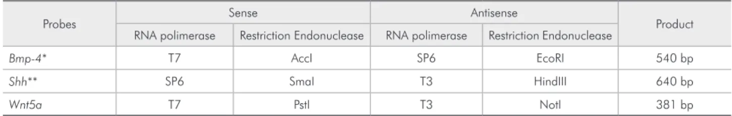
All ISH riboprobes were generated by in vitro transcription labeling with digoxygenin-UTP ac-cording to the manufacturer’s manual (Boehringer Mannhein). Probe size and yield were determined by electrophoresis on a 1.5% agarose gel with a RNA standard (Table 1). Hybridization with transcripts de-rived from the sense orientation of each probe result-ed in no signal above background levels (Figure 1).
Table 1 -In situ hibridization probes according to orientation, RNA polimerase and restriction endonuclease.
Probes Sense Antisense Product
RNA polimerase Restriction Endonuclease RNA polimerase Restriction Endonuclease
Bmp-4* T7 AccI SP6 EcoRI 540 bp
Shh** SP6 SmaI T3 HindIII 640 bp
Wnt5a T7 PstI T3 NotI 381 bp
Synthesis of DNA template
Total RNA was extracted from E9-E10 mice heads by using Trizol (Invitrogen, Carlsbad, CA, USA) ac-cording to the manufacturer’s instruction. Reverse transcription was performed with SuperscriptTM
(In-vitrogen) and oligoDT primers. cDNA was synthe-sized with 1 µg of total RNA treated with DNAse I in a volume of 20 µl. Wnt-5a primers were designed spanning intron-exon boundary using the human and mouse mRNA sequence (forward 5’ GGAGAAGGC-GCGAAGACAG 3’; reverse 5’ GGGCGTCCAC-GAACTCCT 3’) and GeneTool 1.0 Software (BioTo-ols Incorporated, Edmonton, Alberta, Canada). PCR was conducted in 25 µl reactions using 50 pM of each primer, 1.5 mM MgCl2, 1 X PCR Buffer, 100 µM of each dNTP, 20 ng cDNA, and 0.02 U/ µl Taq (Invi-trogen). The thermal proile consisted of an initial denaturation step for 4 min at 93°C, followed by 25 cycles of ampliication. Each round consisted of denaturation for 45 s/94°C, annealing for 1 min 30 s/55°C, extension for 2 min/72°C, and an additional 7 min/72°C for terminal elongation. Ampliication products were analyzed on a 1% agarose gel with ethidium bromide, where a single band of 381 bp was visualized. Speciicity of the amplicons was con-irmed by cloning and sequencing.
ISH of frozen sections, alkaline phosphatase staining
Frozen sections of mouse embryos were processed for ISH as described previously5,26 with some
chang-es dchang-escribed bellow. Embryos from E11.5 to E14.5 were ixed by immersion in 4% paraformaldehyde in phosphate buffered saline (PBS, pH 7.4) over-night, dehydrated to 30% sucrose, embedded in Tis-sue-Tek OCT (Sakura Finetek, Torrance, CA, USA), and frozen at –80°C. Coronal plane serial sections of 10 µm were then collected on silane-coated glass
slides. Sections were permeabilized with 10 µg/ml proteinase K for 2 min. Hybridizations were carried out in “seal-a-meal” bags, overnight/70°C, in 5 ml of hybridization solution (50% formamide, 5 X SSC (pH 4.5), heparin 50 µg/ml, yeast RNA 50 µg/ml, 1% SDS) with a probe concentration of ~0.2 µg/ ml. Washes were as follows: three 15 min changes of 50% formamide, 30% 20 X SSC (pH 4.5) and 10% 10 X SDS at 70°C, and three 15 min changes of 50% formamide and 12% 20 X SSC (pH 4.5) at 65°C. Detection of bound probe was performed using anti-digoxigenin antibody and NBT/BCIP as color substrate. Slides were examined on a Nikon SMZ-2T microscope and digital pictures were taken with an Axiophot 2 Zeiss microscope (Carl Zeiss MicroImaging, Thornwood, NY, USA) and a 3CCD MTI camera (Dage-MTI, Michigan City, IN, USA). Images were captured and stored on a Macintosh computer using Adobe Photoshop 5.5 software.
Results
The results described below were separated ac-cording to the stage of tooth development.
Initiation stage of tooth development (IS)
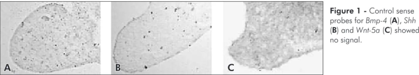
During the initiation of odontogenesis the devel-oping teeth can be visualized as localized thicken-ings of the oral epithelium. At E11.5, Bmp-4 was ex-pressed in the underlying mesenchyme (Figure 2A). When the dental lamina was formed and could be distinguished as an epithelial thickening, Bmp-4 was expressed transiently in epithelial cells (data not shown) and the underlying mesenchyme (Figures 2A and 2B), in the same stage as Shh (Figure 2D) and Wnt-5a (Figure 2G). Shh expression was also seen and strongly maintained in the epithelial thicken-ing, which represents the irst morphological mani-festation of the developing tooth (Figures 2C and
A B C
Figure 1 - Control sense probes for Bmp-4 (A), Shh
2D), although not uniformly throughout the epi-thelium. Shh transcripts were restrictedly expressed in dental epithelium, including incisor and molar germs (Figures 2C and 2D, respectively). Wnt-5a expression domains exhibited widespread expres-sion in the mandibular and maxillary mesenchyme (Figure 2G).
Bud stage (BS)
By E12.5, the dental epithelium had invaginated to form the epithelial tooth bud. From this stage on, Bmp-4 was expressed in the condensed mesenchyme around the bud (data not shown). Shh expression was not seen either in the epithelium or in the mes-enchyme (Figure 2E), although signals could be seen
A
D
C
F
H
B
E
G I
Figure 2 -Bmp-4 (A and B), Shh (C, D, E and F) and Wnt-5a (G, H and I) expression during early tooth development. A: At E12.5, Bmp-4 transcripts are expressed in the mandible mesenchyme before dental epithelium is thickened (initiation stage - IS).
B:Bmp-4 signal is broader and weaker than in A at the mandible mesenchyme of an E12.5 embryo. In the maxilla mesenchyme expression is adjacent to the thickened dental epithelium, where transcripts are seen in a small population of cells. C:Shh tran-scripts are localized in the mandible incisors dental epithelium at the IS. D: At E11.5, Shh transcripts are seen in a small popula-tion of cells in the dental epithelium. Shh signal in the maxilla epithelium is not related to odontogenesis. E: At 12.5, Shh signal is absent in the developing dental organ in the bud stage (delimited by arrows, to be compared to H). Shh medial signal in the maxilla epithelium is not related to odontogenesis. F:Shh is strongly expressed in a population of cells of the stellate reticulum in a developing dental organ in the cap stage (CS). G: An adjacent, more ventral section than A, showing broader Wnt-5a
throughout the sections. Expression of Wnt-5a was now well localized around the mesenchyme of the bud (Figure 2H).
Cap stage (CS)
At the CS (E14.5), the developing dental pa-pilla became visible, where a subset of epithelial cells formed the enamel knot, a transient cluster of non-proliferative epithelial cells supposed to act as a signaling center that directs further tooth devel-opment. Shh expression was seen as a strong signal in the tooth epithelium (Figure 2F) in the region corresponding to the enamel knot. Wnt-5a expres-sion was localized in the mesenchyme around the dental follicle, and at the tip of the dental papilla (Figure 2I).
There was no noticeable difference between Bmp-4, Shh and Wnt-5a expression in incisors or molars germs in the developing maxilla or mandible.
Discussion
The present results showed that Bmp-4, Shh and Wnt-5a are expressed at a very early stage when the ectoderm thickens and forms a placode that buds to the underlying neural crest derived mesenchyme. Moreover, when analyzing serial sections, our data suggest that Wnt-5a is expressed in a different cell population than Bmp-4.
The results presented here revealed the expres-sion of Bmp-4 restricted to the underlying mesen-chyme during the initiation of tooth development (E11.5), being transiently expressed in epithelial cells and the underlying mesenchyme when the dental lamina was formed. At the BS (E12.5), Bmp-4 was preferably expressed in the condensed mesenchyme, in accordance with Aberg et al.1 (1997). Conversely,
Nadiri et al.16 (2004), using immunohistochemistry,
found that Bmp-4 was immunolocalized both in the epithelium and mesenchyme at the BS of mouse irst lower molar.
With regards to Shh expression, the signal was intense but restrictedly expressed in the epithelial thickening during the initiation of incisor and mo-lar development, as well as in the tooth epithelium in the region corresponding to the presumptive de-veloping cusps in the CS. In fact, there is evidence
suggesting that Shh acts as a mitogen, inducing pro-liferation, growth and polarization8 as those
thick-enings form a tooth bud.2,19,20 The highly restricted
expression of Shh at sites of tooth formation is likely to be essential for specifying the sites where tooth buds will invaginate and teeth will form.15,19
Accord-ing to our results, there was a lack of Shh signaling during the BS with subsequent increase in the CS, possibly regulating the shape of the tooth crown. In-deed, some authors have reported that the inhibition of Shh signaling in mandibular explants from E10.5 results in a failure of bud formation and an arrest of tooth development.2,20
Several authors have studied the complex regula-tion of the Shh signaling pathway during mice den-tal tissues development using Shh antagonists.3,15
Although our study did not reveal different expres-sion patterns between Shh, Bmp-4 and Wnt-5a, in incisor or molar germs in the developing maxilla or mandible in the CS, according to Miletich et al.15
(2005), Rab23 demonstrated contrasting expression domains in the incisor and molar mice dentition during the CS, restricted to the mesenchymal com-partment of molar teeth and the epithelium of the enamel knot in incisor teeth. These indings provide the irst evidence of distinct regulatory pathways for Shh in teeth of different classes, and suggest that the additional complexity of the molar dentition may require higher levels of Shh signaling activity.
In the CS, Shh transcripts were strongly ex-pressed in the stellate reticulum, possibly including the enamel knot. Indeed, Shh has been previously shown to be expressed in the enamel knot in the CS.6 Our results, however, showed a broader
ex-pression. This inding, in addition to the expression of Shh just prior to bud formation, is consistent with the statement that Shh has dual roles in early odon-togenesis, irst in bud formation by stimulating epi-thelial proliferation, and second in the development of cap-stage tooth germs by increasing epithelial cell survival.2 Furthermore, during tooth development,
this pattern of expression may become restricted to the stratum intermedium, as has been shown in bo-vine tooth germs.12
chyme, becoming well localized around the mesen-chyme of the bud as well as around the dental follicle and at the tip of the dental papilla in the CS. Inter-actions between WNT and HH signaling pathways were irst described as playing a role in establishing boundaries between ectodermal cells in Drosophila segmentation.13 These molecules share the principle
of keeping potent transcriptional activators in check in the absence of receptor ligand.11 A relationship
between Wnt-5a and Shh signals, as seen for WNT-7B and Shh,19 cannot be suggested for now, although
our data revealed that transcripts of both genes were present at the same period in the developing teeth. In other organs, however, this interaction is possible. Reddy et al.18 (2001) identiied Wnt-5a as a target of
Shh in hair follicle morphogenesis. So, it is interest-ing to speculate that the absence of Shh transcripts in the dental epithelium during the BS may be re-lated to Wnt-5a presence in the mesenchyme.
Very little is known about Wnt-5a and Bmp-4 signaling pathways interactions. According to Li et al.14 (2002) Wnt-5a may inhibit Bmp-4 expression
during lung morphogenesis in mice. These results may explain the present indings of Wnt-5a being expressed in a different cell population than Bmp-4 during early tooth development.
Interactions between the ectoderm and under-lying mesenchyme constitute a central mechanism regulating the morphogenesis of several organs.23
Tooth development is considered an important mod-el to study epithmod-elial-mesenchymal interactions and, although many questions are still unanswered, genes that regulate tooth development are being identiied with increasing speed. Understanding how these genes regulate tooth formation will help us to un-derstand how speciic genes cause dental defects, and possibly the mechanisms underlying odonto-genic tumor formation.
Conclusion
The present results showed that Bmp-4, Shh and Wnt-5a are expressed since the very early stages of tooth development, and they suggest that Wnt-5a is expressed in a different cell population than Bmp-4 and Shh.
Acknowledgements
The authors thank Dr. Brigid Hogan (Vanderbilt University, Nashville, TN) and Dr. Andrew McMa-hon for kindly providing Bmp-4 and Shh probes, respectively. This work was supported by FAPESP grants (97/13228-5).
References
1. Aberg T, Wozney J, Thesleff I. Expression patterns of bone morphogenetic proteins (Bmps) in the developing mouse tooth suggest roles in morphogenesis and cell differentiation. Dev Dyn. 1997;210(4):383-96.
2. Cobourne MT, Hardcastle Z, Sharpe PT. Sonic hedgehog regulates epithelial proliferation and cell survival in the de-veloping tooth germ. J Dent Res. 2001;80(11):1974-9. 3. Cobourne MT, Miletich I, Sharpe PT. Restriction of sonic
hedgehog signalling during early tooth development. Develop-ment. 2004;131(12):2875-85.
4. Cobourne MT, Sharpe PT. Tooth and jaw: molecular mechan-isms of patterning in the first branchial arch. Arch Oral Biol. 2003;48(1):1-14.
5. Cole LK, Le Roux I, Nunes F, Laufer E, Lewis J, Wu DK. Sensory organ generation in the chicken inner ear: contribu-tions of bone morphogenetic protein 4, serrate1, and lunatic fringe. J Comp Neurol. 2000;424(3):509-20.
6. Dassule HR, Lewis P, Bei M, Maas R, McMahon AP. Sonic hedgehog regulates growth and morphogenesis of the tooth. Development. 2000;127(22):4775-85.
7. Epstein DJ, McMahon AP, Joyner AL. Regionalization of Sonic hedgehog transcription along the anteroposterior axis of the mouse central nervous system is regulated by Hnf3-dependent and -inHnf3-dependent mechanisms. Development. 1999;126(2):281-92.
8. Gritli-Linde A, Bei M, Maas R, Zhang XM, Linde A, McMa-hon AP. Shh signaling within the dental epithelium is neces-sary for cell proliferation, growth and polarization. Develop-ment. 2002;129(23):5323-37.
9. Ingham PW, McMahon AP. Hedgehog signaling in ani-mal development: paradigms and principles. Genes Dev. 2001;15(23):3059-87.
neurogenesis in the mouse. Development. 1991;111(2):531-42.
11. Kalderon D. Similarities between the Hedgehog and Wnt sig-naling pathways. Trends Cell Biol. 2002;12(11):523-31. 12. Koyama E, Wu C, Shimo T, Iwamoto M, Ohmori T, Kurisu
K et al. Development of stratum intermedium and its role as a Sonic hedgehog-signaling structure during odontogenesis. Dev Dyn. 2001;222(2):178-91.
13. Lawrence PA, Struhl G. Morphogens, compartments, and pat-tern: lessons from drosophila? Cell. 1996;85(7):951-61. 14. Li C, Xiao J, Hormi K, Borok Z, Minoo P. Wnt5a participates
in distal lung morphogenesis. Dev Biol. 2002;248(1):68-81. 15. Miletich I, Cobourne MT, Abdeen M, Sharpe PT. Expression
of the Hedgehog antagonists Rab23 and Slimb/betaTrCP dur-ing mouse tooth development. Arch Oral Biol. 2005;50(2):147-51.
16. Nadiri A, Kuchler-Bopp S, Haikel Y, Lesot H. Immunolo-calization of BMP-2/-4, FGF-4, and WNT10b in the de-veloping mouse first lower molar. J Histochem Cytochem. 2004;52(1):103-12.
17. Nunes FD, de Almeida FC, Tucci R, de Sousa SC. Homeobox genes: a molecular link between development and cancer. Pesqui Odontol Bras. 2003;17(1):94-8.
18. Reddy S, Andl T, Bagasra A, Lu MM, Epstein DJ, Morrisey EE
et al. Characterization of Wnt gene expression in developing and postnatal hair follicles and identification of Wnt5a as a
target of Sonic hedgehog in hair follicle morphogenesis. Mech Dev. 2001;107(1/2):69-82.
19. Sarkar L, Cobourne M, Naylor S, Smalley M, Dale T, Sharpe PT. Wnt/Shh interactions regulate ectodermal boundary for-mation during mammalian tooth development. Proc Natl Acad Sci USA. 2000;97(9):4520-4.
20. Sarkar L, Sharpe PT. Inhibition of Wnt signaling by exog-enous Mfrzb1 protein affects molar tooth size. J Dent Res. 2000;79(4):920-5.
21. Theiler K. The house mouse; development and normal stages from fertilization to 4 weeks of age. New York: Springer-Verlag; 1972.
22. Thesleff I. Epithelial-mesenchymal signalling regulating tooth morphogenesis. J Cell Sci. 2003;116(Pt 9):1647-8.
23. Thesleff I. Homeobox genes and growth factors in regulation of craniofacial and tooth morphogenesis. Acta Odontol Scand. 1995;53(3):129-34.
24. Tucker AS, Matthews KL, Sharpe PT. Transformation of tooth type induced by inhibition of BMP signaling. Science. 1998;282(5391):1136-8.
25. Uusitalo M, Heikkila M, Vainio S. Molecular genetic studies of Wnt signaling in the mouse. Exp Cell Res. 1999;253(2):336-48.