Contusion Injury in Adult Rats
Carmen Marı´a Ferna´ndez-Martos1, Carlos Gonza´lez-Ferna´ndez1, Pau Gonza´lez1, Alfredo Maqueda1, Ernest Arenas2, Francisco Javier Rodrı´guez1*
1Laboratorio de Neurologı´a Molecular, Hospital Nacional de Paraple´jicos (HNP), Toledo, Spain,2Molecular Neurobiology Unit, MBB, Karolinska Institute, Stockholm, Sweden
Abstract
Background:Spinal cord injury is a major cause of disability that has no clinically accepted treatment. Functional decline following spinal cord injury is caused by mechanical damage, secondary cell death, reactive gliosis and a poor regenerative capacity of damaged axons. Wnt proteins are a family of secreted glycoproteins that play key roles in different developmental processes although little is known of the expression patterns and functions of Wnts in the adult central nervous system in normal or diseased states.
Findings: Using qRT-PCR analysis, we demonstrate that mRNA encoding most Wnt ligands and soluble inhibitors are constitutively expressed in the healthy adult spinal cord. Strikingly, contusion spinal cord injury induced a time-dependent increase in Wnt mRNA expression from 6 hours until 28 days post-injury, and a narrow peak in the expression of soluble Wnt inhibitors between 1 and 3 days post-injury. These results are consistent with the increase in the migration shift, from day 1 to 7, of the intracellular Wnt signalling component, Dishevelled-3. Moreover, after an initial decrease by 1 day, we also found an increase in phosphorylation of the Wnt co-receptor, low-density lipoprotein receptor-related protein 6, and an increase in activeb-catenin protein, both of which suffer a dramatic change, from a homogeneous expression pattern in the grey matter to a disorganized injury-induced pattern.
Conclusions:Our results suggest a role for Wnts in spinal cord homeostasis and injury. We demonstrate that after injury Wnt signalling is activated via the Wnt/b-catenin and possibly other pathways. These findings provide an important foundation to further address the function of individual Wnt proteins in vivo and the pathophysiology of spinal cord injury.
Citation:Ferna´ndez-Martos CM, Gonza´lez-Ferna´ndez C, Gonza´lez P, Maqueda A, Arenas E, et al. (2011) Differential Expression of Wnts after Spinal Cord Contusion Injury in Adult Rats. PLoS ONE 6(11): e27000. doi:10.1371/journal.pone.0027000
Editor:Fabrizio Gelain, University of Milan-Bicocca, Italy
ReceivedApril 14, 2011;AcceptedOctober 7, 2011;PublishedNovember 2, 2011
Copyright:ß2011 Ferna´ndez-Martos et al. This is an open-access article distributed under the terms of the Creative Commons Attribution License, which
permits unrestricted use, distribution, and reproduction in any medium, provided the original author and source are credited.
Funding:This work was supported by: Fundacio´n para la Investigacio´n Sanitaria de Castilla-La Mancha (FISCAM; Grant PI2008-39) and Fondode Investigaciones Sanitarias (FIS; Grant PI08-1475 with Fondo Europeo de Desarrollo Regional (FEDER) co-funding). Carlos Gonzalez-Fernandez was supported by FISCAM (MOV-2009_JI-06). The funders had no role in study design, data collection and analysis, decision to publish, or preparation of the manuscript.
Competing Interests:The authors have declared that no compiting interests exist.
* E-mail: fjrodriguez@sescam.jccm.es
Introduction
Spinal cord injury (SCI) is a major cause of disability with no clinically accepted treatment [1]. The functional impairment following SCI is produced by multi-factorial processes as a result of primary mechanical damage, secondary cell death, reactive gliosis and the poor capacity to regenerate damaged axons. Typically, the epicentre of the SCI is characterized by necrotic neural death, while secondary tissue damage is also evident in the penumbra zone, where processes such as ischemia, hypoxia, excitotoxicity, free radical formation, protease release and inflammation contribute to the expansion of segmental loss of function. Another serious detrimental effect of SCI is the massive death of oligodendrocytes at a distance from the epicentre of the insult, leading to demyelination and deteriorated axon conduction. The final outcome is a pathohistological lesion that is far larger than the initial mechanical wound, consisting of a cyst cavity surrounded by a glial scar that inhibits axon growth [2–4].
The Wnt family of proteins plays key roles during the development of the nervous system, influencing cell proliferation
and patterning, cell polarity and motility, axonal guidance, neuronal survival and connectivity, and cell-cell adhesion [5,6]. This wide range of effects is possible because the specific responses elicited in target cells are dependent on the spatiotemporal distribution of Wnt ligands, modulators and receptors [7–11].
To date, 19 Wnt ligands and 10 Frizzled (Fz) receptors have been identified, which activate at least three different signalling pathways: the canonical or Wnt/b-catenin pathway; and the non-canonical Planar Cell Polarity (PCP, Wnt-JNK) and Wnt-Ca2+
been associated with cytoskeletal regulation and cell motility [15-17]. Finally, activation or inhibition of Wnt signalling is further modulated by co-receptors, such as Kremen (Krm1/2), and antagonists, such as the Wnt inhibitory factor 1 (Wif1), Dickkopf (Dkk) and secreted Frizzled-Related Proteins (sFRPs) [18,19].
Although even the earliest reports suggested that Wnt expression in the nervous system may be prolonged into adulthood [20,21], little is known about the expression or function of Wnt at these stages. Functional studies in the adult have been hampered by the labile nature of Wnt proteins, the embryonic lethality of mutants and by a lack of selective pharmacological tools [9]. Otherwise, the literature provides ample evidence implicating Wnt signalling pathways in adult CNS homeostasis and disease [13,22–35], including SCI [32,33,36–41]. In this way, experimental modulation of Wnt-dependent pathways has pro-duced promising results in different neuropathological situations [22,23,25,29], such as in stroke, where Dkk1 is expressed by neurons with pro-apoptotic effects, a process that can be rescued by lithium [42]. Furthermore, Glycogen Synthase Kinase-3b (GSK3-b) inhibition [36], lithium [36,43] or Wnt3a [41], all of which activate Wnt/b-catenin signalling, exert neuroprotective effects in SCI.
However, to our knowledge, the expression of Wnts has been reported as undetectable byin situhybridization (ISH) in the adult spinal cord of mice, while after SCI onlyWnt1, Wnt4andWnt5a together withFrizzled-1andRykreceptors were transiently induced [32], with no effect on ß-catenin mediated transcription [35]. On the other hand, in rats, the only Wnt protein shown to be expressed after SCI is Wnt5a, which is expressed by astrocytes from the glial scar and inhibits corticospinal regeneration through non-canonical Ryk activation [33].
Therefore, to investigate the role of the Wnt family of proteins in the adult spinal cord, we decided to profile by quantitative Real Time-PCR (qRT-PCR) the temporal pattern of mRNA expression for all Wnt ligands and inhibitors, followed by Western-blot and immunohistochemistry analysis of key members of the canonical pathway, in both non-injured and a moderate contusion model of SCI in rats. In agreement with previous indirect findings [22,29,44,45], our results demonstrate that most Wnt ligands and inhibitors are constitutively expressed in the non-injured adult spinal cord, with activation of at least the canonical pathway in the grey matter. After injury, this pattern of expression was dramatically altered with significant activa-tion of the canonical pathway in a large proporactiva-tion of cells around the epicentre of the wound. The reasons for the difference between our results and previous studies in mice might be the higher sensitivity of qRT-PCR versus the ISH technique, but also a different pattern of Wnt expression between mice and rats. Hence, Wnts would appear to participate in the SCI pathophysiology.
Materials and Methods
Animals
Adult female Wistar rats, aged 10–12 weeks and weighing 200– 220 grams were used throughout the study. Animals were obtained from Harlan (Barcelona, Spain) and housed in climate controlled quarters with a 12 hour light cycle. Handling was carried out according to European Union and NIH guidelines for animal experimentation in order to minimize suffering and the number of animals used. All the experimental protocols were approved by the Bioethics Committee of The National Hospital of Paraplegics (Permit numbers 51/2009 and 45/2008).
Surgical procedure and experimental design
Sterile surgical techniques and methods were used throughout this study in a designated room. Briefly, rats were anesthetized with intraperitoneal injections of pentobarbital (40 mg/kg) and xylazine (10 mg/kg). Laminectomy was performed at the level of T8 via a controlled 200-kilodyne contusion injury using an Infinite Horizon Impactor (Precision Systems and Instrumentation LLC), and the overlying muscle and skin layers were sutured. After surgery, rats were allowed to recover on a warmed blanket with access to water and food, and they received daily subcutaneous injections of saline solution containing enrofloxacine (2.5 mg/kg) and buprenorphine (0.03 mg/kg) for the following 5 and 2 days, respectively. Post-operative care also included manual bladder emptying twice daily until recovery of voiding control was achieved, and inspection for signs of infection, dehydration or autophagia.
The Open-Field Locomotor Basso-Beattie-Bresnahan (BBB) scale was applied by two examiners blind to the treatments to determine severity and reproducibility of injury on days 1, 3, 7, 14 and 28 post-injury (dpi) [46]. In order to establish a homogeneous group in which expression levels were strictly due to temporal changes after injury, animals with a functional score 0–3 the day after surgery (at 24 hours) were excluded from the study. In the entire study, only one animal from the 28 dpi group was excluded. A total of 21 animals were randomly distributed between each of the following 7 groups for qRT-PCR analysis (n = 3 per group): Non-Injured Control (C), 6 hours post-injury (hpi), and 1, 3, 7, 14 and 28 dpi. For histological and Western blot studies 30 animals were randomly distributed into each of the following groups (n = 3 per group and technique): C, 1, 7, 14 and 28 dpi.
RNA isolation and qRT-PCR analysis
At each of the time-points chosen for study, animals were terminally anesthetized with pentobarbital (40 mg/kg) and perfused intracardially with heparinized saline (150 ml) to remove blood from the tissue. Total RNA was isolated with the RNeasy Lipid Mini Kit (Qiagen) from a 1 cm long fragment of the spinal cord containing the wound epicentre, according to the manufac-turer’s instructions. Complementary DNA (cDNA) was synthe-sized from DNase-treated RNA (3mg) as described previously [47]. For relative quantification, each gene of interest was first subjected to a serial dilution assay to determine the optimum detection range of Ct values, with a Ct threshold of 35 for undetectable levels of expression. Relative quantitation of all 19 Wnt ligands, the co-receptors of theb-catenin signalling cascade (LRP5/6), the soluble Wnt signalling inhibitors (sFRP-1/5, Wif1, Dkk-1/3) and the intracellular canonical Wnt signalling effectors (GSK-3b and b-catenin) was performed using 10 ng of reverse-transcribed total RNA, 20 pmol/ml of both sense and antisense primers, and the Fast SYBR Green PCR Master Mix (Applied Biosystems) in a final reaction volume of 20ml. The reactions were run on an ABI PRISM 7900 Fast Sequence Detection System instrument and software (Applied Biosystem) according to the manufacturer’s protocol.
SEM of two separate determinations of 3 independent samples per experimental group.
Western blotting
Spinal cords from animals intracardially perfused with hepa-rinized saline were rapidly dissected and a 1 cm long fragment containing the injury epicentre was homogenized in RIPA buffer (Sigma-Aldrich) containing a proteinase inhibitor cocktail (Roche). Denatured protein samples (100mg) from each group (C, 1, 7, 14 and 28 dpi: n = 3 per group) were resolved on a 10% SDS-PAGE gel (BioRad) and transferred to PVDF membranes (Millipore). Membranes were blocked for 1 hour in 5% non-fat dried milk containing TBS-T (0.1 M Tris-HCl [pH 7.4], 0.15 M NaCl and 0.1% Tween 20) and probed overnight at 4uC with a primary mouse Dvl-3 antibody (1:500; Santa Cruz Biotechnology Inc.). After washing, the membranes were incubated for 1 hour at room temperature (RT) with a Horse Radish Peroxidase (HRP)-conjugated anti-mouse secondary antibody (1:5000; Amersham), and the resulting antibody binding was visualized by bathing for 2 minutes in the ECL solution (SuperSignal West Pico Chemilumi-nescent Substrate, Thermo Scientific) and exposing to hyperfilm (Amersham) for 1–10 minutes. The antibodies were subsequently stripped from the membranes by incubating them for 5 minutes in buffer (0.1 M Glycine [pH 2.9]), and after washing in TBS-T for 15 minutes they were re-probed with a mouse anti-GAPDH antibody (1:10.000; Abcam) as a loading control.
Histology
As described above, 3 animals from each experimental group (C, 1, 7, 14 and 28 dpi) were perfused intracardially with a heparinized saline solution followed by 4% paraformaldehyde, and their spinal cords post-fixed for 4 hours in the same fixative. The spinal cords were then cryoprotected by immersion in 30% sucrose for 48 hours, embedded in Neg-50 frozen medium (Richard-Allan Scientific) and stored at220uC. From each spinal cord, 30mm thick parallel cryostat sections were obtained (Microm) from a 3-cm long fragment containing the wound
epicentre, which were mounted on 33 serial slides and stored at
220uC.
To generate a histological time-course of phosphorylated-LRP6 (p-LRP6) and active b-catenin expression in the lesioned spinal cord, a set of parallel sections from each animal was processed by immunohistochemistry. Briefly, endogenous peroxidase activity was inactivated in the sections by incubation with 2% H2O2in a
70% methanol solution at RT for 10 minutes, and they were then blocked for 1 hour at RT in blocking buffer (BB) containing 10% Fetal Bovine Serum (FBS), 0.3% Bovine Serum Albumin (BSA) and 0.3% Triton X-100 in TBS. The sections were then incubated overnight at 4uC with the primary rabbit anti-p-LRP6 (1:100; Cell Signaling) or rabbit anti-active b-catenin (1:500; Millipore) antibodies in BB, plus 1 hour at RT. Antibody binding was visualized by sequential incubations with a biotinylated anti-rabbit secondary antibody (1:500; VectorLabs) and HRP-linked strepta-vidin (1:500; Perkin Elmer), and with the ‘‘Nova Red Kit’’ (VectorLabs) according to the manufacturers instructions. The sections were finally dehydrated in graded ethanol, cleared with xylene and coverslipped with DPX (Panreac).
Statistical analysis
Statistical comparisons were examined by one-way ANOVA (GraphPad Prism 4.0 software), followed by aTukeypost hoc test to determine individual differences between means.Pvalues lower than 0.05 were considered as statistically significant.
Results
Wnt ligands expression is prolonged in the adult spinal cord and is modulated by injury
In order to investigate the role of the Wnt family of proteins in the adult spinal cord, we examined by qRT-PCR the temporal pattern of mRNA expression for all Wnt ligands in both non-injured rats and in SCI rats up to 28 days after moderate spinal cord contusion. Of the 19 Wnt ligands already described in rats, we found that most were expressed in non-injured adult spinal Table 1.List of qRT-PCR primers.
Gene name Forward primer Reverse primer Acc no.
b-catenin 59-GCCAAGTGGGTGGCATAGA-39 59-TCCCTGTCACCAGCACGAA-39 NM_053357.2
Dkk-1 59-CTGTCTGCCTCCGATCATCA-39 59-CAGAAATGTCTTGCACAACACA-39 NM_001106350.1
Dkk-2 59-GCTCGCGGGCCAAAC-39 59-CAACTCCATCAAGTCC-39 NM_001106472.1
Dkk-3 59-CAGCTGTGACATCCAGACAGAAG-39 59-GCACCTGAAACTGTCATCTGAGA-39 NM_138519.1
Dkk-4 59-AGGCCTCTGTGGGCAACTT-39 59-CTAAGGCTGGGCTGCTGAGT-39 NM_001109332.1
GSK-3b 59-TCTGGCCACCATCCTTATCC-39 59-TTGCAGGCGGTGAAGCA-39 NM_032080.1
sFRP-1 59-CTGCCACCAGCTGGACAAC-39 59-ACCTTGCGCCCCATGA-39 XM_224987.4
sFRP-2 59-CGTGAAACGGTGGCAGAAG-39 59-CGGATGCTGCGGGAGAT-39 NM_001100700.1
sFRP-3 59-CTACCCTGGAACATGACCAAGAT-39 59-TGGCGTTAGCCTGGGTACTG-39 NM_001100527.1
TCF-1 59-GCTGCTTCAGGGCTAAGATTGT-39 59-TGTGACCTTGGCATGAGTTACG-39 NM_012669.1
Wnt6 5`-GCGGTCACTCAGGCCTGTT-39 59-GGGTGCCTGACAACCACACT-39 NM_001108226.1
Wnt8 59-CCTGGGAGCGGTGGAACT-39 59-CCTGGTGTGGGTTGAAAACTG-39 NM_001106155.1
Wnt10b 5 -CCTCAAGCGCGGTTTCC-39 59-CAGCAGCCAGCATGGAGAA-39 NM_001108111.1
Wnt15 59-CACCCATGTGGGCATCAA-39 59-CCATGACACTTGCAGGTTGTTC-39 NM_001107055.1
Wif1 59-TGCGGTGCCCATGGA-39 59-CTGCCACGAACCCA-39 NM_053738.1
Primers used for qRT-PCR analysis of the genes assessed here, including the gene symbol, primer sequence (forward and reverse sequence respectively) and GenBank accession number. The primers used to assess the expression of Wnt ligands and inhibitors not included in the list were obtained elsewhere [47].
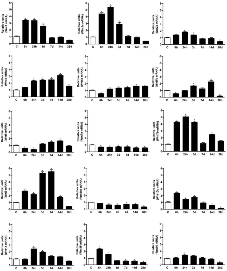
cord, onlyWnt-3, 3a, 8aand8bmRNAs remained below the level of detection of the qRT-PCR protocol (Ct over 35). After injury, the Wnt ligand mRNA expression was regulated distinctly at the different time-points analyzed (Figure 1). The relative increases and decreases observed with respect to the non-injured basal value, allowed the ligands to be grouped in function of three distinct patterns: i) no change or a slight reduction (Wnt7aand 10a); ii) early induction from 6 hours up to 3 dpi (Wnt1, 2a, 2b, 7b, 10b, 15, 16); and iii) late induction from 1–3 up to 7–14 dpi (Wnt4, 5a, 5b, 6, 9, 11). For instance,Wnt1and2are good examples of early induced genes that are markedly up-regulated up to 3 dpi, after which their mRNA expression decreases to control levels by 7 dpi. Conversely,Wnt 5a, 5band6exhibited a 50% reduction in mRNA expression in the first 6 hpi, followed by a steady increase from 1–3 dpi to 14 dpi, when maximal expression was observed (Figure 1). Finally, none of the Wnt ligands with undetectable or null levels in non-injured spinal cords were expressed following injury.
Wnt inhibitors and modulators are differentially expressed in the healthy and injured spinal cord
We next assessed the expression of secreted Wnt inhibitors and modulators, key regulators of extracellular signalling by the large and complex family of Wnt ligands. We focused our analysis on the families with well described roles in the nervous system: Dkk-1/4, sFRP-1/5 and Wif1. As observed for Wnt ligands, most of the Wnt inhibitors and modulators were expressed in the non-injured adult spinal cord, exceptsFRP-4andsFRP-5 (Figure 2). Interest-ingly, the specific inhibitors of the canonical pathway, Dkk-1/4, were up-regulated early, with a striking peak of expression at 24 hpi that returned to basal levels at 3 dpi. The sFRPs are generally considered as broad and non-specific Wnt inhibitors, yet sFRP-1 and 2exhibited a similar but slightly delayed pattern of expression, with a narrow peak at 3 dpi, whilesFRP-3remained stable. Finally, Wif1, a broad inhibitor of Wnt signalling, was dramatically down-regulated from 24 hpi until the end of the study.
Canonical Wnt signalling is active in the adult spinal cord and in cells around the wound epicentre after SCI
At the cellular level, Wnt binding to Fz or non-conventional receptors activates three distinct downstream signalling cascades: the canonical or Wnt/b-catenin; and the non-canonical Planar Cell Polarity (PCP, Wnt-JNK) and Wnt-Ca2+
pathways. As the non-canonical pathways remain poorly characterized, we focused on a representative set of components in the main canonical pathway. Irrespective of the Fz receptor expressed by a specific cell, activation of the canonical pathway requires the expression and recruitment (via serine phosphorylation) of LRP5/6 co-receptors [10,50,51]. Importantly, both co-receptors were expressed in non-injured adult spinal cord, a pattern which changed little for mRNA after SCI (Figure 3). Moreover, active LRP6 was homogenously distributed in the grey matter, although was also observed in several cells around the wound epicentre at 24 hpi, and it was even more prominent in the cells surrounding the cyst and in the grey matter from 7 dpi (Figure 4). Dvl is thought to be a key transducer of Wnt signalling [17] and it was expressed in a similar pattern to LRP6, with an increase in the active phosphorylated isoform 3 at 24 hpi peaking at 7 dpi (Figure 4).
Canonical activation relies on the dephosphorylation of b -catenin by GSK-3ß and the eventual translocation of the activeb -catenin to the nucleus where it promotes transcription of a set of target genes after interacting with members of the TCF/LEF
family [7,9]. b-catenin mRNA expression was up-regulated following SCI, peaking at 6-24 hpi (a 1.8-fold increase when compared to the controls) and returning to basal levels at 14 dpi. In parallel, its negative regulatorGSK-3bwas also up-regulated at 6–24 hpi, its expression decreasing thereafter until the end of the study (Figure 3). Strikingly, activeb-catenin was expressed strongly in the grey matter of the non-injured spinal cord with a highly suggestive vascular pattern. SCI altered this pattern, with cells in the white matter around the epicentre of the wound expressingb -catenin, increasing in number and occupying the centre of the wound at 7 dpi, and finally concentrating around the developing cyst at 14–28 dpi (Figure 4).
Discussion
In the present study, we provide evidence that the expression of Wnt ligands, their inhibitors and components of their intracellular signalling pathways is prolonged in the adult spinal cord, as is the activation of the canonical pathway, suggesting that the Wnt family of proteins play a role in spinal cord function and physiology. More importantly, in a clinically relevant rat model of SCI we demonstrate that trauma induces a dramatic and time-dependent change in the physiological pattern of Wnt mRNA expression (Figure 5A). Furthermore, we describe a concomitant activation of Dvl-3, the downstream intracellular signalling transducer of Wnt, and activation of the canonical pathway in cells around the wound core in a pattern suggesting that it influences glial scar formation. To our knowledge, this is the first report demonstrating constitutive expression of Wnts in the adult spinal cord and their differential regulation after injury. These observations are likely to be highly relevant to understanding the potential role of Wnts in the cell and molecular responses induced by SCI (Figure 5B).
The clinical outcome of SCI can be improved by limiting the extent of secondary tissue damage, which is largely dependent on the inflammatory response induced during both the acute and chronic phases [2,3,52]. Minutes after injury, a massive inflam-matory response is induced that is characterized by a variety of complex and interrelated events and cellular responses, in particular those involving astroglial and microglial reactivity or leukocyte infiltration. It was recently suggested that canonical Wnt signalling is involved in microglial proinflammatory instruction [28], while inhibitory and inductive roles have also been proposed for canonical and non-canonical signalling in proinflammation, respectively [53–56]. Indeed, canonical activation by lithium, Wnt1 or Wnt3a inhibits several inflammatory events, including endothelial activation [57], transendothelial migration of mono-cytes [58], and proinflammatory cytokine production by activated macrophages [54,59]. By contrast, TLR/NFkB signalling pro-motes the production of proinflammatory cytokines and Wnt5a in an in vitro model of macrophage activation [53,55,56,60,61]. In turn, these factors exert an autocrine effect through the Fz5-mediated activation of the Wnt-Ca2+ non-canonical pathway,
Figure 1. Wnt ligands expression in the spinal cord of adult rats following SCI.All Wnt ligands except forWnt3,3a,8aand8bwere constitutively
expressed in the non-injured adult spinal cord. After SCI, 3 main patterns of Wnt ligand expression were observed: i)No change or slight reduction(Wnt7aand
10a); ii)early induction(Wnt1, 2a, 2b, 7b, 10b, 15and16); and iii)late induction(Wnt4, 5a, 5b, 6, 9and11). All analyses were performed using total RNA samples isolated from a 1 cm long fragment of the spinal cord from non-lesioned control animals (C) and fragments containing the wound from contused animals at different times post-injury (6 and 24 hpi and 3, 7, 14 and 28 dpi). Expression of rat Wnt ligand genes was assessed by qRT-PCR using specific primers (Table 1
and [47]) and normalized to ribosomal18Sexpression. Values for each experimental group and day are expressed as mean6SEM, n = 3. Each animal/sample
(‘‘n’’) was measured in triplicate in two occasions (2 independent technical triplicates, 6 measurements per sample). *p,0.05 compared with C.
Figure 2. Short term up-regulation of Wnt inhibitors expression after SCI.Expression of the Wnt inhibitor genesDkk-1/4,sFRP(sFRP-1/5)
matter of the wound core, concomitant with an early peak in the expression of mRNA encoding canonical inhibitors like Dkkand GSK-3b, and the later expression of Wnt5a mRNA, which was previously shown to be expressed in reactive astrocytes of the glial scar [33]. Wnt5a expression has also been reported in other cell types, including fibroblasts [68] and endothelial cells [69], suggesting a cross-talk between all the cells involved in restoring tissue homeostasis after SCI, which based on our results may involve a larger number of ligands, receptors, and modulators of the Wnt family of proteins.
A serious consequence of SCI is the large-scale death of neurons and oligodendrocytes due to excitotoxic and inflammatory apoptosis [2,3]. In this regard, in different neuropathological situations inhibition and activation of the canonical Wnt pathway has been shown to induce neuronal death and survival, respec-tively, including circumstances of excitotoxicity [23,25,70–72], brain ischemia [25,43,73] and Parkinson’s [74] and Alzheimers diseases [23,45,71,75–77]. Indeed, lithium is a non-specific
GSK-3ß inhibitor that is employed in the pharmacotherapy of bipolar diseases and it is a potent inhibitor of apoptotic neuronal deathin vitro, as well as that associated with various neurodegenerative conditions in vivo [73,78–81], including CNS stroke and SCI [36,43]. This effect may be partly caused by the preservation and/ or reinduction of the barrier properties of brain microvessels in the injured area. Importantly, acute administration of Wnt3a after moderate SCI provokes a significant recovery of motor function in association with a moderate neuroprotective effect [41]. Thus, Wnts would appear to fulfil a role of in adult CNS physiology as well as representing potential therapeutic targets.
Another critical impairment to functional recovery following SCI is the generation of a glial scar around the epicentre of the wound, which strongly inhibits axonal regeneration [3,4]. As during development [6,82,83], Wnt proteins are critical factors governing axonal growth after CNS injury. Both endogenous Wnt2b and exogenous Wnt3a directly promote b -catenin-dependent CNS regeneration in the retina of adult mammals
Figure 3. SCI alters the mRNA expression of canonical Wnt signalling components.Expression of the canonical co-receptorLRP5/6, the
downstream intracellular inhibitory transducerGSK3-ßand theß-catenintranscription factor were quantified by qRT-PCR as described in Figure1. All
signalling components were expressed in non-injured spinal cord. SCI did not alter the transcription of theLRP5/6co-receptors, although there was a
striking increase inGSK3-bexpression that coincided with that of theDkkinhibitors, as well as a mild up-regulation ofb-cateninafter 6 hours that
gradually decreased until the end of the study. Values for each experimental group and day are expressed as mean6SEM, n = 3. Each animal/sample
(‘‘n’’) was measured in triplicate in two occasions (2 independent technical triplicates, 6 measurements per sample). *p,0.05 compared with
non-injured control animals (C).
doi:10.1371/journal.pone.0027000.g003
spinal cord. After SCI, theDkkswere strikingly up-regulated with a narrow and specific peak after 24 hours, followed by a slightly delayed peak at
3 dpi forsFRP-1and2. By contrast,Wif1was dramatically down-regulated 24 hpi until the end of the study. Values for each experimental group and
day are expressed as mean6SEM, n = 3. Each animal/sample (‘‘n’’) was measured in triplicate in two occasions (2 independent technical triplicates, 6
measurements per sample). *p,0.05 compared with non-injured control animals (C).
[84,85], while transplantation of Wnt3a-secreting fibroblasts one week after SCI improves locomotor recovery by promoting axonal regeneration in rats [86]. Conversely, recent studies reported that SCI-induced Wnt5a expression around the injury site inhibited corticospinal axonal growth via non-canonical activation of the Ryk receptor in both mice [32] and rats [33]. This effect was overcome by intrathecal administration of Ryk neutralizing antibodies, enhancing the functional recovery. Accordingly, GSK-3binhibition by lithium increases the intrinsic growth capacity of damaged neurons after SCI, permitting significant sprouting of descending corticospinal and serotonin-ergic axons in the caudal spinal cord and promoting functional recovery [37,40]. On the other hand, glial scar is triggered by BSCB disruption and microglia/macrophage activation [4]. Indeed, this process appears to correlate with the areas of maximum BSCB breakdown and greatest number of activated microglia/macrophages [87]. Therefore, we can not exclude that at least in part Wnt promotion of axonal growth could be mediated by indirect action on BSCB restoration and inflamma-tory response modulation.
Wnts are also crucial physiological regulators of stem cells [13,88,89], which is significant as the adult spinal cord has been described to contain slow-dividing neural precursors that prolif-erate and differentiate into NG2+glial progenitors after SCI, and they migrate around the lesion core to mainly form reactive astrocytes [90,91]. b-catenin signalling is active in progenitor populations from adult neurogenic regions like the hippocampus, subventricular zone (SVZ) and olfactory bulb, and it is known to participate in the injury response of various tissues, including the
CNS [26,35,84,92]. Indeed, b-catenin has been shown to be responsible for SVZ/striatal proliferation after brain ischemia [93] and to be transcriptionally active in NG2 precursors associated to glial scar formation after traumatic brain injury [35]. However, the same authors describe a total lack ofb-catenin transcriptional activation after SCI in mice, what could be derived of differences between CNS regions in front of the same type of insult [35] and/ or versus to our results, a clear activation of both the canonical LRP6 co-receptor andb-catenin in cells around the injury core in a specific pattern that was highly suggestive of a role in glial scar formation, the reflect of a distinct physiopathology in rats [94] or intrinsic limitations of TCF-dependent transcriptional reporters [95,96]. Intriguingly, a proportion of NG2+positive precursors at a distance from the wound epicentre can differentiate into Olig2+ oligodendroglial precursors (OPC). b-catenin induced transcrip-tion is required for OPC instructranscrip-tion but following the inhibitranscrip-tion for differentiation into mature oligodendrocytes during develop-ment and importantly adult remyelination [26,97,98]. Actually, there is considerable interest in Wnts with regards the develop-ment of novel stem cell based therapies [99], as Wnt3a increased both exogenous [100] and endogenous [41] neuronal differenti-ation of adult neural precursors.
In summary, our results provide compelling evidence that Wnts are expressed and transcriptionally regulated by SCI in adulthood. These novel findings provide an important foundation to further address the function of individual Wnt proteinsin vivo, by loss and gain of function experiments, on the different cell populations of the healthy and injured adult spinal cord. In support, evidence in the literature already indicates that Wnts are not just mere Figure 4. Active canonical Wnt signalling pathway in adult spinal cord before and after SCI.(A) Western blot of Dvl-3 with protein
samples isolated from a 1 cm long spinal cord fragment from non-lesioned control animals (C) and SCI animals at 1, 7 and 14/28 dpi. SCI induced
hyperphosphorylation (upper arrowhead) and thus, the activation of Dvl-3 24 hours post-injury, which peaked at 7 dpi. GAPDH levels were used as a
control for protein loading. (B) Representative histological images of the expression of the active LRP6 canonical co-receptor (phosphorylated at
serine 1490) andb-catenin (dephosphorylated at serine 37 and threonine 41) before SCI and at 1, 7 and 14/28 dpi. Active LRP6 andb-catenin were
both expressed in the grey matter of the non-injured spinal cord, the latter exhibiting a vascular-like pattern. After SCI, active LRP6 andb-catenin was
expressed in cells located in the white matter of the wound epicentre, with a clear increase from 7 dpi and a final location suggestive of a role in
tissue response. Scale bars = 50mm.
bystanders of SCI. For instance, Wnt5a has been shown to be expressed by reactive astrocytes of the glial scar and play an inhibitory role on corticospinal regeneration through non-canonical Ryk activation [32,33], while lithium [36,43] or Wnt3a [41] induced Wnt/b-catenin signalling exerts neuroprotective
effects in SCI. These findings, together with our data and increasing evidence linking Wnt signalling with neurodegenerative diseases in the adult [29] and in the development of CNS [6], suggest that the Wnt family of proteins might play a role in the pathophysiology of the SCI.
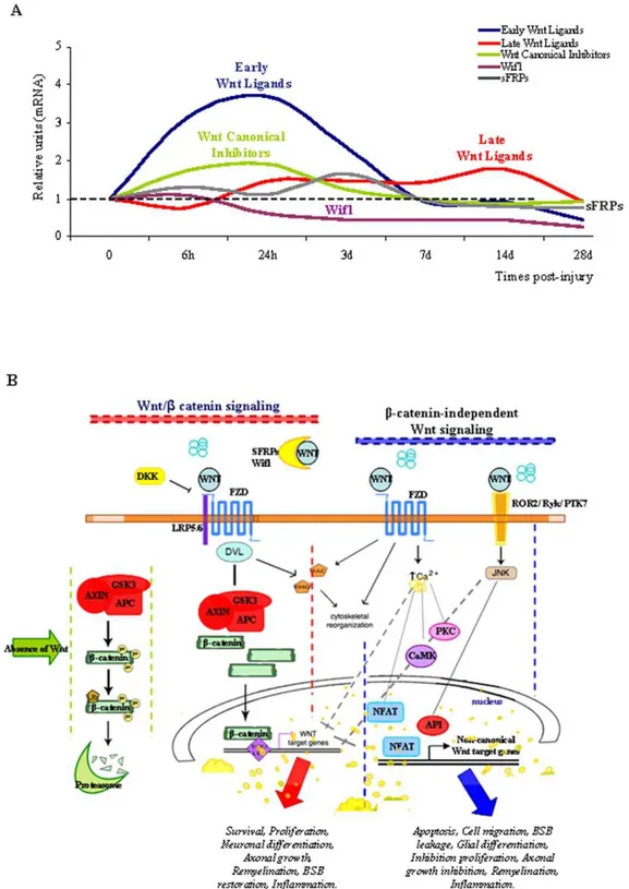
Figure 5. Temporal expression of Wnt mRNAs, and a schematic representation of Wnt signalling elements.(A) Summary of the
integrated mRNA expression for early (Wnt1, Wnt2a, Wnt2b) and late Wnt Ligands (Wnt4, Wnt5a/5b, Wnt6, Wnt7a, Wnt11), as well as the Wnt Canonical
Inhibitors (Dkk-1/4), sFRP (sFRP-1/3) andWif1. (B) Representation of the Wnt/b-catenin andb-catenin-independent signalling pathways and their
putative roles in SCI.
Acknowledgments
We would like to thank the members of the group of Dr Arenas for contributing primer sequence information, and Virginia Pe´rez and Sandra Va´zquez for their technical and experimental support.
Author Contributions
Conceived and designed the experiments: CMF-M FJR. Performed the experiments: CMF-M CG-F PG AM FJR. Analyzed the data: CMF-M PG FJR. Contributed reagents/materials/analysis tools: EA. Wrote the paper: FJR.
References
1. Knafo S, Choi D (2008) Clinical studies in spinal cord injury: moving towards successful trials. Br J Neurosurg 22: 3–12.
2. Hausmann ON (2003) Post-traumatic inflammation following spinal cord injury. Spinal Cord 41: 369–378.
3. Profyris C, Cheema SS, Zang D, Azari MF, Boyle K, et al. (2004) Degenerative and regenerative mechanisms governing spinal cord injury. Neurobiol Dis 15: 415–436.
4. Silver J, Miller JH (2004) Regeneration beyond the glial scar. Nat Rev Neurosci 5: 146–156.
5. Curinga G, Smith GM (2008) Molecular/genetic manipulation of extrinsic axon guidance factors for CNS repair and regeneration. Exp Neurol 209: 333–342. 6. Ciani L, Salinas PC (2005) WNTs in the vertebrate nervous system: from
patterning to neuronal connectivity. Nat Rev Neurosci 6: 351–362. 7. van Amerongen R, Mikels A, Nusse R (2008) Alternative wnt signalling is
initiated by distinct receptors. Sci Signal 1: re9.
8. Angers S, Moon RT (2009) Proximal events in Wnt signal transduction. Nat Rev Mol Cell Biol 10: 468–477.
9. Chien AJ, Conrad WH, Moon RT (2009) A Wnt survival guide: from flies to human disease. J Invest Dermatol 129: 1614–1627.
10. Komiya Y, Habas R (2008) Wnt signal transduction pathways. Organogenesis 4: 68–75.
11. Mikels AJ, Nusse R (2006) Wnts as ligands: processing, secretion and reception. Oncogene 25: 7461–7468.
12. Brembeck FH, Rosario M, Birchmeier W (2006) Balancing cell adhesion and Wnt signalling, the key role of beta-catenin. Curr Opin Genet Dev 16: 51–59. 13. Michaelidis TM, Lie DC (2008) Wnt signalling and neural stem cells: caught in
the Wnt web. Cell Tissue Res 331: 193–210.
14. Pishvaian MJ, Byers SW (2007) Biomarkers of WNT signalling. Cancer Biomark 3: 263–274.
15. Fradkin LG, Dura JM, Noordermeer JN (2010) Ryks: new partners for Wnts in the developing and regenerating nervous system. Trends Neurosci 33: 84–92. 16. Minami Y, Oishi I, Endo M, Nishita M (2010) Ror-family receptor tyrosine
kinases in noncanonical Wnt signalling: their implications in developmental morphogenesis and human diseases. Dev Dyn 239: 1–15.
17. Montcouquiol M, Crenshaw EB, 3rd, Kelley MW (2006) Noncanonical Wnt signalling and neural polarity. Annu Rev Neurosci 29: 363–386.
18. Bovolenta P, Esteve P, Ruiz JM, Cisneros E, Lopez-Rios J (2008) Beyond Wnt inhibition: new functions of secreted Frizzled-related proteins in development and disease. J Cell Sci 121: 737–746.
19. Kawano Y, Kypta R (2003) Secreted antagonists of the Wnt signalling pathway. J Cell Sci 116: 2627–2634.
20. Gavin BJ, McMahon JA, McMahon AP (1990) Expression of multiple novel Wnt-1/int-1-related genes during fetal and adult mouse development. Genes Dev 4: 2319–2332.
21. Shimogori T, VanSant J, Paik E, Grove EA (2004) Members of the Wnt, Fz, and Frp gene families expressed in postnatal mouse cerebral cortex. J Comp Neurol 473: 496–510.
22. Boonen RA, van Tijn P, Zivkovic D (2009) Wnt signalling in Alzheimer’s disease: up or down, that is the question. Ageing Res Rev 8: 71–82. 23. Caraci F, Busceti C, Biagioni F, Aronica E, Mastroiacovo F, et al. (2008) The
Wnt antagonist, Dickkopf-1, as a target for the treatment of neurodegenerative disorders. Neurochem Res 33: 2401–2406.
24. Chong ZZ, Li F, Maiese K (2005) Oxidative stress in the brain: novel cellular targets that govern survival during neurodegenerative disease. Prog Neurobiol 75: 207–246.
25. Chong ZZ, Shang YC, Hou J, Maiese K (2010) Wnt1 neuroprotection translates into improved neurological function during oxidant stress and cerebral ischemia through AKT1 and mitochondrial apoptotic pathways. Oxid Med Cell Longev 3: 153–165.
26. Fancy SP, Baranzini SE, Zhao C, Yuk DI, Irvine KA, et al. (2009) Dysregulation of the Wnt pathway inhibits timely myelination and remyelination in the mammalian CNS. Genes Dev 23: 1571–1585.
27. Gogolla N, Galimberti I, Deguchi Y, Caroni P (2009) Wnt signalling mediates experience-related regulation of synapse numbers and mossy fiber connectivities in the adult hippocampus. Neuron 62: 510–525.
28. Halleskog C, Mulder J, Dahlstrom J, Mackie K, Hortobagyi T, et al. (2010) WNT signalling in activated microglia is proinflammatory. Glia 59: 119–131. 29. Inestrosa NC, Arenas E (2010) Emerging roles of Wnts in the adult nervous
system. Nat Rev Neurosci 11: 77–86.
30. Lein ES, Hawrylycz MJ, Ao N, Ayres M, Bensinger A, et al. (2007) Genome-wide atlas of gene expression in the adult mouse brain. Nature 445: 168–176. 31. Lie DC, Colamarino SA, Song HJ, Desire L, Mira H, et al. (2005) Wnt signalling
regulates adult hippocampal neurogenesis. Nature 437: 1370–1375.
32. Liu Y, Wang X, Lu CC, Kerman R, Steward O, et al. (2008) Repulsive Wnt signalling inhibits axon regeneration after CNS injury. J Neurosci 28: 8376–8382.
33. Miyashita T, Koda M, Kitajo K, Yamazaki M, Takahashi K, et al. (2009) Wnt-Ryk signalling mediates axon growth inhibition and limits functional recovery after spinal cord injury. J Neurotrauma 26: 955–964.
34. Rowe WB, Blalock EM, Chen KC, Kadish I, Wang D, et al. (2007) Hippocampal expression analyses reveal selective association of immediate-early, neuroenergetic, and myelinogenic pathways with cognitive impairment in aged rats. J Neurosci 27: 3098–3110.
35. White BD, Nathe RJ, Maris DO, Nguyen NK, Goodson JM, et al. (2010) Beta-catenin signalling increases in proliferating NG2+progenitors and astrocytes during post-traumatic gliogenesis in the adult brain. Stem Cells 28: 297–307. 36. Cuzzocrea S, Genovese T, Mazzon E, Crisafulli C, Di Paola R, et al. (2006)
Glycogen synthase kinase-3 beta inhibition reduces secondary damage in experimental spinal cord trauma. J Pharmacol Exp Ther 318: 79–89. 37. Dill J, Wang H, Zhou F, Li S (2008) Inactivation of glycogen synthase kinase 3
promotes axonal growth and recovery in the CNS. J Neurosci 28: 8914–8928. 38. Su H, Chu TH, Wu W (2007) Lithium enhances proliferation and neuronal differentiation of neural progenitor cells in vitro and after transplantation into the adult rat spinal cord. Exp Neurol 206: 296–307.
39. Wong YW, Tam S, So KF, Chen JY, Cheng WS, et al. (2010) A three-month, open-label, single-arm trial evaluating the safety and pharmacokinetics of oral lithium in patients with chronic spinal cord injury. Spinal Cord 49: 94–98. 40. Yick LW, So KF, Cheung PT, Wu WT (2004) Lithium chloride reinforces the
regeneration-promoting effect of chondroitinase ABC on rubrospinal neurons after spinal cord injury. J Neurotrauma 21: 932–943.
41. Yin ZS, Zu B, Chang J, Zhang H (2008) Repair effect of Wnt3a protein on the contused adult rat spinal cord. Neurol Res 30: 480–486.
42. Mastroiacovo F, Busceti CL, Biagioni F, Moyanova SG, Meisler MH, et al. (2009) Induction of the Wnt antagonist, Dickkopf-1, contributes to the development of neuronal death in models of brain focal ischemia. J Cereb Blood Flow Metab 29: 264–276.
43. Ren M, Senatorov VV, Chen RW, Chuang DM (2003) Postinsult treatment with lithium reduces brain damage and facilitates neurological recovery in a rat ischemia/reperfusion model. Proc Natl Acad Sci U S A 100: 6210–6215. 44. Kim H, Won S, Hwang DY, Lee JS, Kim M, et al. (2010) Downregulation of
Wnt/beta-catenin signalling causes degeneration of hippocampal neurons in vivo. Neurobiol Aging In press.
45. Rosi MC, Luccarini I, Grossi C, Fiorentini A, Spillantini MG, et al. (2010) Increased Dickkopf-1 expression in transgenic mouse models of neurodegener-ative disease. J Neurochem 112: 1539–1551.
46. Basso DM, Beattie MS, Bresnahan JC (1995) A sensitive and reliable locomotor rating scale for open field testing in rats. J Neurotrauma 12: 1–21.
47. Rawal N, Castelo-Branco G, Sousa KM, Kele J, Kobayashi K, et al. (2006) Dynamic temporal and cell type-specific expression of Wnt signalling components in the developing midbrain. Exp Cell Res 312: 1626–1636. 48. Fernandez CM, del Arco A, Gallardo N, Aguado L, Rodriguez M, et al. (2009)
S-resistin inhibits adipocyte differentiation and increases TNFalpha expression and secretion in 3T3-L1 cells. Biochim Biophys Acta 1803: 1131–1141. 49. Livak KJ, Schmittgen TD (2001) Analysis of relative gene expression data using
real-time quantitative PCR and the 2(-Delta Delta C(T)) Method. Methods 25: 402–408.
50. Cadigan KM, Liu YI (2006) Wnt signalling: complexity at the surface. J Cell Sci 119: 395–402.
51. Gordon MD, Nusse R (2006) Wnt signalling: multiple pathways, multiple receptors, and multiple transcription factors. J Biol Chem 281: 22429–22433. 52. Ankeny DP, Popovich PG (2009) Mechanisms and implications of adaptive
immune responses after traumatic spinal cord injury. Neuroscience 158: 1112–1121.
53. Blumenthal A, Ehlers S, Lauber J, Buer J, Lange C, et al. (2006) The Wingless homolog WNT5A and its receptor Frizzled-5 regulate inflammatory responses of human mononuclear cells induced by microbial stimulation. Blood 108: 965–973.
54. Neumann J, Schaale K, Farhat K, Endermann T, Ulmer AJ, et al. (2010) Frizzled1 is a marker of inflammatory macrophages, and its ligand Wnt3a is involved in reprogramming Mycobacterium tuberculosis-infected macrophages. Faseb J 24: 4599–4612.
55. Pereira C, Schaer DJ, Bachli EB, Kurrer MO, Schoedon G (2008) Wnt5A/ CaMKII signalling contributes to the inflammatory response of macrophages and is a target for the antiinflammatory action of activated protein C and interleukin-10. Arterioscler Thromb Vasc Biol 28: 504–510.
57. Eto M, Kouroedov A, Cosentino F, Luscher TF (2005) Glycogen synthase kinase-3 mediates endothelial cell activation by tumor necrosis factor-alpha. Circulation 112: 1316–1322.
58. Tickenbrock L, Schwable J, Strey A, Sargin B, Hehn S, et al. (2006) Wnt signalling regulates transendothelial migration of monocytes. J Leukoc Biol 79: 1306–1313.
59. Chong ZZ, Li F, Maiese K (2007) Cellular demise and inflammatory microglial activation during beta-amyloid toxicity are governed by Wnt1 and canonical signalling pathways. Cell Signal 19: 1150–1162.
60. Kigerl KA, Lai W, Rivest S, Hart RP, Satoskar AR, et al. (2007) Toll-like receptor (TLR)-2 and TLR-4 regulate inflammation, gliosis, and myelin sparing after spinal cord injury. J Neurochem 102: 37–50.
61. Sen M, Ghosh G (2008) Transcriptional outcome of Wnt-Frizzled signal transduction in inflammation: evolving concepts. J Immunol 181: 4441–4445. 62. Popovich PG, Hickey WF (2001) Bone marrow chimeric rats reveal the unique
distribution of resident and recruited macrophages in the contused rat spinal cord. J Neuropathol Exp Neurol 60: 676–685.
63. Schnell L, Fearn S, Klassen H, Schwab ME, Perry VH (1999) Acute inflammatory responses to mechanical lesions in the CNS: differences between brain and spinal cord. Eur J Neurosci 11: 3648–3658.
64. Daneman R, Agalliu D, Zhou L, Kuhnert F, Kuo CJ, et al. (2009) Wnt/beta-catenin signalling is required for CNS, but not non-CNS, angiogenesis. Proc Natl Acad Sci U S A 106: 641–646.
65. Goodwin AM, Kitajewski J, D’Amore PA (2007) Wnt1 and Wnt5a affect endothelial proliferation and capillary length; Wnt2 does not. Growth Factors 25: 25–32.
66. Liebner S, Corada M, Bangsow T, Babbage J, Taddei A, et al. (2008) Wnt/beta-catenin signalling controls development of the blood-brain barrier. J Cell Biol 183: 409–417.
67. Liebner S, Plate KH (2010) Differentiation of the brain vasculature: the answer came blowing by the Wnt. J Angiogenes Res 2: 1.
68. Sen M, Lauterbach K, El-Gabalawy H, Firestein GS, Corr M, et al. (2000) Expression and function of wingless and frizzled homologs in rheumatoid arthritis. Proc Natl Acad Sci U S A 97: 2791–2796.
69. Goodwin AM, Sullivan KM, D’Amore PA (2006) Cultured endothelial cells display endogenous activation of the canonical Wnt signalling pathway and express multiple ligands, receptors, and secreted modulators of Wnt signalling. Dev Dyn 235: 3110–3120.
70. Chen RW, Qin ZH, Ren M, Kanai H, Chalecka-Franaszek E, et al. (2003) Regulation of c-Jun N-terminal kinase, p38 kinase and AP-1 DNA binding in cultured brain neurons: roles in glutamate excitotoxicity and lithium neuroprotection. J Neurochem 84: 566–575.
71. Dinamarca MC, Sagal JP, Quintanilla RA, Godoy JA, Arrazola MS, et al. (2010) Amyloid-beta-Acetylcholinesterase complexes potentiate neurodegenera-tive changes induced by the Abeta peptide. Implications for the pathogenesis of Alzheimer’s disease. Mol Neurodegener 5: 4.
72. Wei H, Qin ZH, Senatorov VV, Wei W, Wang Y, et al. (2001) Lithium suppresses excitotoxicity-induced striatal lesions in a rat model of Huntington’s disease. Neuroscience 106: 603–612.
73. Bian Q, Shi T, Chuang DM, Qian Y (2007) Lithium reduces ischemia-induced hippocampal CA1 damage and behavioral deficits in gerbils. Brain Res 1184: 270–276.
74. L’Episcopo F, Tirolo C, Testa N, Caniglia S, Morale MC, et al. (2011) Reactive astrocytes and Wnt/beta-catenin signalling link nigrostriatal injury to repair in 1-methyl-4-phenyl-1,2,3,6-tetrahydropyridine model of Parkinson’s disease. Neu-robiol Dis 41: 508–527.
75. De Ferrari GV, Chacon MA, Barria MI, Garrido JL, Godoy JA, et al. (2003) Activation of Wnt signalling rescues neurodegeneration and behavioral impairments induced by beta-amyloid fibrils. Mol Psychiatry 8: 195–208. 76. Zhang Z, Hartmann H, Do VM, Abramowski D, Sturchler-Pierrat C, et al.
(1998) Destabilization of beta-catenin by mutations in presenilin-1 potentiates neuronal apoptosis. Nature 395: 698–702.
77. Zenzmaier C, Marksteiner J, Kiefer A, Berger P, Humpel C (2009) Dkk-3 is elevated in CSF and plasma of Alzheimer’s disease patients. J Neurochem 110: 653–661.
78. Aghdam SY, Barger SW (2007) Glycogen synthase kinase-3 in neurodegener-ation and neuroprotection: lessons from lithium. Curr Alzheimer Res 4: 21–31. 79. Chen G, Zeng WZ, Yuan PX, Huang LD, Jiang YM, et al. (1999) The mood-stabilizing agents lithium and valproate robustly increase the levels of the neuroprotective protein bcl-2 in the CNS. J Neurochem 72: 879–882. 80. Chen RW, Chuang DM (1999) Long term lithium treatment suppresses p53 and
Bax expression but increases Bcl-2 expression. A prominent role in neuropro-tection against excitotoxicity. J Biol Chem 274: 6039–6042.
81. Nunes PV, Forlenza OV, Gattaz WF (2007) Lithium and risk for Alzheimer’s disease in elderly patients with bipolar disorder. Br J Psychiatry 190: 359–360. 82. Charron F, Tessier-Lavigne M (2005) Novel brain wiring functions for classical morphogens: a role as graded positional cues in axon guidance. Development 132: 2251–2262.
83. Fenstermaker AG, Prasad AA, Bechara A, Adolfs Y, Tissir F, et al. (2010) Wnt/ planar cell polarity signalling controls the anterior-posterior organization of monoaminergic axons in the brainstem. J Neurosci 30: 16053–16064. 84. Kubo F, Nakagawa S (2008) Wnt signalling in retinal stem cells and
regeneration. Dev Growth Differ 50: 245–251.
85. Osakada F, Ooto S, Akagi T, Mandai M, Akaike A, et al. (2007) Wnt signalling promotes regeneration in the retina of adult mammals. J Neurosci 27: 4210–4219.
86. Suh HI, Min J, Choi KH, Kim SW, Kim KS, et al. (2011) Axonal regeneration effects of Wnt3a-secreting fibroblast transplantation in spinal cord-injured rats. Acta Neurochir (Wien) In press.
87. Preston E, Webster J, Small D (2001) Characteristics of sustained blood-brain barrier opening and tissue injury in a model for focal trauma in the rat. J Neurotrauma 18: 83–92.
88. Katoh M (2008) WNT signalling in stem cell biology and regenerative medicine. Curr Drug Targets 9: 565–570.
89. Nusse R, Fuerer C, Ching W, Harnish K, Logan C, et al. (2008) Wnt signalling and stem cell control. Cold Spring Harb Symp Quant Biol 73: 59–66. 90. Horky LL, Galimi F, Gage FH, Horner PJ (2006) Fate of endogenous stem/
progenitor cells following spinal cord injury. J Comp Neurol 498: 525–538. 91. Meletis K, Barnabe-Heider F, Carlen M, Evergren E, Tomilin N, et al. (2008)
Spinal cord injury reveals multilineage differentiation of ependymal cells. PLoS Biol 6: e182.
92. Clevers H (2006) Wnt/beta-catenin signalling in development and disease. Cell 127: 469–480.
93. Lei ZN, Zhang LM, Sun FY (2008) Beta-catenin siRNA inhibits ischemia-induced striatal neurogenesis in adult rat brain following a transient middle cerebral artery occlusion. Neurosci Lett 435: 108–112.
94. Sroga JM, Jones TB, Kigerl KA, McGaughy VM, Popovich PG (2003) Rats and mice exhibit distinct inflammatory reactions after spinal cord injury. J Comp Neurol 462: 223–240.
95. Barolo S (2006) Transgenic Wnt/TCF pathway reporters: all you need is Lef? Oncogene 25: 7505–7511.
96. Olson LE, Tollkuhn J, Scafoglio C, Krones A, Zhang J, et al. (2006) Homeodomain-mediated beta-catenin-dependent switching events dictate cell-lineage determination. Cell 125: 593–605.
97. Rosenberg SS, Chan JR (2009) Modulating myelination: knowing when to say Wnt. Genes Dev 23: 1487–1493.
98. Shimizu T, Kagawa T, Wada T, Muroyama Y, Takada S, et al. (2005) Wnt signalling controls the timing of oligodendrocyte development in the spinal cord. Dev Biol 282: 397–410.
99. Eftekharpour E, Karimi-Abdolrezaee S, Fehlings MG (2008) Current status of experimental cell replacement approaches to spinal cord injury. Neurosurg Focus 24: E19.
100. Yin ZS, Zhang H, Wang W, Hua XY, Hu Y, et al. (2007) Wnt-3a protein promote neuronal differentiation of neural stem cells derived from adult mouse spinal cord. Neurol Res 29: 847–854.