pseudomallei
-Specific Real-Time PCR Assays for Clinical,
Environmental or Forensic Detection Applications
Erin P. Price1,2, Julia L. Dale1, James M. Cook1, Derek S. Sarovich1,2, Meagan L. Seymour1, Jennifer L. Ginther1, Emily L. Kaufman1, Stephen M. Beckstrom-Sternberg1,3, Mark Mayo2, Mirjam Kaestli2, Mindy B. Glass4, Jay E. Gee4, Vanaporn Wuthiekanun5, Jeffrey M. Warner6, Anthony Baker6, Jeffrey T. Foster1, Patrick Tan7, Apichai Tuanyok1, Direk Limmathurotsakul5, Sharon J. Peacock5,8,9, Bart J. Currie2, David M. Wagner1, Paul Keim1,3, Talima Pearson1*
1Center for Microbial Genetics and Genomics, Northern Arizona University, Flagstaff, Arizona, United States of America,2Menzies School of Health Research, Casuarina, Northern Territory, Australia,3Translational Genomics Research Institute, Phoenix, Arizona, United States of America,4Centers for Disease Control and Prevention, Atlanta, Georgia, United States of America,5Mahidol-Oxford Tropical Medicine Research Unit, Faculty of Tropical Medicine, Mahidol University, Bangkok, Thailand, 6Microbiology and Immunology, School of Veterinary and Biomedical Sciences, James Cook University, Townsville, Queensland, Australia,7Genome Institute of Singapore, Singapore, Singapore,8Department of Microbiology and Immunology, Faculty of Tropical Medicine, Mahidol University, Bangkok, Thailand,9Department of Medicine, University of Cambridge, Cambridge, United Kingdom
Abstract
The bacteriumBurkholderia pseudomallei causes melioidosis, a rare but serious illness that can be fatal if untreated or misdiagnosed. Species-specific PCR assays provide a technically simple method for differentiatingB. pseudomalleifrom near-neighbor species. However, substantial genetic diversity and high levels of recombination within this species reduce the likelihood that molecular signatures will differentiate allB. pseudomalleifrom other Burkholderiaceae. Currently available molecular assays forB. pseudomalleidetection lack rigorous validation across largein silicodatasets and isolate collections to test for specificity, and none have been subjected to stringent quality control criteria (accuracy, precision, selectivity, limit of quantitation (LoQ), limit of detection (LoD), linearity, ruggedness and robustness) to determine their suitability for environmental, clinical or forensic investigations. In this study, we developed two novelB. pseudomalleispecific assays, 122018 and 266152, using a dual-probe approach to differentiateB. pseudomalleifromB. thailandensis,B. oklahomensisand B. thailandensis-like species; other species failed to amplify. Species specificity was validated across a large DNA panel
(.2,300 samples) comprising Burkholderia spp. and non-Burkholderia bacterial and fungal species of clinical and
environmental relevance. Comparison of assay specificity to two previously published B. pseudomallei-specific assays,
BurkDiff and TTS1, demonstrated comparable performance of all assays, providing between 99.7 and 100% specificity against our isolate panel. Last, we subjected 122018 and 266152 to rigorous quality control analyses, thus providing
quantitative limits of assay performance. Using B. pseudomallei as a model, our study provides a framework for
comprehensive quantitative validation of molecular assays and provides additional, highly validatedB. pseudomalleiassays for the scientific research community.
Citation:Price EP, Dale JL, Cook JM, Sarovich DS, Seymour ML, et al. (2012) Development and Validation ofBurkholderia pseudomallei-Specific Real-Time PCR Assays for Clinical, Environmental or Forensic Detection Applications. PLoS ONE 7(5): e37723. doi:10.1371/journal.pone.0037723
Editor:Paul J. Planet, Columbia University, United States of America
ReceivedFebruary 24, 2012;AcceptedApril 23, 2012;PublishedMay 18, 2012
Copyright:ß2012 Price et al. This is an open-access article distributed under the terms of the Creative Commons Attribution License, which permits unrestricted use, distribution, and reproduction in any medium, provided the original author and source are credited.
Funding:This project was funded by the United States Department of Homeland Security (HSHQDC-10-C-00139). DL and VW were supported by the Wellcome Trust. The funders had no role in study design, data collection and analysis, decision to publish, or preparation of the manuscript.
Competing Interests:The authors have declared that no competing interests exist.
* E-mail: [email protected]
Introduction
TheBurkholderiagenus contains over 60 species, some of which are of environmental, clinical or forensic importance. With the exception of the obligate mammalian pathogen, B. mallei, the
Burkholderiaspp. reside in many different environmental niches that include fresh and salt water, soil, and the plant rhizosphere [1,2]. Certain Burkholderia spp. including B. ambifaria, B. anthina, B. cenocepacia, B. cepacia, B. dolosa, B. mallei, B. multivorans, B. oklahomensis,B. pseudomallei,B. pyrrocinia, B. stabilis, B. thailandensis,
B. ubonensis and B. vietnamiensis have been shown to cause opportunistic infections in humans [1,2,3,4,5]. Of these species,
B. pseudomallei is of greatest clinical relevance, being the most common cause of fatal community-acquired bacteremia in northeast Thailand [6] and fatal community-acquired bacteremic pneumonia in Northern Australia [7].B. pseudomalleiandB. mallei
are important from a forensic standpoint due to the disease severity caused by these species and their bioweaponization potential, with both species listed as Category B Select Agents by the Centers for Disease Control and Prevention (http://www.bt. cdc.gov/agent/agentlist-category.asp).
multiple morphotypes exist for this species, even within the same strain [8,9]. Further, many Burkholderia spp. co-reside with B. pseudomallei in the environment and can appear morphologically and serologically similar to B. pseudomallei, even when using selective culture media, or biochemical and serological tests designed to solely detectB. pseudomallei[10,11]. Latex agglutination methods are routinely used in endemic areas such as Thailand and northern Australia and have shown good, but not perfect, specificity for B. pseudomallei [12]. Accurate identification of B. pseudomalleiis particularly difficult in non-endemic regions where selective media are typically not used to isolateB. pseudomalleiand technicians lack the experience required to identify putative B. pseudomalleiisolates. Therefore, positiveB. pseudomalleiidentification cannot be based solely on phenotypic characteristics and molecular characterization is a necessary component of definitive species assignment [13].
Two striking features of B. pseudomallei are its genetic and genomic heterogeneity [14,15,16,17] and high rates of recombi-nation [18]. These factors render accurate B. pseudomallei
identification using molecular methods a non-trivial endeavor. A number ofB. pseudomallei-specific molecular signatures have been described in the literature [13,19,20,21,22,23,24,25,26]. The vast majority of these signatures, however, have been identified using limitedin silicocomparative genomic data; the likelihood of false-positive (i.e. shared with neighboring species) and false-negative (i.e. not universally found within the target species) signatures is therefore reasonably high. Compounding this issue, few signatures have been tested against Burkholderia and non-Burkholderia spp. panels that adequately sample existing genetic diversity and, therefore, more accurately validate specificity. Indeed, one promising species-specific B. pseudomallei signature [24] gave multiple false-positive results following screening across a more diverse species panel [27]. It is thus difficult to develop 100% accurate B. pseudomallei-specific assays despite the importance of this bacterium from a clinical, environmental and forensic stance. The current ‘gold standard’ species-specific assay for B. pseudomallei relies on amplification of orf2 of the type three secretion system 1 (TTS1) cluster, which is only present in B. pseudomallei[13]. More recently, the BurkDiff assay was developed as a dual-probe TaqMan assay to differentiateB. pseudomalleifrom
B. mallei[27]. Both TTS1 and BurkDiff have been tested against
Burkholderiaand non-Burkholderiaspp. strain panels of moderate size and have shown promising speciation accuracy. However, although the TTS1 and BurkDiff assays appear to be highly reliable for identification of B. pseudomallei [13,23,26,27,28,29], both assays give null results for other Burkholderia spp. that can phenotypically resembleB. pseudomallei, such asB. thailandensis, B. thailandensis-like species [30],B. oklahomensis, B. vietnamiensisor B. ubonensis [11,31,32,33], meaning that these other Burkholderia
species often go unidentified and thus their true incidence is largely unknown. In addition, neither assay has been comprehen-sively validated against a wide range of rigorous performance criteria [34], although both assays have demonstrated an impressive limit of detection [13,27], and for TTS1, high selectivity in complex clinical and environmental specimens [13,26,35].
Based on these existing knowledge gaps, the high predicted likelihood that any B. pseudomallei specific assay will sometimes produce false results and the importance of robust detection assays for clinical, environmental and forensic purposes, our aims were as follows. First, to identify B. pseudomallei-specific single nucleotide polymorphisms (SNPs) using whole genome sequence (WGS) data, with a view to providing additional speciation markers that enable differentiation of B. pseudomalleifromB. mallei, B. thailandensis, B.
oklahomensisandB. thailandensis-like species. Second, to develop real-time PCR assays for these targets using the robust dual-probe TaqMan [36] format. Third, to screen our TaqManB. pseudomallei
assays, and the TTS1 and BurkDiff assays, across an extensive panel of 2,332 Burkholderia spp. and non-Burkholderia DNA to determine specificity. Last, to quantitatively assess the accuracy, specificity, precision, selectivity, limit of quantitation (LoQ), limit of detection (LoD), linearity, ruggedness and robustness of our TaqMan assays by pushing them to their performance limits, which provides important information on assay performance for downstream applications.
Materials and Methods
Bacterial growth conditions and DNA preparation All Burkholderia spp., with the exception of B. mallei, were cultured on Luria Bertani (LB) agar (Becton Dickinson, Franklin Lakes NJ);B. malleiLB plates were further supplemented with 4% vol/vol glycerol (Thermo Fisher Scientific, Pittsburgh PA) [37].
BurkholderiaDNA extractions were performed from pure cultures as previously described [38]. For non-Burkholderia bacterial species, cultures were grown using appropriate agar and atmospheric conditions (Hardy Diagnostics, Santa Maria, CA; Becton Dick-inson) and extracted using either the positive or Gram-negative protocols of the DNeasy Blood and Tissue kit (Qiagen, Valencia CA), as appropriate. ForStaphylococcus and Streptococcus
species, lysostaphin or mutanolysin (Sigma-Aldrich, St Louis, MO) was added to the DNeasy lysis buffer, respectively, to improve extraction efficiency. For yeast and fungal species, we used the DNeasy Blood and Tissue kit (Qiagen) as per manufacturer’s instructions for yeast extraction. All DNA samples were quantified using a NanoDrop 8000 spectrophotometer (Thermo Fisher Scientific) and normalized to either 1 or 2 ng/mL in 16TE (pH 8.0; Thermo Fisher Scientific) for direct use in PCR.
SNP discovery
B. pseudomallei-specific SNP signatures were identified using a two-pronged approach. First, we used an in-house pipeline [18] to identify orthologous SNP loci among 35Burkholderiagenomes (24
B. pseudomallei (1026b, 1106a, 1106b, 112, NCTC 13177, 14, MSHR1655, 1710a, 1710b, 22, MSHR305, 406e, 576, 668, 7894, 9, 91, B7210, BCC215, DM98, E208, K96243, Pasteur 52237 and S13), sixB. thailandensis (381, 700388, Bt4, TXDOH, E254 and E264), twoB. oklahomensis(C6786 and EO147), and one each ofB. dolosa(AUO158),B. ubonensis(Bu) andB. thailandensis-like MSMB 43 [30]. BLAST [39] analysis was performed on candidate SNP loci to identify SNPs in genetic regions absent inB. mallei. This additional filter was performed because theB. pseudomallei clade includesB. mallei[40]. As a result, many evolutionarily stable SNP alleles shared among allB. pseudomalleiwill also be shared withB. malleiand, therefore, will not beB. pseudomallei-specific. Second, we validated species specificity by comparing potentialB. pseudomallei -specific signaturesin silico against all availableB. pseudomallei, B. mallei, B. thailandensis, B. thailandensis-like, B. vietnamiensis, B. oklahomensis, B. ubonensisandB. cepacia genomes (as of September 2010). Of five shortlisted signatures (chosen for the conserved nature of their surrounding sequence), only two (122018 and 266152) were investigated further as the others lacked specificity for B. pseudomallei upon BLAST analysis across these other
Burkholderiaspp.
Assay design and PCR conditions
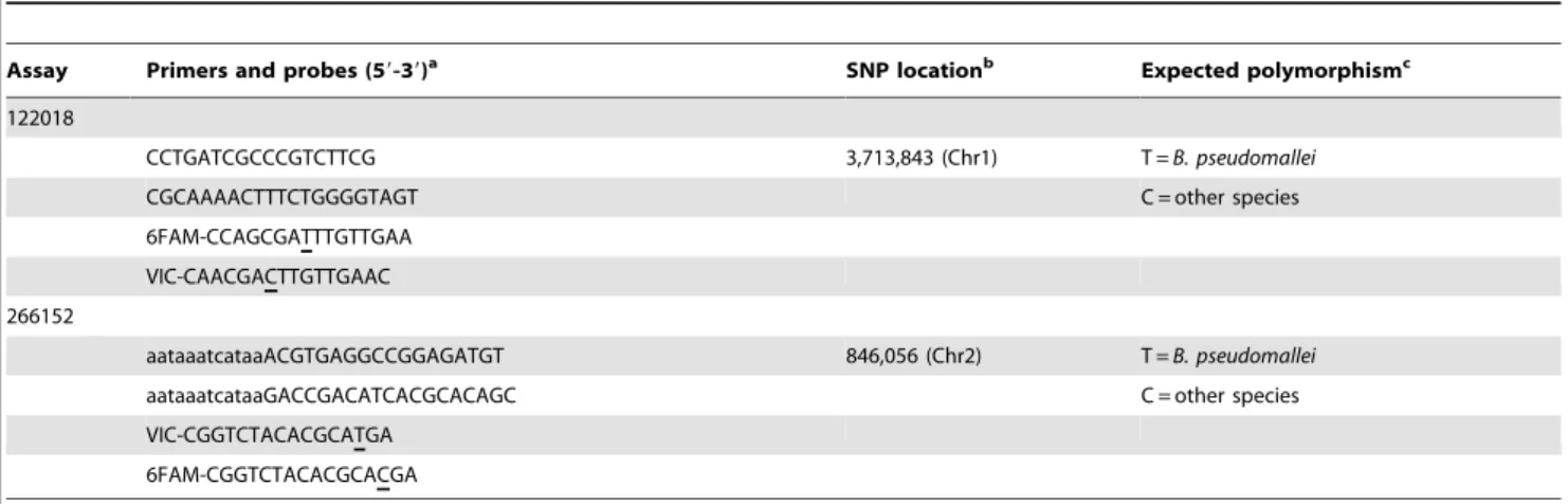
and B. thailandensis,B. oklahomensis and B. thailandensis-like species containing the alternate state. Other Burkholderiaceae possess additional SNPs or indels that would adversely affect binding of the B. pseudomallei-specific probe according to in silico analysis. Oncein silico B. pseudomallei specificity was determined, the SNP signatures were converted to TaqMan MGB probe format [36]. TaqMan probes and primers (Table 1) were designed using Primer Express v3.0 software (Applied Biosystems, Foster City CA). All primers and probes were subject to BLAST analysis to confirm specificity. PCRs were performed in 384-well optical plates using 16TaqMan Universal PCR Master Mix (Applied Biosystems), primers and probes, and molecular-grade H2O (Invitrogen). One
mL DNA template (equating to 2 ng, or 2.56105 genomic equivalents) was added per reaction to a final volume of 10mL. All reactions were carried out in dual-probe format and in duplicate using 2 ng DNA template (1 ng template was used for specificity screening), unless otherwise specified. For the 122018 assay, primer and probe concentrations were 0.3mM and 0.1mM, respectively, whereas the 266152 assay used concentrations at 0.3mM and 0.2mM, respectively. Thermocycling was conducted using default conditions (2 min at 50uC, 10 min at 95uC followed by 40 cycles of denaturation for 15 s at 95uC and annealing and extension for 1 min at 60uC) on a 7900HT Real-Time PCR System (Applied Biosystems).
Previously establishedB. pseudomallei-specific assays Genotyping calls for the 122018 and 266152 assays were compared with two established B. pseudomallei-specific real-time PCR assays, BurkDiff [27] and TTS1 [13], to determine the specificity of all four assays (see ‘Assay quality performance’ below for details). BurkDiff is a dual-probe TaqMan assay that differentiates B. pseudomallei and B. mallei; other Burkholderia spp. fail to amplify due to high levels of sequence diversity. We made a modification to the BurkDiff assay (0.2mM each primer/reaction rather than the 0.9mM previously reported) to improve assay efficiency. TTS1 is a single probe assay that detects the presence or absence ofB. pseudomallei; the gene targeted by this assay is absent in other Burkholderia spp. [13]. We performed TTS1 detection essentially as described elsewhere [41] but with the following alterations: we maintained the originally described primer and probe concentrations [13], and used default thermocycling
parameters on the AB7900HT instrument for consistency with the 122018 and 266152 assays.
Assay quality performance
To determine the suitability of our newB. pseudomallei-specific assays over a wide range of conditions, we tested the performance of the 122018 and 266152 assays across several criteria; accuracy, specificity, precision, selectivity, LoQ, LoD, linearity, ruggedness and robustness (Methods S1). We designed quality performance experiments based on standardized definitions of these parameters [34]. Two representative samples, B. pseudomallei 104 and B. thailandensis-like MSMB 43, were used to test parameters due to inherent differences in probe efficiencies between these different species. Species specificity for the 122018 and 266152 assays was determined by screening them across our entireBurkholderiaDNA collection, which comprises 2,205 Burkholderia spp. samples (Table 2), normalized to 1 ng/mL using the NanoDrop 8000 instrument. We also tested these assays across 127 common soil, water or clinically important prokaryotic and eukaryotic species to confirm specificity (Table S2). A 466 bp real-time SYBR Green 16 S PCR [42] was used to confirm DNA integrity in instances where no amplification with the B. pseudomallei assays was observed, including fungal and yeast species, which amplified (albeit less robustly, but above NTC signal) with this primer set, probably due to non-specific amplification of other rRNA regions. NTCs were included as cycles-to-threshold controls due to delayed but positive amplification using AmpliTaq Gold polymerase, which contains endogenousE. coli16 S RNA.
DNA Sequencing
When species determination discrepancies among the 122018, 266152, BurkDiff and TTS1 assays were observed, samples were subjected to multilocus sequence typing (MLST) [40] or 16 S sequencing [42]. Sequencing of the 266152 locus inB. pseudomallei
Bp5706 was carried out by amplifying a,400 bp fragment that encompassed the 68 bp PCR product generated by the 266152 TaqMan assay to examine primer- or probe-binding mutations. Big Dye v3.1 chemistry (Applied Biosystems) was used for cycle sequencing. Sequencing products were denatured in Hi-Di
Table 1.B. pseudomallei122018 and 266152 assays designed in this study.
Assay Primers and probes (59-39)a SNP locationb Expected polymorphismc
122018
CCTGATCGCCCGTCTTCG 3,713,843 (Chr1) T =B. pseudomallei
CGCAAAACTTTCTGGGGTAGT C = other species
6FAM-CCAGCGATTTGTTGAA
VIC-CAACGACTTGTTGAAC
266152
aataaatcataaACGTGAGGCCGGAGATGT 846,056 (Chr2) T =B. pseudomallei
aataaatcataaGACCGACATCACGCACAGC C = other species
VIC-CGGTCTACACGCATGA
6FAM-CGGTCTACACGCACGA
aUnderlined nucleotides indicate the position of the SNP in the TaqMan probe; lowercase nucleotides indicate a deliberately incorporated 59flap to enhance amplification efficiency [43].
bBased onB. pseudomalleiK96243 genome (GenBank Accession numbers BX571965 and BX571966 for chromosomes 1 and 2, respectively) [14].
c‘Other species’ refers specifically toB. thailandensis,B. oklahomensisandB. thailandensis-like spp.B. malleiand otherBurkholderiaspp. (e.g.B. vietnamiensis,B. ubonensis
formamide (Applied Biosystems) prior to electrophoresis on a 3130 or 3730xlDNA Analyzer (Applied Biosystems).
Results and Discussion
Accuracy of 122018 and 266152 assays
Accuracy is the measure of exactness of an analytical method, or the closeness of agreement between the measured value and the value that is accepted as a conventional true value or an accepted reference value [34], and is thus distinct from specificity and precision (see Methods S1 for definitions). We subjected the 122018 and 266152 SNP signatures to bothin silicoand laboratory screening to determine their accuracy towards B. pseudomallei. BLAST analysis was carried out at the 122018 and 266152 loci against all available B. pseudomallei, B. mallei, B. thailandensis, B. thailandensis-like,B. vietnamiensis, B. oklahomensis,B. ubonensisand B. cepaciagenomes, which confirmed that onlyB. pseudomalleistrains possessed theB. pseudomallei-specific allele at these loci. We then compared in silico and wet-bench genotypes using a panel of 13 whole genome-sequenced Burkholderia strains (Table S1). As predicted from in silico analysis, both assays amplified the B. pseudomallei-specific allele in allB. pseudomalleiDNA samples, with no detectable amplification in the fourB. malleisamples (results not shown). For 122018, both probes were specific to the appropriate species, with no cross-hybridization of the alternate probes (Figure 1). For 266152, B. oklahomensis, B. thailandensis and B. thailandensis-like species possessed some cross-hybridization with the B. pseudomallei probe but were distinguishable from B. pseudomallei due to preferential amplification of the non-B. pseudomalleiprobe (Figure 2).
Specificity of 122018, 266152, BurkDiff and TTS1 assays Following confirmation of assay accuracy, we screened DNA panels comprising 2,205Burkholderiaspp. (Table 2) and 127
non-Burkholderiaspecies (Figure S1; Table S2) with the 122018, 266152, BurkDiff and TTS1 assays to determine their specificity. BurkDiff and TTS1 have previously demonstrated specificity for B. pseudomallei across moderately large DNA panels [13,27]. As expected, the TTS1 assay showed excellent specificity for B. pseudomallei, although we detected one false-negativeB. pseudomallei
isolate, Bp1186 (original ID: SBCT-RF80-BP1, isolated from soil in Northeast Thailand). Further investigation of Bp1186 showed that it possessed a smaller genome than otherB. pseudomalleistrains, and lacked certain virulence loci, includingbimAand other TTS1 loci besidesorf2. These findings suggest that Bp1186 is probably unable to establish human infection (A. Tuanyok, unpublished data). Assay 266152 gave a single ambiguous call inB. pseudomallei
Bp5706 (original ID: MSHR 1559) (0.05% of total samples) in which both probes amplified at the same time, albeit poorly. DNA sequence analysis of this isolate uncovered a second SNP (T/C) 6 bp upstream of the targeted SNP (result not shown). This SNP was within the probe binding site and thus altered probe-binding efficiency. No false-positives were detected using the TTS1 or 266152 assays.
BurkDiff was the only assay we examined that yielded no false-positives or false-negatives across our DNA panels. In contrast, using 122018, we observed six positives (0.3%) and no false-negatives. None of the other B. pseudomallei-specific assays gave detectable amplification of these six samples, indicating incorrect species assignment by 122018. Sequencing for 16 S rDNA and MLST confirmed one isolate as B. vietnamiensis and five as B. ubonensis. Twenty-seven other B. ubonensis and one other B. vietnamiensis did not amplify with the 122018 assay, suggesting variable prevalence of a B. pseudomallei-like locus within these species. All non-Burkholderiaisolates were PCR-negative using the four assays.
Although BurkDiff provided the best speciation performance across our DNA panel, it was the most difficult assay to interpret due to heavy cross-hybridization between probes. Using pureB. pseudomallei templates, we observed a difference of CT (DCT) of
approximately 1, even with optimization measures employed for improving amplification efficiency (results not shown). Despite this very lowDCTwe did not encounter an inconsistent genotyping
call, indicating that this assay is robust in the presence of pure templates. Unlike BurkDiff, TTS1-positive genotypes were readily identifiable due to the single-probe format and amplification efficiency of this assay. However, one drawback of this single-probe format is that low-level cross contamination ofB. pseudomallei
DNA in non-B. pseudomalleitemplates can cause false-positive PCR results, and thus positive results must be interpreted with caution, especially when high CT values are obtained. The 122018 and
266152 assays in their dual-probe format were not influenced by low-level B. pseudomallei contamination in B. thailandensis, B. oklahomensis and B. thailandensis-like templates, although low-level contamination ofB. pseudomalleiDNA in e.g.B. ubonensissamples remains problematic. Coupled with large DCT values, the competitive dual-probe format enabled the most facile differenti-ation between B. pseudomallei and non-B. pseudomallei templates (Figures 1 and 2; Figures S2 and S3, Panel G).
Our results indicate that, with the exception of BurkDiff, no single genotyping method was 100% effective at speciating B. pseudomallei. Many promising molecular markers inBurkholderiaspp. are homoplastic [18,27]. Homoplastic markers may not be apparent when screening assays across relatively small (,1,000) isolate collections but can lead to false-positive and false-negative Table 2.Burkholderiaspp. DNA specificity panel used in this
study.
Burkholderiaspeciesa No. samples
B. pseudomallei 1,954
B. thailandensis 86
B. mallei 76
B. ubonensis 32
B. thailandensis-like/B. thailandensis/B. oklahomensisb 28
B.spp.c 13
B. cepacia 6
B. oklahomensis 4
B. vietnamiensis 2
B. thailandensis-like 2
B. cenocepacia 1
B. phytofirmans 1
Total 2,205
aAccording to genotyping results generated in the current study, 16 S
sequencing or MLST.
bPreviously identified as ‘Burkholderiaspp.’ and renamed
Burkholderia thailandensis/B. thailandensis-like/B. oklahomensisbased on their genotyping outcomes in this study. However, we could not accurately differentiate these three species as they all amplify the non-B. pseudomalleiprobe in the 122018 and 266152 assays. Neither TTS1 nor BurkDiff assays amplifyB. thailandensis,B. thailandensis-like orB. oklahomensisspecies, and thus could not be used for species assignment.
cRuled out as beingB. pseudomallei,B. thailandensis,B. thailandensis-like, B. oklahomensis,B. vietnamiensisandB. ubonensisbut strongly suspected to be Burkholderiaspp. due to their amplification usingB. vietnamiensisandB. ubonensis-specific assays (Price et al., unpublished data).
(as shown by our 122018 assay and the TTS1 assay) genotyping calls when assays are screened across larger and more diverse DNA panels. Additional mutations in probe- or primer-binding sites, such as observed with 266152, can cause aberrant genotyping results in otherwise promising species-specific signa-tures. In our study, we used a large amount of WGS data to minimize the probability of including homoplastic markers or SNPs with polymorphic flanking regions. As more WGS data for Burkholderiaceae are generated, this approach will continue to provide the most accurate speciation targets. Based on our findings, it is our recommendation that speciation ofB. pseudomallei
be based on at least two independent molecular markers, or a single molecular marker when latex agglutination testing is used, to ensure that false-negative and false-positive genotyping calls do not lead to erroneous species designations.
Selectivity
The potential for near-neighbor contamination of DNA is a concern in complex specimens, such as those of environmental, clinical or forensic origin. We therefore performed a selectivity experiment (Methods S1) on the 122018 and 266152 assays to quantitatively assess their ability to detect minor B. pseudomallei
components in the presence of near-neighbor DNA. We mixedB. pseudomallei and B. thailandensis-like templates in known ratios of 0:100, 10:90, 25:75, 50:50, 75:25, 90:10 and 100:0, respectively. Both assays amplified theB. pseudomallei-specific probe (i.e. pureB. pseudomalleitemplate) more efficiently than the alternate probe (i.e. pureB. thailandensis-like template) (Table S3; Figures 1 and 2). As expected, both alleles amplified when in the presence of mixed template (Table S3; Figures S2 and S3).
For the 122018 assay, all mixtures of bothB. pseudomalleiandB. thailandensis-like templates could be reliably distinguished from pure template at the lowest tested limit of selectivity (10%), and the 266152 assay was able to discriminate between pureB. pseudomallei
andB. thailandensis-like template present at 10%. However, theB. thailandensis-like:B. pseudomalleimixtures at 50:50, 25:75 and 10:90 ratios were indistinguishable from pure B. pseudomallei template
using the 266152 assay (Figures S2 and S3; Table S3), indicating that the 266152 assay is insensitive to detectingB. thailandensis-like template when in the presence of 50% or greaterB. pseudomallei
template. Given the primary focus onB. pseudomalleiin our study, we do not consider this result a failure in selectivity as we demonstrated that both assays yielded a significant difference (DCT s.2) between pure B. pseudomallei template and B. pseudomallei containing B. thailandensis-like template at a minor component of#10%.
Our experiments outline a rudimentary protocol for determin-ing selectivity usdetermin-ing TaqMan assays. Although beyond the scope of the current study, future studies should ideally examine lower minor component mixtures below 10% to determine the limit of selectivity for the 122018 and 266152 assays. Selectivity experi-ments using spiked environmental or clinical specimens, such as soil or sputum samples, would shed further light on the true selectivity performance of the 266152 and 122018 assays in the presence of PCR inhibitors or complex DNA constituents. Use of a single-probe approach may provide better detection of minor components than dual-probe format, although we do not recommend using the 266152 assay in a non-competitive format due to cross-hybridization of the probes, which may result in false-positive results.
Limits of quantitation and detection (LoQ and LoD) We calculated the lower LoQ and LoD (Tables S4 and S5) using pure DNA template for B. pseudomallei and B. thailandensis-like species. The LoQ was defined as the lowest level of DNA detected that provided an acceptable level of precision (i.e. 8/8 replicates amplified with a CTstandard deviation (s),0.8 from the mean
CT), whereas LoD was measured as the concentration of analyte
that gave rise to a signal significantly different from the negative control (i.e. at least 2/8 replicates amplified, irrespective ofs) [34]. We were not able to establish the upper LoD or LoQ as these values were not reached using our highest DNA amount of 40 ng/ PCR. For the 122018 assay, the lower LoQ was$461025ng ($40 fg, or 5 genomic equivalents (GEs)) and $400 fg (50 GEs)
Figure 1. 122018 TaqMan dual probe assay.Red, theB. pseudomallei-specific TaqMan probe amplifies onlyB. pseudomalleitemplate; the
non-B.pseudomalleiTaqMan probe (green) amplifies well withB. thailandensisandB. thailandensis-like species and weakly withB. oklahomensistemplates but notB. pseudomallei. OtherBurkholderiaspp. do not amplify with either probe.
for B. pseudomalleiand B. thailandensis-like templates, respectively, whereas the 266152 assay yielded LoQ values at $4 fg (,0.5 GEs) and $4 ng (56105GEs), respectively. The poor LoQ value forB. thailandensis-like template using the 266152 assay was surprising given that all eight replicates amplified at$400 fg (50 GEs). Using our LoQ criteria, this assay is unreliable for quantitating B. thailandensis-like DNA. The LoD of the 122018 assay was$4 fg (,0.5 GEs) and$40 fg (5 GEs) forB. pseudomallei
and B. thailandensis-like templates, respectively, and $4 fg total DNA (,0.5 GEs) for both templates using the 266152 assay (Table S5). These results contrast with LoD values previously reported for the 266152 assay [35], which demonstrated a LoD of 10 GEs (see ‘Linearity’ below for further discussion on the 266152 results).
Although it is difficult to compare LoD values between studies due to experimental design differences (e.g. number of replicates
tested, or differences in mastermix constituents, DNA quantita-tion, instruments, or thermal conditions), TaqMan probes theoretically have the capacity to reach 0.5 GEs (the equivalent of a single PCR target) in a well-designed assay. The TTS1 assay reportedly provides a LoD of 76 fg, or 10 GEs, in PCR and 122 fg (16 GEs) in spiked human blood [13], and the BurkDiff assay provides a LoD of 10 GEs in PCR [27]. Therefore, the LoD of our dual-probe assays are similar in performance to TTS1 and BurkDiff, particularly in the presence ofB. pseudomallei template. As expected, the LoD and LoQ of theB. thailandensis-like template were less sensitive. Although not tested in the current study, the LoD and LoQ of near-neighbor templates, such asB.
thailandensis-like species MSMB 43, could potentially be increased by using the non-B. pseudomalleiprobe by itself to avoid competition issues with theB. pseudomallei-specific probe.
Figure 2. 266152 TaqMan dual probe assay.Green, theB. pseudomallei-specific TaqMan probe preferentially amplifiesB. pseudomalleitemplate;
the non-B.pseudomalleiTaqMan probe (red) amplifies well inB. thailandensis-like species and weakly inB. thailandensisandB. oklahomensis. Other
Linearity
We tested linearity of the 122018 and 266152 assays under controlled conditions (see Methods S1 for details) to establish the range of DNA amounts that enable accurate quantification [34]. Such information is valuable for quantifying the concentration of uncharacterized samples, or for determining the lowest concen-tration at which reliable genotyping data can be attained, particularly when DNA is limiting.
We did not reach the upper limit of linearity using the highest concentration of 40 ng DNA for either B. pseudomallei or B. thailandensis-like templates, indicating that the range of linearity for our assays is close to or greater than this amount.B. thailandensis-like template linearity lacked precision across DNA concentrations, particularly for assay 266152 (Table S6), indicating that the 266152 assay should not be used to quantifyB. thailandensis-like DNA. The lower-limit of linearity for B. pseudomallei, based on 100% amplification across eight replicates ands,0.8 between replicates, was 40 fg (5 GEs) and 4 fg (,0.5 GEs) for the 122018 and 266152 assays, respectively. In other words, our data indicate that B. pseudomalleitemplate can be accurately quantified down to 0.5 GEs (as also observed in ‘LoQ’ above), which is equivalent to a single PCR target, the theoretical limit of PCR detection. We have also determined the LoD of the 266152 assay as 10 GEs [35]; however, this value was based on 95% amplification success across 64 replicates, whereas the current study used only 25% amplification success across eight replicates and was thus less stringent. DNA quantitation varied between these two studies (NanoDrop spectro-photometric quantitation vs. normalization against a 16 S rDNA target), and it is possible that minor variations in DNA quantitation or dilution preparation influenced our quantitation results. Never-theless, we have demonstrated that these dual-probe assays possess a large linearity range in the presence of pureB. pseudomalleitemplates and can thus be used to precisely quantify unknown samples across a wide range of DNA concentrations (Figure S4 and Table S6).
Determining linearity also allowed us to calculate the PCR efficiency of the 122018 and 266152 assays. For the 122018 assay, PCR efficiency was 91% and 89% for B. pseudomallei and B. thailandensis-like templates, respectively. The efficiency of the 266152 assay was higher, at 97% and 94% for B. pseudomallei
and B. thailandensis-like templates, respectively (Table S6). In contrast, the efficiency of the TTS1 assay has been reported at 99% forB. pseudomallei[13]. However, the linear range used in this study was much more restrictive than ours due to the smaller number of DNA concentrations included in the linear dynamic range, so comparison of PCR efficiencies between studies must be prudently interpreted. It remains to be determined whether PCR efficiency for the 122018 and 266152 assays could be improved by using single-probe format, as is used in the TTS1 assay.
Robustness and Ruggedness
Robustness and ruggedness are oft-neglected aspects of assay performance, despite their inter-laboratory importance. We therefore assessed these criteria for the 122018 and 266152 assays using multiple AB7900HT instruments (ruggedness), and TaqMan probe and Universal Master Mix (Applied Biosystems) reagent lots, types of commercial mastermixes (Universal and Genotyping Master Mixes; Applied Biosystems) and annealing/extension temperatures (robustness) to determine those features most critical to inter-laboratory assay transfer.
Comparison of commercial mastermixes with the 266152 and 122018 assays yielded unexpected results. The TaqMan Geno-typing Master Mix (Applied Biosystems) showed poor amplifica-tion with the 122018 and 266152 assays (results not shown), despite its purported suitability for SNP genotyping applications
(http://products.invitrogen.com/ivgn/product/4371355). Due to the proprietary nature of most commercial mastermixes, we were unable to establish the component difference between the Genotyping and Universal Master Mixes. However, the BurkDiff assay does not amplify using the TaqMan Universal Master Mix (results not shown), suggesting that neither mix is ideal for all SNP genotyping applications. In contrast, we did not observe differences in precision or accuracy of amplification using three different AB7900HT instruments, nor did we identify perfor-mance differences among two probe and four TaqMan Universal Master Mix reagent lots (results not shown), suggesting that our TaqMan assays can be reliably reproduced on this platform using our reaction conditions. We did not test the assays on different real-time PCR platforms. Thus, further studies are required to determine their suitability on different real-time PCR instruments and across other commercial mastermixes.
Although our TaqMan probes were specifically designed with an optimal annealing/extension temperature of 60uC, inter-laboratory differences in thermal block temperatures can potentially influence genotyping calls. We therefore tested the robustness of the 122018 and 266152 assays across multiple annealing/extension temperatures and observed their effect on accurately and precisely calling correct genotypes. Overall, annealing/extension temperatures at 57.5 and 62.5uC still amplified templates and gave correct genotyping calls, although we did observe differences in robustness at these altered temperatures, particularly when theB. thailandensis-like template was assessed (Table S7). For the 122018 assay, the 57.5uC annealing/ extension temperature was comparable in performance to 60uC, whereas 62.5uC exhibited more amplification failures than at the lower annealing/extension temperatures. In contrast, the 62.5uC and 60uC annealing/extension temperatures for assay 266152 were comparable in performance, whereas 57.5uC replicates gave poorer replicate success, especially in the presence ofB. thailandensis-like template. The DCTvalues at 57.5uC and 62.5uC were smaller than those at 60uC,
demonstrating that 60uC is indeed the optimal temperature (Table S7) although they did not result in an increase in cross-hybridization. Importantly, no erroneous genotypes were observed at either the reduced or elevated temperature for either assay, indicating that these assays are tolerant to thermal block variations, but deviations from 60uC will result in a reduction in assay robustness and thus assays should ideally be optimized on each individual instrument.
Conclusions
B. pseudomallei is an important pathogen from a clinical, environmental and forensic stance. Correct identification of B. pseudomalleirequires molecular characterization due to shortcomings with phenotypic speciation techniques. Identification and quantifica-tion ofB. pseudomalleifrom pure cultures through to complex soil or sputum samples is dependent on a thorough understanding of the limits of species-specific assays. Despite the plethora ofB. pseudomallei
As these four assays become more widely used, we have no doubt that false-positives and false-negatives will be encountered; however, our work suggests that this will be a rare occurrence and false conclusions will be further minimized by including more than one of these assays for speciation. Additional specificity testing of TTS1, BurkDiff, 122018 and 266152 assays on different
Burkholderiaspecies is important as such strains will provide more informative false-positive rates due to their genetic relatedness to
B. pseudomallei. Likewise, further testing of B. pseudomalleiisolates using these four assays will increase the accuracy of false-negative rates. Our assays demonstrated comparable LoD and LoQ performance to the current ‘gold standard’B. pseudomallei typing techniques. Although assay parameters like ruggedness, robustness and selectivity are not typically examined when developing and validating molecular assays, we anticipate that our methods will provide a framework for future studies where quantitative measures of comparative assay performance are paramount. Last, accurate standardization of input DNA is a crucial component of assay performance yet is difficult for complex environmental or clinical specimens whereB. pseudomalleiis usually isolated, due to the non-homogeneous nature of these samples, the presence of PCR inhibitors or the abundance of non-BurkholderiaDNA. While the tests of assay performance included here measure the effects of many potential variables, this list is not comprehensive. As such, users should be aware that other untested factors that might be encountered when samples are extracted from complex environ-ments may have an impact on assay performance.
Supporting Information
Methods S1 Supplemental materials and methods. (DOC)
Figure S1 266152 assay specificity results against non- Burk-holderia yeast, fungal and bacterial species and theB. pseudomallei
sample, 104 (see Table S2 for the list of organisms). Only theB. pseudomalleisample, 104, amplified with this assay (red and green amplification curves). All samples were run in duplicate. TTS1 and 122018 assays performed identically to 266152, with no amplification in non-Burkholderiaspecies (results not shown). (DOC)
Figure S2 Selectivity performance of the 122018 assay using varying mixtures ofBurkholderia pseudomallei104 (Bp; green) andB. thailandensis-like MSMB 43 (Bh; red) DNA. A, 0:100 Bp:Bh; B, 10:90 Bp:Bh; C, 25:75 Bp:Bh, D, 50:50 Bp:Bh, E, 75:25 Bp:Bh; F, 90:10 Bp:Bh, G, 100:0 Bp:Bh. All mixture ratios could be differentiated from pure Bp or Bh template (see Table S3 for details).
(DOC)
Figure S3 Selectivity performance of the 266152 assay using varying mixtures ofBurkholderia pseudomallei104 (Bp; green) andB. thailandensis-like MSMB 43 (Bh; red) DNA. A, 0:100 Bp:Bh; B, 10:90 Bp:Bh; C, 25:75 Bp:Bh, D, 50:50 Bp:Bh, E, 75:25 Bp:Bh; F, 90:10 Bp:Bh, G, 100:0 Bp:Bh. Note that in D, E and F, the standard deviation (s) between curves falls below our accepted threshold (see Table S3 for details), indicating that DNA mixtures containing up to 50% Bh DNA cannot reliably be differentiated from pure Bp template (G) with this assay.
(DOC)
Figure S4 Range of linearity for B. pseudomallei 122018 and 266152 TaqMan real-time PCR assays.
(DOC)
Table S1 Determination of 122018 and 266152 assay accuracy by comparison of in silico and TaqMan real-time PCR ‘‘wet-bench’’ single-nucleotide polymorphism results.
(DOC)
Table S2 Non-Burkholderiaspecies of clinical, environmental or forensic importance tested against the TTS1, 122018 and 266152 TaqMan assays for specificity (also see Figure S1). BurkDiff was not tested with this panel as it has been previously screened against a similar panel of 390 non-Burkholderiaspecies [27]. Bacteria, no shading; fungi, light grey; yeasts, dark grey.
(DOC)
Table S3 Selectivity results for B. pseudomallei 122018 and 266152 assays.
(DOC)
Table S4 Limit of Quantitation (LoQ) for the 122018 and 266152 assays.
(DOC)
Table S5 Limit of Detection (LoD) for the 122018 and 266152 assays.
(DOC)
Table S6 Range of linearity for B. pseudomallei 122018 and 266152 TaqMan assays.
(DOC)
Table S7 Robustness summary for 122018 and 266152 Taq-Man assays.
(DOC)
Acknowledgments
We thank Shalamar Georgia, Lindsey Watson, Alex Von Schulze, Travis Mullins, Heidie O’Neill, Christopher Allender and Stephanie Warrington (Northern Arizona University) for assistance with extracting, organizing or arraying DNA samples used for this study. Amy Vogler, Dawn Birdsell, Judy Lee, Christina Allen, Jodi Beaudry and James Schupp (Northern Arizona University) designed similar quality control parameters for other bacterial species that were helpful in the design of our Burkholderia
experiments. John Gillece (Translational Genomics Research Institute) produced some of the whole genome sequence data used in this study. John Pemberton (University of Queensland) kindly donatedRalstonia eutrophaand
Pseudomonas stutzeristrains for our non-Burkholderiaspecificity panels. Lastly, we gratefully acknowledge the contribution of otherBurkholderia pseudomallei
researchers (Don Woods, David De Shazer, Mohammad Saqib, Naureen Abeera and Steven P. Harvey), many of whom providedBurkholderiaDNA and isolates that populate our collections.
Author Contributions
Conceived and designed the experiments: EPP JLD JMC SMB-S JTF DMW PK TP. Performed the experiments: EPP JLD JMC MLS JLG ELK SMB-S MM MK MBG JEG VW JMW AB PT AT DL SJP BJC. Analyzed the data: EPP JLD JMC DSS MLS MK DMW PK TP. Contributed reagents/materials/analysis tools: EPP JMC DSS JLG SMB-S MM MBG JEG VW JMW AB JTF PT AT DL SJP BJC PK. Wrote the paper: EPP JLD DSS TP.
References
1. Coenye T, Vandamme P (2003) Diversity and significance ofBurkholderiaspecies occupying diverse ecological niches. Environ Microbiol 5: 719–729.
3. Glass MB, Gee JE, Steigerwalt AG, Cavuoti D, Barton T, et al. (2006) Pneumonia and septicemia caused by Burkholderia thailandensisin the United States. J Clin Microbiol 44: 4601–4604.
4. Glass MB, Steigerwalt AG, Jordan JG, Wilkins PP, Gee JE (2006)Burkholderia oklahomensissp. nov., aBurkholderia pseudomallei-like species formerly known as the Oklahoma strain of Pseudomonas pseudomallei. Int J Syst Evol Microbiol 56: 2171–2176.
5. Lieberman TD, Michel JB, Aingaran M, Potter-Bynoe G, Roux D, et al. (2011) Parallel bacterial evolution within multiple patients identifies candidate pathogenicity genes. Nat Genet 43: 1275–1280.
6. Suputtamongkol Y, Hall AJ, Dance DA, Chaowagul W, Rajchanuvong A, et al. (1994) The epidemiology of melioidosis in Ubon Ratchatani, northeast Thailand. Int J Epidemiol 23: 1082–1090.
7. Currie B (1993) Medicine in tropical Australia. Med J Aust 158: 609, 612–605. 8. Chantratita N, Wuthiekanun V, Boonbumrung K, Tiyawisutsri R, Vesaratchavest M, et al. (2007) Biological relevance of colony morphology and phenotypic switching byBurkholderia pseudomallei. J Bacteriol 189: 807–817. 9. Wiersinga WJ, van der Poll T, White NJ, Day NP, Peacock SJ (2006) Melioidosis: insights into the pathogenicity ofBurkholderia pseudomallei. Nat Rev Microbiol 4: 272–282.
10. Dance DA (2000) Ecology of Burkholderia pseudomallei and the interactions between environmentalBurkholderiaspp. and human-animal hosts. Acta Trop 74: 159–168.
11. Levy A, Merritt AJ, Aravena-Roman M, Hodge MM, Inglis TJ (2008) Expanded range ofBurkholderiaspecies in Australia. Am J Trop Med Hyg 78: 599–604. 12. Amornchai P, Chierakul W, Wuthiekanun V, Mahakhunkijcharoen Y,
Phetsouvanh R, et al. (2007) Accuracy ofBurkholderia pseudomalleiidentification using the API 20NE system and a latex agglutination test. J Clin Microbiol 45: 3774–3776.
13. Novak RT, Glass MB, Gee JE, Gal D, Mayo MJ, et al. (2006) Development and evaluation of a real-time PCR assay targeting the type III secretion system of Burkholderia pseudomallei. J Clin Microbiol 44: 85–90.
14. Holden MT, Titball RW, Peacock SJ, Cerdeno-Tarraga AM, Atkins T, et al. (2004) Genomic plasticity of the causative agent of melioidosis, Burkholderia pseudomallei. Proc Natl Acad Sci U S A 101: 14240–14245.
15. U’Ren JM, Schupp JM, Pearson T, Hornstra H, Friedman CL, et al. (2007) Tandem repeat regions within theBurkholderia pseudomalleigenome and their application for high resolution genotyping. BMC Microbiol 7: 23.
16. Tu mapa S, Holden MT, V es aratch avest M , W uthiekanun V, Limmathurotsakul D, et al. (2008) Burkholderia pseudomallei genome plasticity associated with genomic island variation. BMC Genomics 9: 190.
17. Tuanyok A, Leadem BR, Auerbach RK, Sternberg SM, Beckstrom-Sternberg JS, et al. (2008) Genomic islands from five strains ofBurkholderia pseudomallei. BMC Genomics 9: 566.
18. Pearson T, Giffard P, Beckstrom-Sternberg S, Auerbach R, Hornstra H, et al. (2009) Phylogeographic reconstruction of a bacterial species with high levels of lateral gene transfer. BMC Biol 7: 78.
19. Thibault FM, Valade E, Vidal DR (2004) Identification and discrimination of Burkholderia pseudomallei,B. mallei, andB. thailandensisby real-time PCR targeting type III secretion system genes. J Clin Microbiol 42: 5871–5874.
20. Supaprom C, Wang D, Leelayuwat C, Thaewpia W, Susaengrat W, et al. (2007) Development of real-time PCR assays and evaluation of their potential use for rapid detection ofBurkholderia pseudomalleiin clinical blood specimens. J Clin Microbiol 45: 2894–2901.
21. Dharakul T, Songsivilai S, Viriyachitra S, Luangwedchakarn V, Tassaneetritap B, et al. (1996) Detection ofBurkholderia pseudomalleiDNA in patients with septicemic melioidosis. J Clin Microbiol 34: 609–614. 22. Gal D, Mayo M, Spencer E, Cheng AC, Currie BJ (2005) Short report:
application of a polymerase chain reaction to detectBurkholderia pseudomalleiin clinical specimens from patients with suspected melioidosis. Am J Trop Med Hyg 73: 1162–1164.
23. Meumann EM, Novak RT, Gal D, Kaestli ME, Mayo M, et al. (2006) Clinical evaluation of a type III secretion system real-time PCR assay for diagnosing melioidosis. J Clin Microbiol 44: 3028–3030.
24. U’Ren JM, Van Ert MN, Schupp JM, Easterday WR, Simonson TS, et al. (2005) Use of a real-time PCR TaqMan assay for rapid identification and differentiation ofBurkholderia pseudomalleiandBurkholderia mallei. J Clin Microbiol 43: 5771–5774.
25. Tomaso H, Pitt TL, Landt O, Al Dahouk S, Scholz HC, et al. (2005) Rapid presumptive identification ofBurkholderia pseudomalleiwith real-time PCR assays using fluorescent hybridization probes. Mol Cell Probes 19: 9–20.
26. Trung TT, Hetzer A, Gohler A, Topfstedt E, Wuthiekanun V, et al. (2011) Highly sensitive direct detection and quantification ofBurkholderia pseudomallei bacteria in environmental soil samples by using real-time PCR. Appl Environ Microbiol 77: 6486–6494.
27. Bowers JR, Engelthaler DM, Ginther JL, Pearson T, Peacock SJ, et al. (2010) BurkDiff: a real-time PCR allelic discrimination assay forBurkholderia pseudomallei andB. mallei. PLoS One 5: e15413.
28. Kaestli M, Mayo M, Harrington G, Ward L, Watt F, et al. (2009) Landscape changes influence the occurrence of the melioidosis bacterium Burkholderia pseudomalleiin soil in northern Australia. PLoS Negl Trop Dis 3: e364. 29. Kaestli M, Mayo M, Harrington G, Watt F, Hill J, et al. (2007) Sensitive and
specific molecular detection ofBurkholderia pseudomallei, the causative agent of melioidosis, in the soil of tropical northern Australia. Appl Environ Microbiol 73: 6891–6897.
30. Gee JE, Glass MB, Novak RT, Gal D, Mayo MJ, et al. (2008) Recovery of a Burkholderia thailandensis-like isolate from an Australian water source. BMC Microbiol 8: 54.
31. Glass MB, Beesley CA, Wilkins PP, Hoffmaster AR (2009) Comparison of four selective media for the isolation ofBurkholderia malleiandBurkholderia pseudomallei. Am J Trop Med Hyg 80: 1023–1028.
32. Marshall K, Shakya S, Greenhill AR, Padill G, Baker A, et al. (2010) Antibiosis ofBurkholderia ubonensisagainistBurkholderia pseudomallei, the causative agent for melioidosis. Southeast Asian J Trop Med Public Health 41: 904–912. 33. Yabuuchi E, Kosako Y, Arakawa M, Hotta H, Yano I (1992) Identification of
Oklahoma isolate as a strain ofPseudomonas pseudomallei. Microbiol Immunol 36: 1239–1249.
34. Araujo P (2009) Key aspects of analytical method validation and linearity evaluation. J Chromatogr B Analyt Technol Biomed Life Sci 877: 2224–2234. 35. Kaestli M, Richardson LJ, Colman RE, Tuanyok A, Price EP, et al. (2012) Comparison of TaqMan PCR Assays for Detection of the Melioidosis Agent Burkholderia pseudomalleiin Clinical Specimens. J Clin Microbiol: EPub ahead of print.
36. Livak KJ (1999) Allelic discrimination using fluorogenic probes and the 59
nuclease assay. Genet Anal 14: 143–149.
37. Ulrich RL, DeShazer D (2004) Type III secretion: a virulence factor delivery system essential for the pathogenicity ofBurkholderia mallei. Infect Immun 72: 1150–1154.
38. Currie BJ, Gal D, Mayo M, Ward L, Godoy D, et al. (2007) Using BOX-PCR to exclude a clonal outbreak of melioidosis. BMC Infect Dis 7: 68.
39. Altschul SF, Gish W, Miller W, Myers EW, Lipman DJ (1990) Basic local alignment search tool. J Mol Biol 215: 403–410.
40. Godoy D, Randle G, Simpson AJ, Aanensen DM, Pitt TL, et al. (2003) Multilocus sequence typing and evolutionary relationships among the causative agents of melioidosis and glanders,Burkholderia pseudomalleiandBurkholderia mallei. J Clin Microbiol 41: 2068–2079.
41. Engelthaler DM, Bowers J, Schupp JA, Pearson T, Ginther J, et al. (2011) Molecular investigations of a locally acquired case of melioidosis in Southern AZ, USA. PLoS Negl Trop Dis 5: e1347.
42. Nadkarni MA, Martin FE, Jacques NA, Hunter N (2002) Determination of bacterial load by real-time PCR using a broad-range (universal) probe and primers set. Microbiology 148: 257–266.